

A new bioinspired collagen-hydroxyapatite bone graft substitute in adult scoliosis surgery: results at 3-year follow-up
- PMID: 28604992
- PMCID: PMC6379889
- DOI: 10.5301/jabfm.5000366
A new bioinspired collagen-hydroxyapatite bone graft substitute in adult scoliosis surgery: results at 3-year follow-up
Abstract
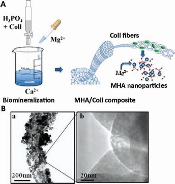
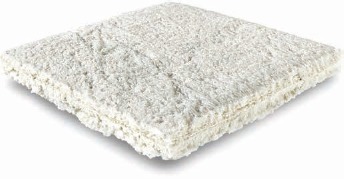
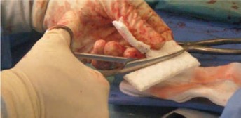
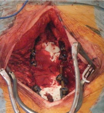
Background: Spinal fusion is a common procedure used for surgical treatment of spinal deformity. In recent years, many bone graft substitutes (BGS) have been developed to provide good arthrodesis when the available autologous bone harvested from the patient is not enough. The aim of this study was to analyze the use of a new-generation composite material (RegenOss) made of Mg-hydroxyapatite nanoparticles nucleated on type I collagen to obtain long posterolateral fusion in adult scoliosis surgery.
Methods: A total of 41 patients who underwent spinal fusion for the treatment of adult scoliosis were retrospectively analyzed. According to Lenke classification, visual analog scale (VAS) score and Oswestry Disability Index (ODI) score, radiographic rates of bone union were evaluated before surgery and at 6, 12 and 36 months of follow-up. Fusion was considered to be successful when criteria for Lenke grade A or B were satisfied. Patient-related risk factors were considered for the evaluation of the final outcome.
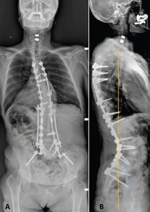
Results: At 36-month follow-up, radiographic evidence of spinal fusion was present in the majority of patients (95.1%). A time-dependent statistically significant improvement was evidenced after surgery for all clinical outcomes evaluated. Based on the demographic data collected, there were no statistically significant factors determining fusion. The correction of deformity was maintained at different time points. No intraoperative or postoperative complications were recorded.
Conclusions: The present study demonstrated that RegenOss can safely be used to achieve good arthrodesis when associated with autologous bone graft to obtain long spinal fusion in the treatment of adult scoliosis.
Conflict of interest statement
Conflict of interest: None.
Figures
Similar articles
-
Clinical outcomes and fusion rates following anterior lumbar interbody fusion with bone graft substitute i-FACTOR, an anorganic bone matrix/P-15 composite.J Neurosurg Spine. 2014 Dec;21(6):867-76. doi: 10.3171/2014.9.SPINE131151. Epub 2014 Oct 17. J Neurosurg Spine. 2014. PMID: 25325176
-
Healos graft carrier with bone marrow aspirate instead of allograft as adjunct to local autograft for posterolateral fusion in degenerative lumbar scoliosis: a minimum 2-year follow-up study.J Neurosurg Spine. 2010 Aug;13(2):211-5. doi: 10.3171/2010.3.SPINE09603. J Neurosurg Spine. 2010. PMID: 20672956 Clinical Trial.
-
Natural hydroxyapatite as a bone graft extender for posterolateral spine arthrodesis.Int Orthop. 2016 Sep;40(9):1875-82. doi: 10.1007/s00264-016-3140-4. Epub 2016 Mar 10. Int Orthop. 2016. PMID: 26961192
-
[Bone substitutes in scoliosis surgery].Orthopade. 2009 Feb;38(2):181-8. doi: 10.1007/s00132-008-1369-3. Orthopade. 2009. PMID: 19093096 Review. German.
-
Efficacy of using autologous cells with graft substitutes for spinal fusion surgery: A systematic review and meta-analysis of clinical outcomes and imaging features.JOR Spine. 2024 Jun 28;7(3):e1347. doi: 10.1002/jsp2.1347. eCollection 2024 Sep. JOR Spine. 2024. PMID: 38947860 Free PMC article. Review.
Cited by
-
Innovative Options for Bone Metastasis Treatment: An Extensive Analysis on Biomaterials-Based Strategies for Orthopedic Surgeons.Front Bioeng Biotechnol. 2020 Oct 6;8:589964. doi: 10.3389/fbioe.2020.589964. eCollection 2020. Front Bioeng Biotechnol. 2020. PMID: 33123519 Free PMC article. Review.
-
Differences in osteogenic induction of human mesenchymal stem cells between a tailored 3D hybrid scaffold and a 2D standard culture.J Mater Sci Mater Med. 2019 Dec 4;30(12):136. doi: 10.1007/s10856-019-6346-3. J Mater Sci Mater Med. 2019. PMID: 31802234
-
3D Bone Biomimetic Scaffolds for Basic and Translational Studies with Mesenchymal Stem Cells.Int J Mol Sci. 2018 Oct 13;19(10):3150. doi: 10.3390/ijms19103150. Int J Mol Sci. 2018. PMID: 30322134 Free PMC article. Review.
-
An Observational Prospective Clinical Study for the Evaluation of a Collagen-Hydroxyapatite Composite Scaffold in Hip Revision Surgery.J Clin Med. 2022 Oct 28;11(21):6372. doi: 10.3390/jcm11216372. J Clin Med. 2022. PMID: 36362601 Free PMC article.
-
Overcoming the Design Challenge in 3D Biomimetic Hybrid Scaffolds for Bone and Osteochondral Regeneration by Factorial Design.Front Bioeng Biotechnol. 2020 Jul 7;8:743. doi: 10.3389/fbioe.2020.00743. eCollection 2020. Front Bioeng Biotechnol. 2020. PMID: 32775321 Free PMC article.
References
-
- Aebi M The adult scoliosis. Eur Spine J 200514(10)925–948. - PubMed
-
- Berjano P Lamartina C Classification of degenerative segment disease in adults with deformity of the lumbar or thoracolumbar spine. Eur Spine J 201423(9)1815–1824. - PubMed
-
- Park JJ Hershman SH Kim YH Updates in the use of bone grafts in the lumbar spine. Bull Hosp Jt Dis 201371(1)39–48. - PubMed
-
- Dimar JR II Glassman SD Burkus JK Pryor PW Hardacker JW Carreon LY Two-year fusion and clinical outcomes in 224 patients treated with a single-level instrumented posterolateral fusion with iliac crest bone graft. Spine J 20099(11)880–885. - PubMed
-
- Gruskay JA Basques BA Bohl DD Webb ML Grauer JN Short-term adverse events, length of stay, and readmission after iliac crest bone graft for spinal fusion. Spine 201439(20)1718–1724. - PubMed
MeSH terms
Substances
LinkOut - more resources
Full Text Sources
Other Literature Sources
Medical
