. 2018 Aug;38(8):1354-1370.
doi: 10.1177/0271678X17713105. Epub 2017 Jun 13.

Dimethyl fumarate improves white matter function following severe hypoperfusion: Involvement of microglia/macrophages and inflammatory mediators

Affiliations

Dimethyl fumarate improves white matter function following severe hypoperfusion: Involvement of microglia/macrophages and inflammatory mediators

Jill H Fowler et al. J Cereb Blood Flow Metab. 2018 Aug.

Abstract

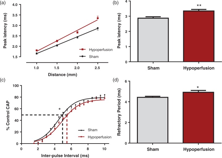
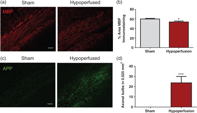
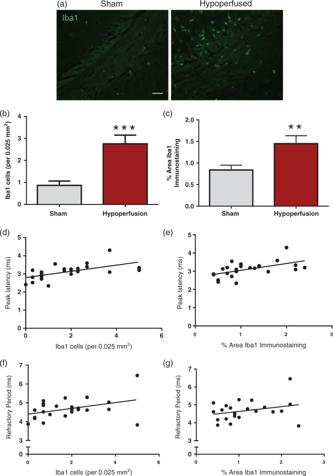
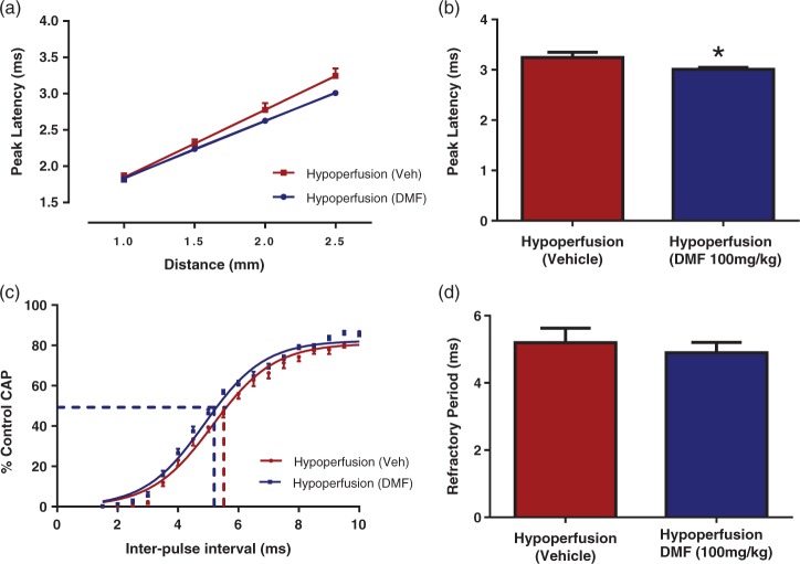
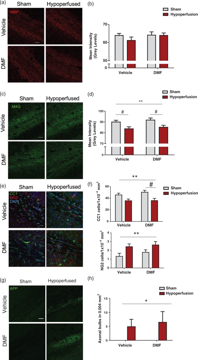
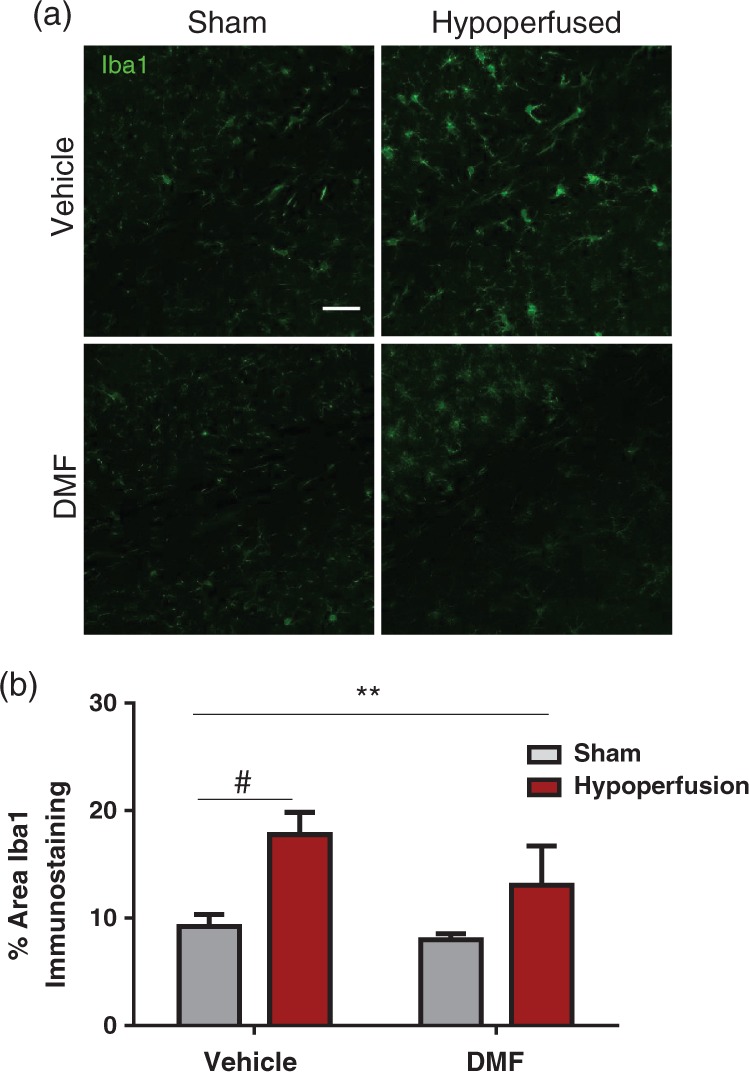
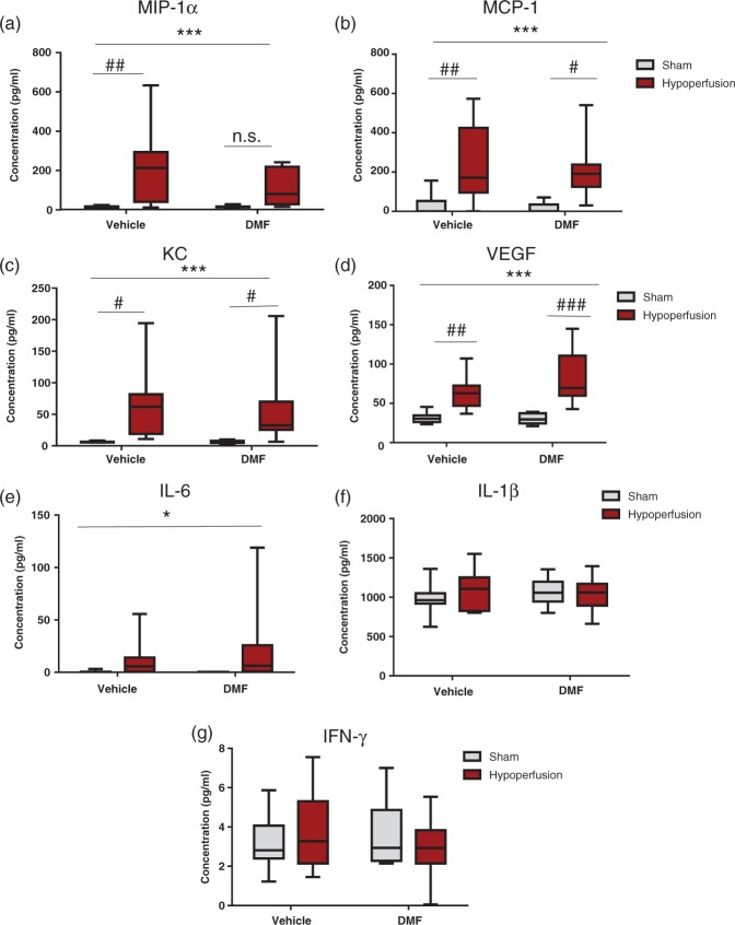
The brain's white matter is highly vulnerable to reductions in cerebral blood flow via mechanisms that may involve elevated microgliosis and pro-inflammatory pathways. In the present study, the effects of severe cerebral hypoperfusion were investigated on white matter function and inflammation. Male C57Bl/6J mice underwent bilateral common carotid artery stenosis and white matter function was assessed at seven days with electrophysiology in response to evoked compound action potentials (CAPs) in the corpus callosum. The peak latency of CAPs and axonal refractoriness was increased following hypoperfusion, indicating a marked functional impairment in white matter, which was paralleled by axonal and myelin pathology and increased density and numbers of microglia/macrophages. The functional impairment in peak latency was significantly correlated with increased microglia/macrophages. Dimethyl fumarate (DMF; 100 mg/kg), a drug with anti-inflammatory properties, was found to reduce peak latency but not axonal refractoriness. DMF had no effect on hypoperfusion-induced axonal and myelin pathology. The density of microglia/macrophages was significantly increased in vehicle-treated hypoperfused mice, whereas DMF-treated hypoperfused mice had similar levels to that of sham-treated mice. The study suggests that increased microglia/macrophages following cerebral hypoperfusion contributes to the functional impairment in white matter that may be amenable to modulation by DMF.

Keywords: Electrophysiology; cerebrovascular disease; inflammation; microglia; white matter.


Figures

Figure 1.
Figure 1.
Deficits in white matter function in response to severe chronic hypoperfusion in myelinated fibres. (a, b) There was a significant increase in peak latency at 2.5 mm from the stimulating electrode (**p = 0.003), indicative of slowed conduction of myelinated fibres, in hypoperfused animals. (c, d) For axonal refractoriness, the interpulse interval resulting in a 50% reduction in the CAP was significantly reduced in hypoperfused mice, indicative of perturbed axonal health. (*p = 0.03), when compared with sham-treated animals. Data are presented as mean ± S.E.M. Student’s t test, *p < 0.05 **p < 0.01, n = 12 per group.
Figure 2.
Figure 2.
White matter pathology in response to severe chronic hypoperfusion. (a) Confocal images from the corpus callosum of animals immunostained with MBP (red), scale bar: 50 µm. (b) There was a significant reduction in the % area of MBP immunostaining following hypoperfusion (*p = 0.016). (c) Confocal images from the corpus callosum of animals immunostained with APP (green), scale bar: 50 µm. (d) There was a significant increase in axonal damage following hypoperfusion (***p = 0.001).
Figure 3.
Figure 3.
Elevated microglia/macrophage number and density in response to severe chronic hypoperfusion. (a) Confocal images from the corpus callosum of animals immunostained with Iba1 (green), scale bar: 50 µm. (b) There was a significant increase in microglial numbers following hypoperfusion (***p = 0.0003). (c) There was a significant increase in % area of Iba1 immunostaining following hypoperfusion (**p = 0.009). (d) There was a significant positive correlation between numbers of microglia and slowing of peak latency (r = 0.59, p = 0.002). (e) There was a signification positive correlation between % area of Iba1 immunostaining and peak latency (r = 0.55, p = 0.006). (f) There was no significant association between microglial numbers and slowing of axonal refractory period (r = 0.4, p = 0.06) (g) and no association between % area of Iba1 immunostaining and peak latency (r = 0.29, p = 0.17). Data are presented as mean ± S.E.M. Student’s t test, *p < 0.05 **p < 0.01, ***p < 0.001; n = 12 per group.
Figure 4.
Figure 4.
DMF treatment improved the peak latency of CAP, but not axonal refractoriness following severe chronic hypoperfusion. (a, b) DMF-treated hypoperfused mice had a reduced peak latency compared to vehicle-treated mice (*p < 0.05) indicating that DMF is able to improve conduction velocity of myelinated fibres. (c, d) Axonal refractoriness was not significantly altered by DMF treatment (p = 0.07). Data are presented as mean ± S.E.M. Student’s t test, *p < 0.05 n = 12 per group.
Figure 5.
Figure 5.
Axon-glial integrity was damaged by severe hypoperfusion but unaffected by DMF administration. (a) Confocal images from the corpus callosum of animals from four experimental groups immunostained with MBP (red), scale bar: 50 µm. (b) Quantification of MBP immunostaining sections showing no significant changes in myelin among the groups. (c) Confocal images from the corpus callosum of animals from four experimental groups immunostained with MAG (green), scale bar: 50 µm. (d) MAG immunostained sections showing changes in myelin in hypoperfused animals. Quantification of MAG mean intensity shows an overall significant effect of surgery (F(1-31) = 11.7; **p = 0.002) Post hoc comparison shows a significant reduction with hypoperfusion in both vehicle (#p < 0.05) and DMF treated (#p < 0.05) groups. (e) Confocal images from the corpus callosum from four experimental groups immunostained for markers of mature oligodendrocytes (CC1) and oligodendrocyte precursor cells (NG2). Green: CC1; red: NG2, blue: DAPI, scale bar: 100 µm. (f) There is a significant reduction in the number of CC1 cells following hypoperfusion (F(1-33) = 13.0; **p = 0.001) Post hoc comparison shows a significant reduction in the number of CC1 immunopositive cells following hypoperfusion in DMF-treated mice (#p < 0.05) but not in vehicle-treated mice (#p > 0.05). There was significant increase in NG2-positive cells following hypoperfusion surgery (F(1-33) = 7.6; **p = 0.009). (g) APP-immunostained sections showing axonal damage in hypoperfused animals while minimal immunostaining is detected in shams. Green: APP. Scale bar: 50 µm. (h) Quantification of axonal bulbs shows an overall significant effect of surgery (F(1-33) = 4.9; *p = 0.04). Data presented as mean ± SEM, Two-way ANOVA followed by Bonferroni post hoc test, sham vehicle n = 7; sham DMF n = 8; hypoperfusion vehicle n = 13; hypoperfusion DMF n = 9.
Figure 6.
Figure 6.
The effect of DMF on the microglial response. (a) Confocal images from the corpus callosum from four experimental groups. Green: Iba1; Scale bar: 50 µm. (b) There was a significant effect of surgery (F (1-33) = 7.9; **p = 0.008) in the % area of Iba1 immunostaining. Notably, post hoc analysis showed that there is a significant increase in Iba1 staining in hypoperfused vehicle treated mice compared with sham vehicle treated mice (#p < 0.05), but no difference between sham and hypoperfused mice treated with DMF (p > 0.05). Data are presented as mean ± SEM, Two-way ANOVA followed by Bonferroni post hoc test, sham vehicle n = 7; sham DMF n = 8; hypoperfusion vehicle n = 13; hypoperfusion DMF n = 9.
Figure 7.
Figure 7.
The effect of DMF on chemokines, growth factors and cytokines. Levels of inflammatory-related proteins were calculated (pg/ml) using a multiplex assay. (a) There was a highly significant effect of surgery (F (1-34) = 13.7; ***p = 0.0008) in the concentration of MIP-1α (pg/ml). Post hoc analysis showed that there was a significant increase in MIP-1α levels in hypoperfused vehicle-treated animals compared with sham vehicle-treated animals (##p < 0.01); however, notably there was no significant increase in MIP-1a levels in the DMF-treated hypoperfused mice compared with the DMF-treated sham mice (p > 0.05). (b) There was a highly significant effect of surgery (F (1-34) = 18.2; ***p = 0.0001) in the concentration of MCP-1 (pg/ml). Post hoc analysis showed that there was a significant increase in MCP-1 levels in hypoperfused vehicle-treated animals compared with sham vehicle-treated animals (##p < 0.01) and a significant increase in MCP-1 levels in hypoperfused DMF-treated animals compared with sham DMF-treated animals, but by a lesser magnitude (#p < 0.05). (c) There was a significant effect of surgery (F (1-33) = 14.3; ***p = 0.0006) in the concentration of KC (pg/ml). Post hoc analysis showed that there was a significant elevation in KC levels following hypoperfusion vehicle compared with sham vehicle treatment (#p < 0.05), and in hypoperfused DMF-treated mice when compared with sham DMF-treated mice (#p < 0.05). (d) There was a highly significant effect of surgery (F (1-34) = 34.8; ***p < 0.0001) in the concentration of VEGF (pg/ml). Post hoc analysis showed that there was a significant increase in VEGF levels in hypoperfused vehicle-treated animals compared with sham vehicle-treated animals (##p < 0.01). Notably, there was also a significant increase in VEGF levels in the DMF-treated hypoperfused mice compared with the DMF-treated sham mice, by a greater magnitude (###p < 0.001) than the increase seen in vehicle treated mice. (e) For IL-6 levels, there was a significant effect of surgery (F (1-34) = 4.4, *p = 0.04). (f and g) There was no significant effect of hypoperfusion or DMF for IL-1β (f) or for IFN-γ (g). Data are presented as mean ± SEM, two-way ANOVA followed by Bonferroni post hoc test, sham vehicle n = 8; sham DMF n = 8; hypoperfusion vehicle n = 11; hypoperfusion DMF n = 11.

References

    1. Wang Y, Liu G, Hong D, et al. White matter injury in ischemic stroke. Prog Neurobiol 2016; 141: 45–60. - PMC - PubMed
    1. Pantoni L, Garcia JH, Gutierrez JA. Cerebral white matter is highly vulnerable to ischemia. Stroke 1996; 27: 1641–1646. discussion 7. - PubMed
    1. Petito CK, Olarte JP, Roberts B, et al. Selective glial vulnerability following transient global ischemia in rat brain. J Neuropathol Exp Neurol 1998; 57: 231–238. - PubMed
    1. Matute C, Ransom BR. Roles of white matter in central nervous system pathophysiologies. ASN Neuro 2012; 4: 89–101. - PMC - PubMed
    1. Fernando MS, Simpson JE, Matthews F, et al. White matter lesions in an unselected cohort of the elderly: molecular pathology suggests origin from chronic hypoperfusion injury. Stroke 2006; 37: 1391–1398. - PubMed

Publication types

MeSH terms

LinkOut - more resources