. 2017 Jun 12;16(1):103.
doi: 10.1186/s12943-017-0675-y.

MicroRNA-1296 inhibits metastasis and epithelial-mesenchymal transition of hepatocellular carcinoma by targeting SRPK1-mediated PI3K/AKT pathway

Affiliations

MicroRNA-1296 inhibits metastasis and epithelial-mesenchymal transition of hepatocellular carcinoma by targeting SRPK1-mediated PI3K/AKT pathway

Qiuran Xu et al. Mol Cancer. .

Abstract

Background: Increasing evidences demonstrate that miRNAs contribute to development and progression of hepatocellular carcinoma (HCC). Underexpression of miR-1296 is recently reported to promote growth and metastasis of human cancers. However, the expression and role of miR-1296 in HCC remain unknown.

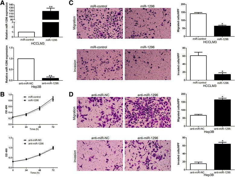
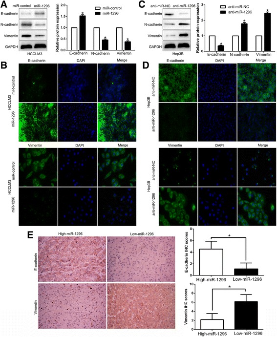
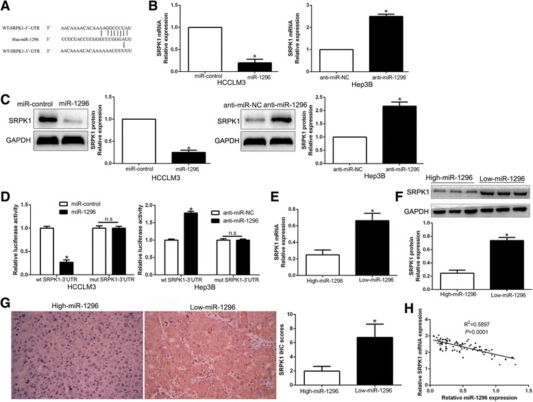
Methods: The levels of miR-1296 in HCC tissues and cells were detected by qRT-PCR. Immunoblotting and immunofluorescence were used for detection of epithelial-to-mesenchymal transition (EMT) progression in HCC cells. Transwell assays were performed to determine migration and invasion of HCC cells. A lung metastasis mouse model was used to evaluated metastasis of HCC in vivo. The putative targets of miR-1296 were disclosed by public databases and a dual-luciferase reporter assay.

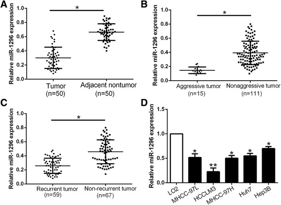
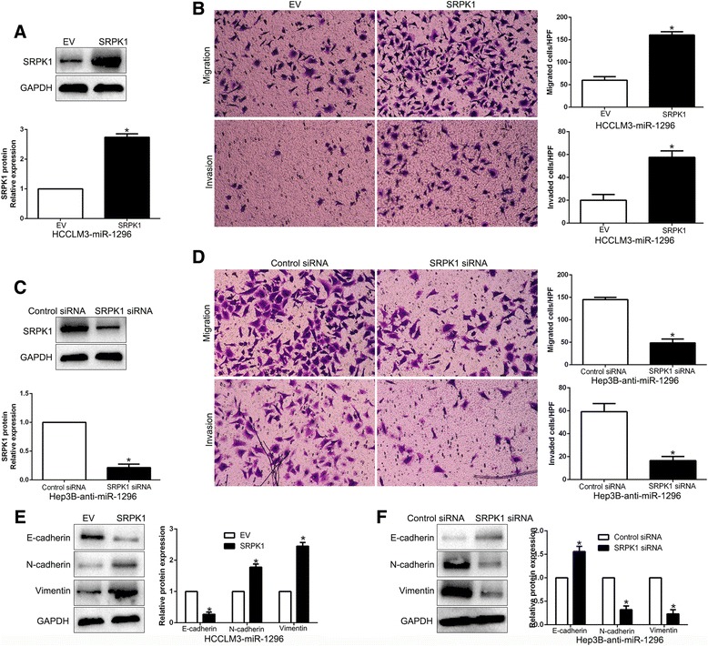
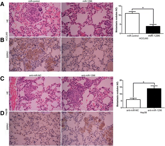
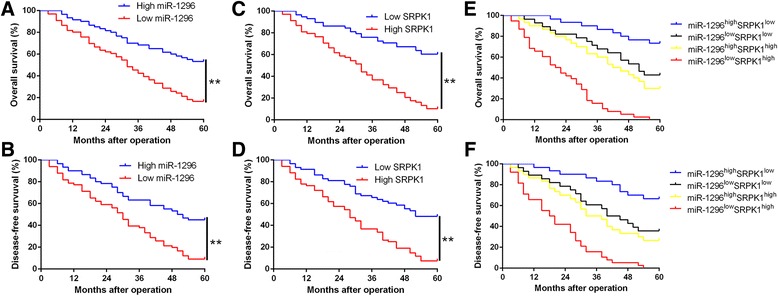
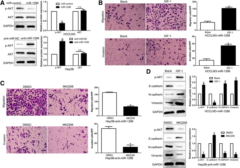
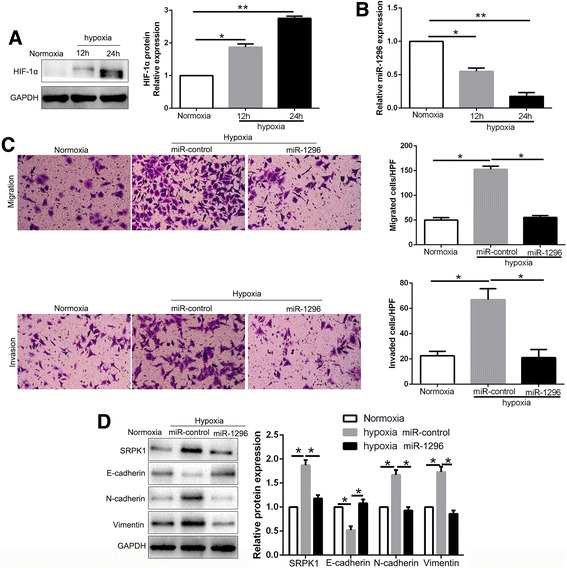
Results: We found that the expression of miR-1296 was reduced in HCC tissues and cell lines, and it was associated with metastasis and recurrence of HCC. Notably, miR-1296 overexpression inhibited migration, invasion and EMT progress of HCCLM3 cells, while miR-1296 loss facilitated these biological behaviors of Hep3B cells in vitro and in vivo. In addition, miR-1296 inversely regulated SRPK1 abundance by directly binding to its 3'-UTR, which subsequently resulted in suppression of p-AKT. Either SRPK1 re-expression or PI3K/AKT pathway activation, at least partially, abolished the effects of miR-1296 on migration, invasion and EMT progress of HCC cells. Furthermore, miR-1296 and SRPK1 expression were markedly correlated with adverse clinical features and poor prognosis of HCC patients. We showed that hypoxia was responsible for the underexpression of miR-1296 in HCC. And the promoting effects of hypoxia on metastasis and EMT of HCC cells were reversed by miR-1296.

Conclusions: Underexpression of miR-1296 potentially serves as a prognostic biomarker in HCC. Hypoxia-induced miR-1296 loss promotes metastasis and EMT of HCC cells probably by targeting SRPK1/AKT pathway.

Keywords: Hepatocellular carcinoma; Metastasis; MicroRNA-1296; PI3K/AKT pathway; SRPK1.


Figures

Fig. 1
Fig. 1
miR-1296 is underexpressed in HCC. Comparing differences in the expressions of miR-1296 between (a) HCC and matched tumor-adjacent tissues; (b) aggressive and nonaggressive tumor tissues; (c) HCC tissues arising from recurrent and non-recurrent groups; and (d) HCC cell lines and the immortalized hepatic cell line LO2. *P < 0.05, **P < 0.01
Fig. 2
Fig. 2
miR-1296 inhibits the migration and invasion of HCC cells in vitro. a HCCLM3 and Hep3B cells that were transfected with corresponding miRNA vectors were subjected to qRT-PCR for miR-1296 expression. b Modulating miR-1296 expression showed no obvious effect on proliferation of HCC cells, as determined by MTT assays. c Cell migration and invasion as measured by Transwell assays were inhibited by miR-1296 overexpression in HCCLM3 cells. d miR-1296 knockdown promoted migration and invasion of Hep3B cells. *P < 0.05, **P < 0.01
Fig. 3
Fig. 3
miR-1296 inhibits the EMT process of HCC cells. a Western blot analysis of epithelial marker E-cadherin, and mesenchymal markers N-cadherin and Vimentin after miR-1296 overexpression in HCCLM3 cells. b IF staining of E-cadherin and Vimentin after miR-1296 overexpression in HCCLM3 cells. c miR-1296 knockdown decreased E-cadherin expression, and increased the levels of N-cadherin and Vimentin in Hep3B cells. d IF staining of E-cadherin and Vimentin after miR-1296 knockdown in Hep3B cells. e Immunohistochemistry of E-cadherin and Vimentin were showed and compared between miR-1296 high expressing HCC tissues and miR-1296 low expressing cases. *P < 0.05
Fig. 4
Fig. 4
SRPK1 is a direct target of miR-1296 in HCC cells. a miR-1296 and its putative binding sequences in the 3′-UTR of SRPK1. The mutant binding site was generated in the complementary site for the seed region of miR-1296. b HCCLM3 and Hep3B cells that were transfected with precursor miR-1296 and miR-1296 inhibitors (anti-miR-1296), respectively, were subjected to qRT-PCR for SRPK1 mRNA expression. c miR-1296 overexpression reduced the expression of SRPK1 protein in HCCLM3 cells and miR-1296 knockdown increased the level of SRPK1 protein in Hep3B cells. d miR-1296 overexpression significantly suppressed, while miR-1296 loss increased the luciferase activity that carried wild-type (wt) but not mutant (mt) 3′-UTR of SRPK1. e and f The expression of SRPK1 in miR-1296 high-expressing tumors was significantly lower than that in miR-1296 low-expressing tumors, as determined by qRT-PCR and immunoblotting. g Representative immunohistochemical staining showed a weak staining of SRPK1 in miR-1296 high-expressing HCC tissue and strong staining of SRPK1 in the miR-1296 low-expressing tumor. h An inverse correlation between the levels of miR-1296 and SRPK1 mRNA was observed in HCC tissues. *P < 0.05
Fig. 5
Fig. 5
Modulation of SRPK1 partially abolishes miR-1296-mediated cellular processes in HCC. a miR-1296-overexpressing HCCLM3 cells that were transfected with empty vector (EV) or SRPK1 overexpression plasmid were subjected to western blot for SRPK1. b SRPK1 restoration promoted migration and invasion of miR-1296-overexpressing HCCLM3 cells. c miR-1296-suppressive Hep3B cells that were transfected with scrambled siRNA or SRPK1 siRNA were subjected to western blot for SRPK1. d SRPK1 knockdown abrogated the effects of miR-1296 loss on migration and invasion of Hep3B cells. e SRPK1 restoration decreased the expression of E-cadherin and increased the levels of N-cadherin and Vimentin in miR-1296 overexpressing HCCLM3 cells. f SRPK1 knockdown abolished the effects of miR-1296 loss on EMT process of Hep3B cells. *P < 0.05
Fig. 6
Fig. 6
miR-1296 inhibits the lung metastasis of HCC cells in nude mice. a Representative HE staining of lung metastases between HCCLM3-miR-1296 cells and control cells. b Immunohistochemistry suggested that SRPK1 expression showed a weaker staining in HCCLM3-miR-1296 cells than control cells. c Representative HE staining of lung metastases between Hep3B-anti-miR-1296 cells and control cells. d Immunohistochemistry revealed that SRPK1 expression in shows a stronger staining in Hep3B-anti-miR-1296 cells than control cells. *P < 0.05
Fig. 7
Fig. 7
The prognostic value of miR-1296 and SRPK1 for HCC patients. a and b Overall survival (OS) and disease-free survival (DFS) were compared between miR-1296 high expressing HCC patients and low expressing cases. c and d OS and DFS were compared between SRPK1 high expressing HCC patients and low expressing cases. e and f OS and DFS were compared between four subgroups of HCC patients (subgroup I: high miR-1296/low SRPK1; subgroup II: low miR-1296 /low SRPK1; subgroup III: high miR-1296/high SRPK1; subgroup IV: low miR-1296/high SRPK1). For each cohort, subgroups were divided according to the cutoff values, which were determined as the median level of miR-1296 and SRPK1 in HCC tissues. **P < 0.01
Fig. 8
Fig. 8
PI3K/AKT signaling is essential for the biological function of miR-1296 in HCC. a HCCLM3 and Hep3B cells that were transfected with corresponding miRNA vectors were subjected to immunoblotting for phosphorylated AKT and AKT. b IGF-1 treatment promoted the migration and invasion of miR-1296 overexpressing HCCLM3 cells. c AKT inhibitor MK2206 treatment abrogated the effect of miR-1296 loss on mobility of Hep3B cells. d Western blot analysis indicated that modulating AKT phosphorylation reversed the effects of miR-1296 alteration on EMT process of HCC cells. *P < 0.05
Fig. 9
Fig. 9
miR-1296 mediates the promoting effects of hypoxia on metastasis and EMT of HCC cells. a The expressions of HIF-1α in different time points in normoxia and hypoxia condition. b The levels of miR-1296 in Hep3B cells cultured in normoxia and hypoxia. c Transwell assays revealed that hypoxia promoted migration and invasion of Hep3B cells, while miR-1296 overexpression abolished the effects of hypoxia. d Hypoxia facilitated the EMT process of Hep3B cells and miR-1296 restoration showed a opposite effect. *P < 0.05, **P < 0.01

References

    1. Torre LA, Bray F, Siegel RL, Ferlay J, Lortet-Tieulent J, Jemal A. Global cancer statistics, 2012. CA Cancer J Clin. 2015;65(2):87–108. doi: 10.3322/caac.21262. - DOI - PubMed
    1. El-Serag HB, Rudolph KL. Hepatocellular carcinoma: epidemiology and molecular carcinogenesis. Gastroenterology. 2007;132(7):2557–2576. doi: 10.1053/j.gastro.2007.04.061. - DOI - PubMed
    1. Forner A, Llovet JM, Bruix J. Hepatocellular carcinoma. Lancet. 2012;379(9822):1245–1255. doi: 10.1016/S0140-6736(11)61347-0. - DOI - PubMed
    1. Maluccio M, Covey A. Recent progress in understanding, diagnosing, and treating hepatocellular carcinoma. CA Cancer J Clin. 2012;62(6):394–399. doi: 10.3322/caac.21161. - DOI - PubMed
    1. Garzon R, Calin GA, Croce CM. MicroRNAs in cancer. Annu Rev Med. 2009;60:167–179. doi: 10.1146/annurev.med.59.053006.104707. - DOI - PubMed

MeSH terms