. 2017 Jun 12;14(1):55.
doi: 10.1186/s12984-017-0254-x.

Modifying upper-limb inter-joint coordination in healthy subjects by training with a robotic exoskeleton

Affiliations

Modifying upper-limb inter-joint coordination in healthy subjects by training with a robotic exoskeleton

Tommaso Proietti et al. J Neuroeng Rehabil. .

Abstract

Background: The possibility to modify the usually pathological patterns of coordination of the upper-limb in stroke survivors remains a central issue and an open question for neurorehabilitation. Despite robot-led physical training could potentially improve the motor recovery of hemiparetic patients, most of the state-of-the-art studies addressing motor control learning, with artificial virtual force fields, only focused on the end-effector kinematic adaptation, by using planar devices. Clearly, an interesting aspect of studying 3D movements with a robotic exoskeleton, is the possibility to investigate the way the human central nervous system deals with the natural upper-limb redundancy for common activities like pointing or tracking tasks.

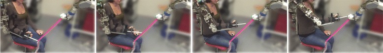
Methods: We asked twenty healthy participants to perform 3D pointing or tracking tasks under the effect of inter-joint velocity dependant perturbing force fields, applied directly at the joint level by a 4-DOF robotic arm exoskeleton. These fields perturbed the human natural inter-joint coordination but did not constrain directly the end-effector movements and thus subjects capability to perform the tasks. As a consequence, while the participants focused on the achievement of the task, we unexplicitly modified their natural upper-limb coordination strategy. We studied the force fields direct effect on pointing movements towards 8 targets placed in the 3D peripersonal space, and we also considered potential generalizations on 4 distinct other targets. Post-effects were studied after the removal of the force fields (wash-out and follow up). These effects were quantified by a kinematic analysis of the pointing movements at both end-point and joint levels, and by a measure of the final postures. At the same time, we analysed the natural inter-joint coordination through PCA.

Results: During the exposition to the perturbative fields, we observed modifications of the subjects movement kinematics at every level (joints, end-effector, and inter-joint coordination). Adaptation was evidenced by a partial decrease of the movement deviations due to the fields, during the repetitions, but it occurred only on 21% of the motions. Nonetheless post-effects were observed in 86% of cases during the wash-out and follow up periods (right after the removal of the perturbation by the fields and after 30 minutes of being detached from the exoskeleton). Important inter-individual differences were observed but with small variability within subjects. In particular, a group of subjects showed an over-shoot with respect to the original unexposed trajectories (in 30% of cases), but the most frequent consequence (in 55% of cases) was the partial persistence of the modified upper-limb coordination, adopted at the time of the perturbation. Temporal and spatial generalizations were also evidenced by the deviation of the movement trajectories, both at the end-effector and at the intermediate joints and the modification of the final pointing postures towards targets which were never exposed to any field.

Conclusions: Such results are the first quantified characterization of the effects of modification of the upper-limb coordination in healthy subjects, by imposing modification through viscous force fields distributed at the joint level, and could pave the way towards opportunities to rehabilitate pathological arm synergies with robots.

Keywords: Force fields adaptation; Motor coordination learning; Motor redundancy; Rehabilitation robotics; Upper-limb robotic exoskeletons.


Figures

Fig. 1
Fig. 1
Example of goal-directed pointing task (GDM). The four pictures show the motion from the starting position to the WAM button, while performing GDM task. In this case the subjects were not asked to follow any specific endpoint trajectory
Fig. 2
Fig. 2
Example of path-constrained tracking task (PCT). The four pictures show the motion from the starting position to the WAM button, while performing PCT exercise. The participants were asked to follow the specific endpoint path shown by the rubber band going from the starting position of the ABLE exoskeleton to the WAM end-effector
Fig. 3
Fig. 3
WAM positions. The eight Experimental Target positions (ET) and the four Generalization Target positions (GT). The asterisks * show the mean position of head and shoulder, and the projection of the starting position of the elbow/end-effector. On the left, x-z frontal plane, on the right, y-z sagittal plane (some targets are coincident on this plane). The frames are consistent with the reference frame of Fig. 1
Fig. 4
Fig. 4
Phases of the experiment. Experimental protocol, showing the four consecutive phases, respectively preliminary, experiment, wash-out and follow up. Before the follow up, the subject was resting, detached from the exoskeleton, for about 30 minutes. The number in front of each phase stands for the number of repetition of each pattern (1 repetition for PRE, 15 for EXP, and 2 for WAS and FOL)
Fig. 5
Fig. 5
Two illustrative cases. For two subjects, during GDM task, we show two different figures: on top, the averaged trajectory of the the shoulder abduction/adduction (dark plots) and the single trajectories (lighter plots), when moving towards ET 4; on bottom, for the same target, the resulting cycloids when considering the ratio between the first two joint velocities (shoulder abduction/adduction versus internal/external rotation). In this case the light plots are the cycloids, while the dark dashed lines are the mean ratio. For the four graphs, data refer to the 5 phases of the experiment, preliminary (PRE), early exposition (E-EXP, first five repetitions of EXP), late exposition (L-EXP, last five repetitions of EXP), wash-out (WAS), and follow up (FOL)
Fig. 6
Fig. 6
Mean joint final displacement on movement towards ET 3. Mean joint final displacement and standard error with respect to the final posture of first movement in PRE, in the two different modes, over the 10 subjects. The target position is ET number 3. Columns are the four joint of the exoskeleton. Horizontal dashed lines represent the joint maximum standard deviation σ for the spontaneous variability experiment
Fig. 7
Fig. 7
Mean motion duration T (a), mean peak velocity v max (b), and mean smoothness η (c). Averaged data over the ten participants and standard error for the pointing tasks towards ET positions for the two different modes. Smoothness, through spectral arc-length, is higher when η is closer to zero
Fig. 8
Fig. 8
Mean trajectory curvature Φ. Mean trajectory curvature and standard error for the ten subjects, over the 8 ET positions, for the two tasks
Fig. 9
Fig. 9
PCs distance from PRE, on ET pointing task, for the two tasks GDM and PCT. PC subspaces mean distance and standard error with respect to first repetition in PRE phase (trial 1) over the 10 participants when pointing toward ET. In red, mean values and standard deviation of spontaneous variability experiment with 5 healthy subjects of Section “Quantification of the human spontaneous variability within the exoskeleton”. Asterisks * mean significant difference w.r.t. spontaneous variability after non parametric one-sample sign test
Fig. 10
Fig. 10
PCs distance from constraining vector, on ET pointing task, for the two tasks GDM and PCT. PC subspaces mean distance and standard error from constraining vector (Eq. 2) (Eq. 2) over the 10 subjects during pointing task towards ET
Fig. 11
Fig. 11
Mean joint final displacement on movement towards GT 3. Mean joint displacement and standard error with respect to final posture in PRE, in the two different mode, over the 10 subjects. The target position is GT number 3. Columns are the four joint of the exoskeleton. Horizontal dashed lines represent the joint maximum standard deviation σ for the spontaneous variability experiment
Fig. 12
Fig. 12
Mean motion duration T (a), mean peak velocity v max (b), and mean smoothness η (c). Averaged data over the ten participants and standard error for the pointing tasks towards GT positions for the two different modes. Smoothness, through spectral arc-length, is higher when η is closer to zero
Fig. 13
Fig. 13
Mean trajectory curvature Φ. Mean trajectory curvature and standard error for the ten subjects, over the 4 GT positions, for the two tasks
Fig. 14
Fig. 14
PCs distance from PRE, on GT pointing task, for the two tasks GDM and PCT. PC subspaces mean distance and standard error with respect to first repetition in PRE phase (trial 1) over the 10 participants when pointing toward GT. In red, spontaneous variability mean values and standard deviation of spontaneous variability experiment with 5 healthy subjects of Section “Quantification of the human spontaneous variability within the exoskeleton”. Asterisks * mean significant difference w.r.t. spontaneous variability after non parametric one-sample sign test
Fig. 15
Fig. 15
PCs distance from constraining vector, on GT pointing task, for the two tasks GDM and PCT. PC subspaces mean distance and standard error from constraining vector (Eq. 2) over the 10 subjects during pointing task towards GT

References

    1. Levin MF, Kleim JA, Wolf SL. What do motor “recovery” and “compensation” mean in patients following stroke? Neurorehabil Neural Repair. 2009;23(4):313–9. doi: 10.1177/1545968308328727. - DOI - PubMed
    1. Taub E, Uswatte G, Mark VW, Morris DM. The learned nonuse phenomenon: implications for rehabilitation. Eura Medicophys. 2006;42(3):241–56. - PubMed
    1. Langhorne P, Bernhardt J, Kwakkel G. Stroke rehabilitation. Lancet. 2011;377(9778):1693–702. doi: 10.1016/S0140-6736(11)60325-5. - DOI - PubMed
    1. Krakauer JW. Motor learning: its relevance to stroke recovery and neurorehabilitation. Current Opin Neurol. 2006;19(1):84–90. doi: 10.1097/01.wco.0000200544.29915.cc. - DOI - PubMed
    1. Winstein CJ, Kay DB. Translating the science into practice: shaping rehabilitation practice to enhance recovery after brain damage. Progress Brain Res. 2015;218:331–60. doi: 10.1016/bs.pbr.2015.01.004. - DOI - PubMed

LinkOut - more resources