. 2017 Jun 13:6:e24634.
doi: 10.7554/eLife.24634.

Effects of myosin variants on interacting-heads motif explain distinct hypertrophic and dilated cardiomyopathy phenotypes

Affiliations

Effects of myosin variants on interacting-heads motif explain distinct hypertrophic and dilated cardiomyopathy phenotypes

Lorenzo Alamo et al. Elife. .

Abstract

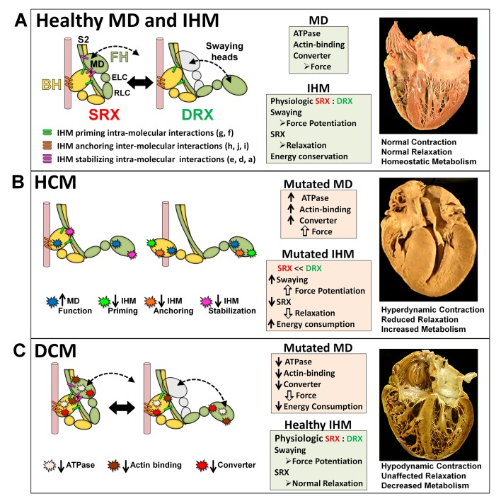
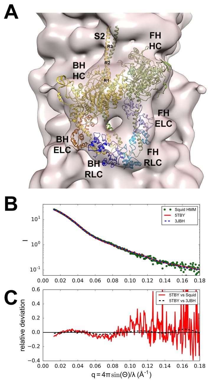
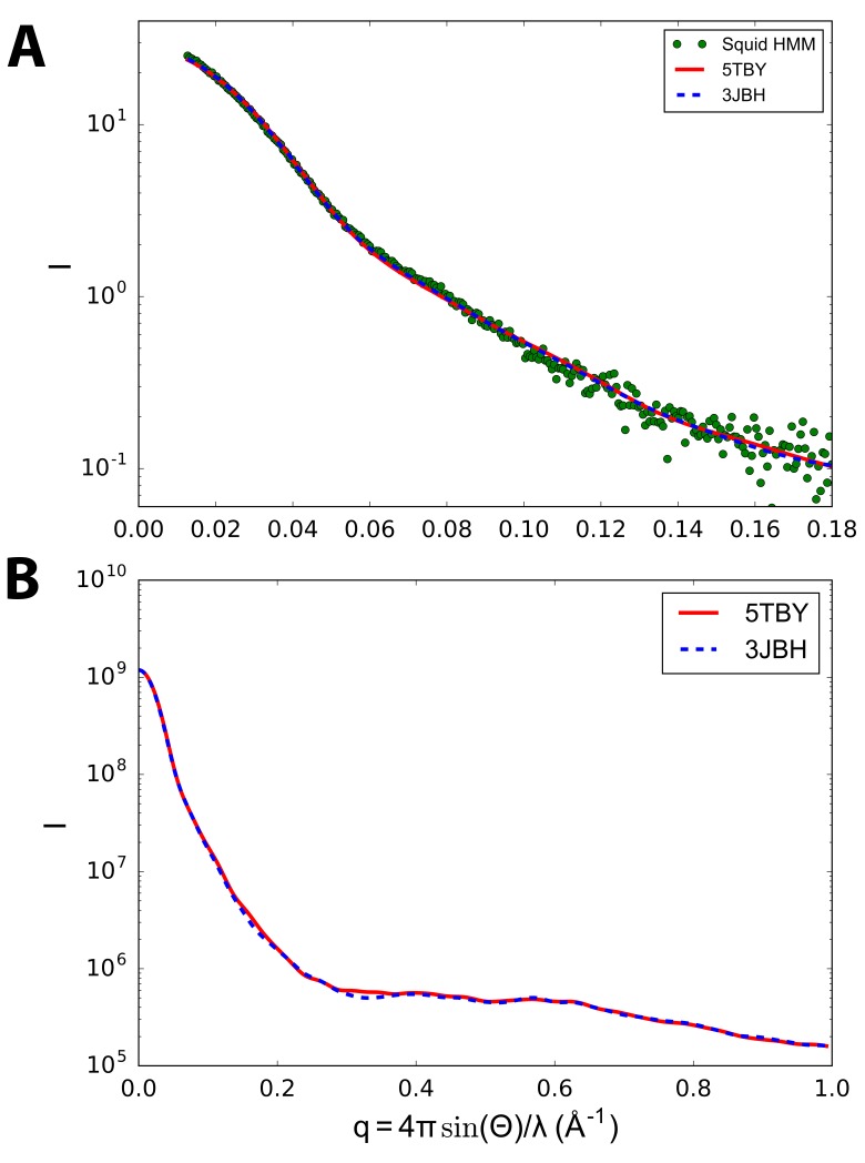
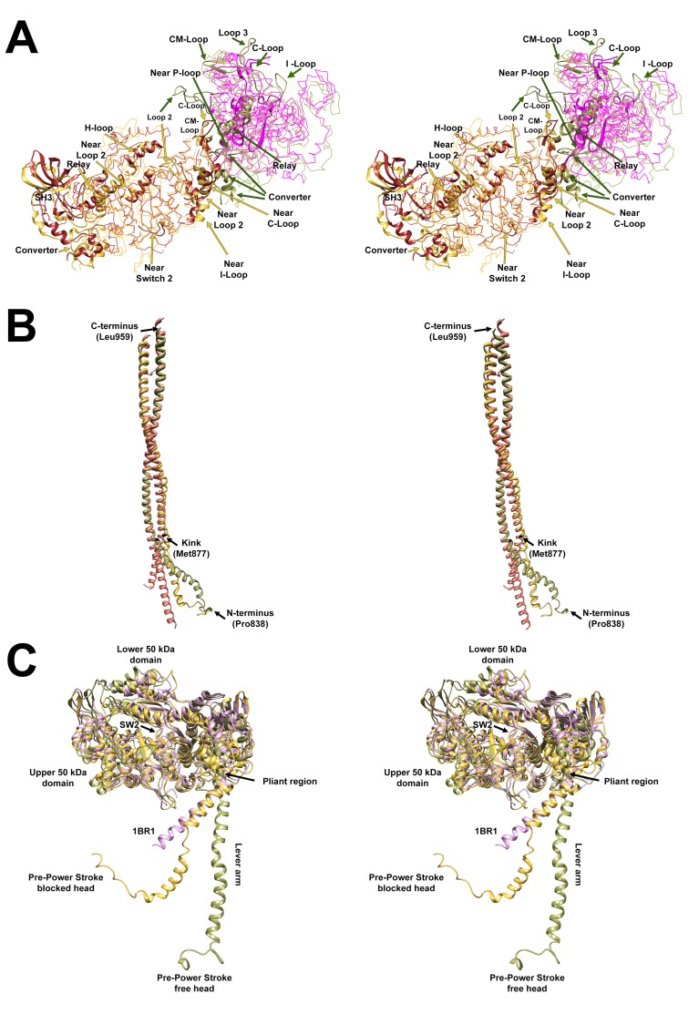
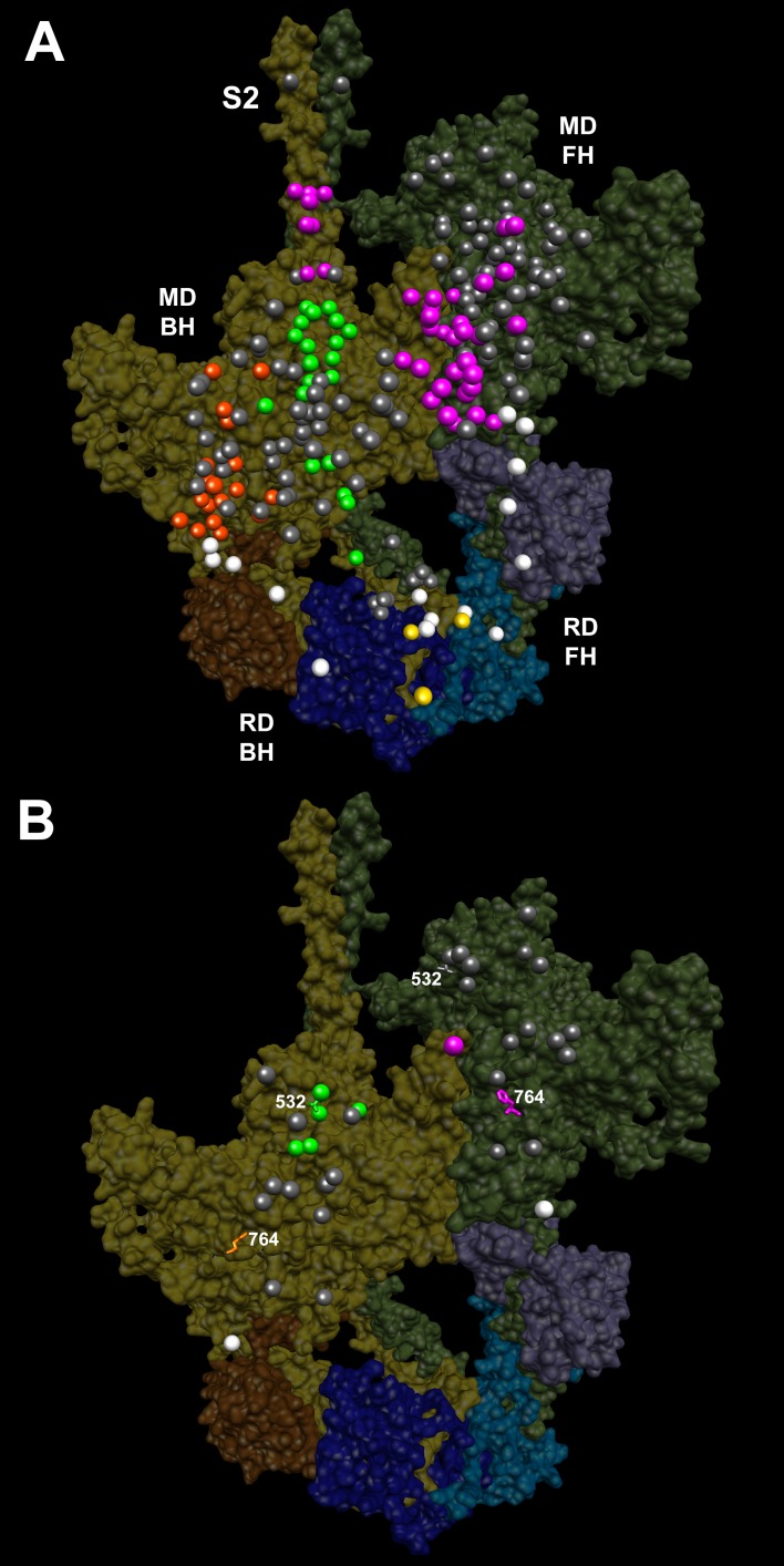
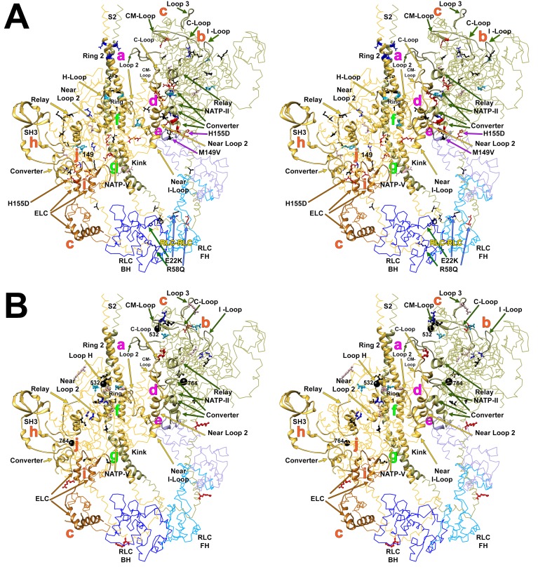
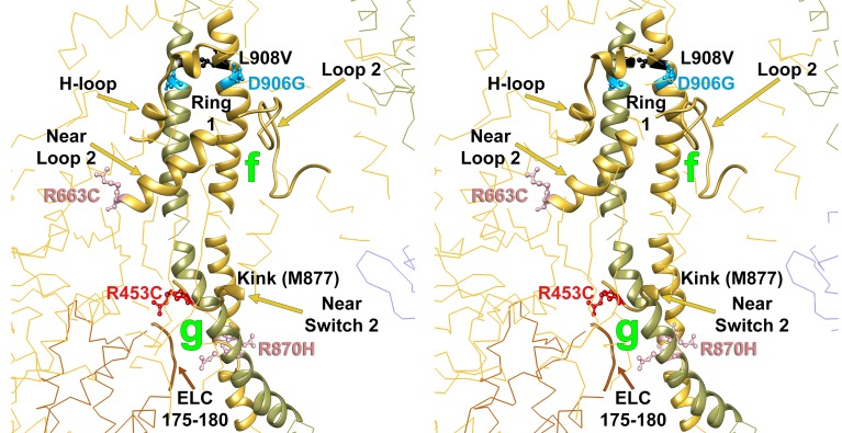
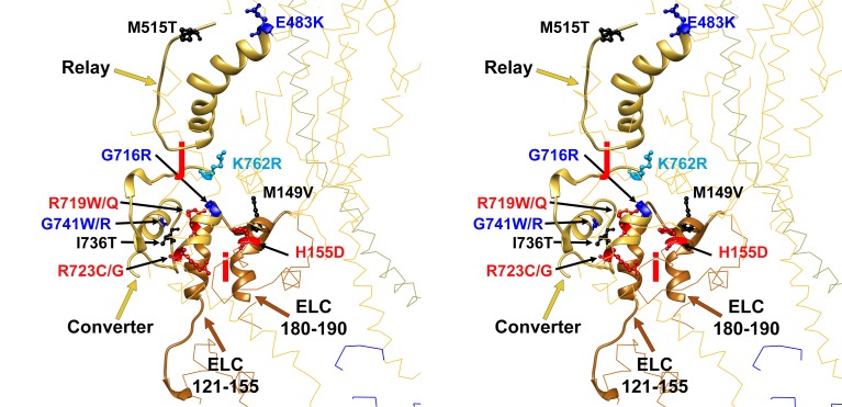
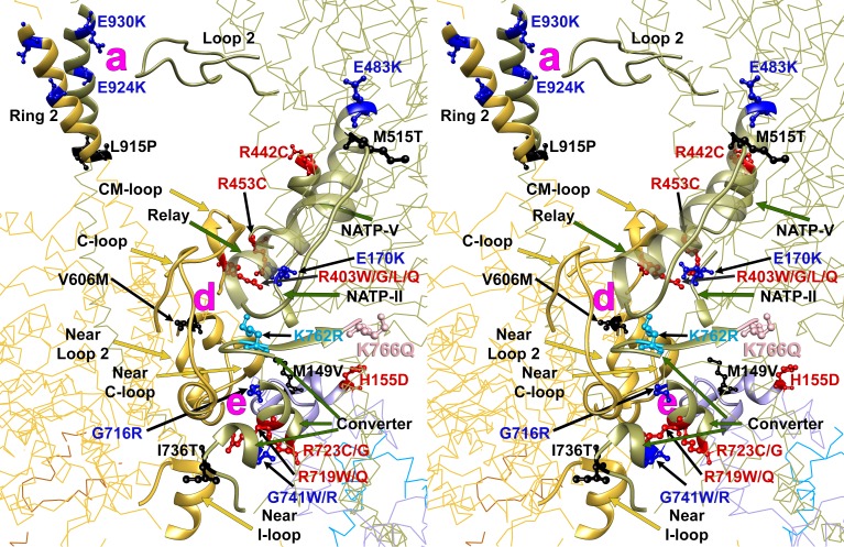
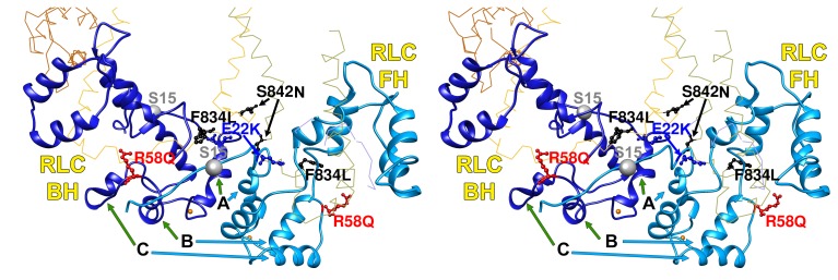
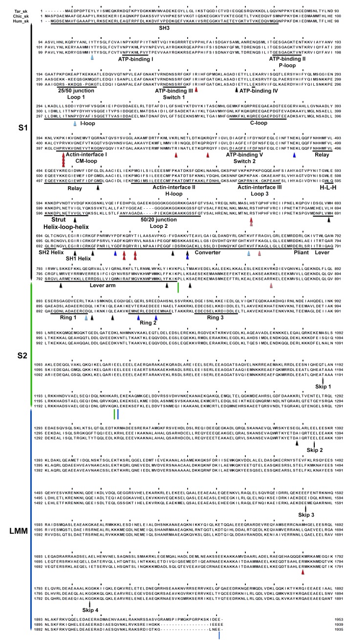
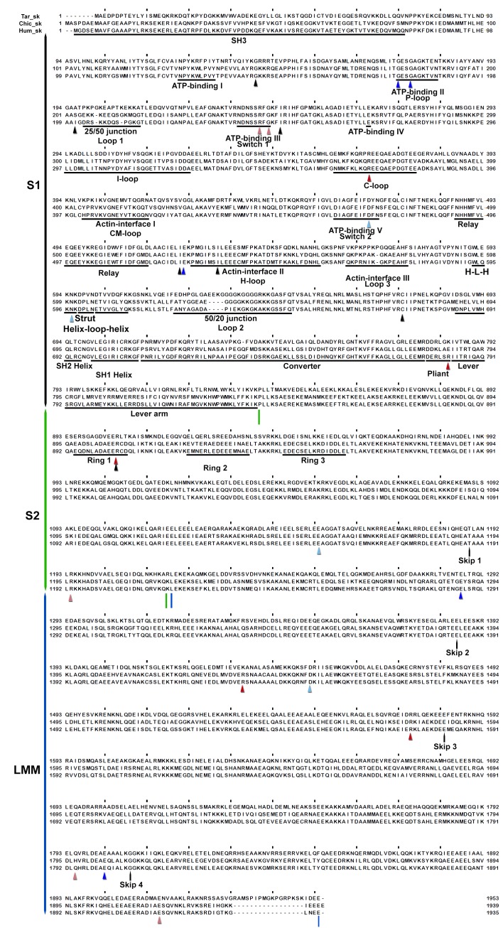
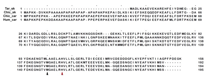
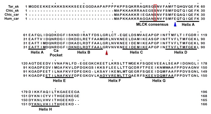
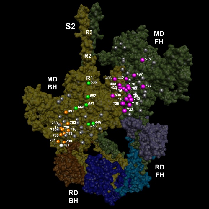
Cardiac β-myosin variants cause hypertrophic (HCM) or dilated (DCM) cardiomyopathy by disrupting sarcomere contraction and relaxation. The locations of variants on isolated myosin head structures predict contractility effects but not the prominent relaxation and energetic deficits that characterize HCM. During relaxation, pairs of myosins form interacting-heads motif (IHM) structures that with other sarcomere proteins establish an energy-saving, super-relaxed (SRX) state. Using a human β-cardiac myosin IHM quasi-atomic model, we defined interactions sites between adjacent myosin heads and associated protein partners, and then analyzed rare variants from 6112 HCM and 1315 DCM patients and 33,370 ExAC controls. HCM variants, 72% that changed electrostatic charges, disproportionately altered IHM interaction residues (expected 23%; HCM 54%, p=2.6×10⁻¹⁹; DCM 26%, p=0.66; controls 20%, p=0.23). HCM variant locations predict impaired IHM formation and stability, and attenuation of the SRX state - accounting for altered contractility, reduced diastolic relaxation, and increased energy consumption, that fully characterizes HCM pathogenesis.

Keywords: cardiomyopathy; diastolic heart disease; human; human biology; human mutation; medicine; myosin interacting-heads motif; super-relaxation; β-cardiac myosin.


Conflict of interest statement

JGS: is a founder and owns shares in Myokardia Inc., a startup company that is developing therapeutics that targets the sarcomere.

CES: is a founder and owns shares in Myokardia Inc., a startup company that is developing therapeutics that targets the sarcomere.

The other authors declare that no competing interests exist.

Figures

Figure 1.
Figure 1.. The molecular pathogenesis of hypertrophic (HCM) and dilated (DCM) cardiomyopathy assessed in the context of the myosin interacting-heads motif (IHM) paradigm.
Myosin interactions involved in IHM assembly and myosin motor domain (MD) functions that are altered by PVs and LPVs are depicted. (A) Relaxed healthy cardiac muscle contains myosin heads populations in the super-relaxed (SRX) state (left) with lowest ATP consumption and a disordered relaxed (DRX) state (right) with swaying free heads that generate force with higher ATP consumption. The population of cardiac myosins in SRX is more stable than in skeletal muscle (Hooijman et al., 2011 and see Material and methods) which supports physiologic contraction and relaxation, energy conservation, and normal cardiac morphology. (B) HCM myosin variants both alter residues involved in MD functions (causing increased biophysical power [Tyska et al., 2000]), and destabilize IHM interactions (particularly those with altered electrostatic charge). Reduced populations of myosins in the SRX state and increased populations of myosins in DRX as well as enhanced MD properties will result in increased contractility, decreased relaxation, and increased ATP consumption, the three major phenotypes observed in HCM hearts. Compensatory signals may promote ventricular hypertrophy. (C) MYH7 DCM variants have modest effects on IHM interactions but substantially reduce MD functions, particularly nucleotide binding, resulting in reduced ATP consumption and sarcomere power (Schmitt et al., 2006), with minimal impact on relaxation and overall diminished contractility. Compensatory signals result in ventricular dilatation to maintain circulatory demands in DCM hearts. DOI: http://dx.doi.org/10.7554/eLife.24634.002
Figure 2.
Figure 2.. Structure of the human β-cardiac myosin interacting-heads motif.
(A) Quasi-atomic homologous model of human β-cardiac myosin interacting-heads motif (IHM) PDB 5TBY composed of blocked (BH) and free (FH) heads, fitted to the human cardiac thick filament 3D-map EMD-2240 (Al-Khayat et al., 2013). (Also see Video 1.) Domains and residue equivalence are provided in Supplementary file 1. Sarcomere proteins depicted are MHC (BH: gold, FH: olive), essential light chain (ELC associated with BH: brown, FH: purple), and regulatory light chain (RLC associated with BH: dark blue, FH: blue). The three negatively-charged rings in the S2 are labeled R1, R2 and R3. (B) Calculated small angle X-ray solution scattering (SAXS) profile of PDB 5TBY (red line) matches the experimental squid heavy meromyosin SAXS profile (green dots) (Gillilan et al., 2013). Integrated scattering intensity (I in arbitrary units) is given as a function of momentum transfer, q = 4π sin(θ)/λ, with a scattering angle of 2θ and a wavelength of λ. (C) Relative deviation between PDB 5TBY scattering and squid HMM (red line) is calculated as (Imodel-Iexp)/Iexp. The corresponding difference in scattering between PDB 5TBY and PDB 3JBH models is also shown on the same scale (black dashed line). Comparison shows that the models cannot be distinguished based on currently available scattering data (See Supplementary Figures). DOI: http://dx.doi.org/10.7554/eLife.24634.003
Figure 2—figure supplement 1.
Figure 2—figure supplement 1.. Calculated small angle X-ray solution scattering (SAXS) profile of PDB 5TBY (red line) matches experimental squid heavy meromyosin (HMM) SAXS profile (green dots) (Gillilan et al., 2013).
The comparison of model-based tarantula PDB 3JBH (blue dashed line) vs. human cardiac ventricular IHM PDB 5TBY (red line) scattering with measured squid HMM scattering profiles (green dots) in (A) shows that the models cannot be distinguished based on the scattering data that is currently available. Computations are performed using the FoXS algorithm (Schneidman-Duhovny et al., 2013) using the default parameters of hydration layer, excluded volume, and background adjustment (Gillilan et al., 2013). In the case of (A) the ‘profile offset’ optimization was performed to obtain best fit with the data at widest angles. The predicted scattering profiles are based on electron microscopy-derived striated tarantula muscle PDB 3JBH (Alamo et al., 2016) (blue dashed line) IHM models. Calculated wide-angle scattering data (B), computed without reference to the squid HMM profile, confirms that the models do not significantly differ in the wide angle X-ray solution scattering (WAXS) region. DOI: http://dx.doi.org/10.7554/eLife.24634.004
Figure 2—figure supplement 2.
Figure 2—figure supplement 2.. Wide-eye stereo pairs that compare the PDB 5TBY model with crystal structures of fragments for human β-cardiac S1 MD fragment (PDB 4DB1) and S2.
(A) Superimposed MDs of S1 from the PDB 5TBY antavd a S1 fragment of human β-cardiac myosin S1 crystal structure PDB 4DB1 (AMPPNP rigor-like structure) See Video 2. Color code: PDB 5TBY: Blocked head (gold), free head (FH) (olive); PDB 4DB1: blocked head (brown), free head (magenta). All chains are shown as wires with the interacting loops highlighted as ribbons. (B) Superimposed S2 from the PDB 5TBY (same color code as A) and the S2 of the human β-cardiac myosin S2 (pink) crystal structure PDB 2FXM (Blankenfeldt et al., 2006) See Video 3. (C) Free and blocked head structures of PDB 5TBY vs. PDB 1BR1 structures (See Video 4). The free and blocked head MDs of PDB 5TBY (same color code as A) fits well the pre-powerstroke PDB 1BR1 crystal structure (magenta). The ELC and RLC were removed to highlight their lever arms, which are in the same plane but have different angles. DOI: http://dx.doi.org/10.7554/eLife.24634.005
Figure 3.
Figure 3.. IHM PDB 5TBY models depicting pathogenic (PVs) and likely pathogenic variants (LPVs), in HCM and DCM (listed in Tables 1–3).
Each variant appears as a pair, one located on or associated with the blocked head (BH, olive) and one on the free head (FH, green). Associated proteins are the essential light chain (ELC interacting with BH, brown; FH, purple) and regulatory light chain (RLC interacting with BH, dark blue; FH, light blue). (A) HCM PVs and LPVs that alter residues involved in IHM interactions (73/135 variants, 54%) are represented by colored balls: priming, green (‘f’ and ‘g’, Figure 3—figure supplement 2); anchoring, orange (‘i’ and ‘j’, Figure 3—figure supplement 3); stabilizing, pink (‘a’, ‘d’, and ‘e’, Figure 3—figure supplement 4); scaffolding, white (ELC-MHC and RLC-MHC); RLC-RLC interface, yellow (Figure 3—figure supplement 5). For variants with multiple IHM interactions, only one is depicted. PVs and LPVs that do not alter residues involved in IHM interactions are grey. (B) DCM PV and LPV (7/27; 26%) defined here are colored as described above, along with two prior PVs (S532P and F764L, denoted by side chains) that alter IHM interacting residues. Detailed IHM PDB 5TBY models of variants involved in specific interactions are provided in Supplemental Files and Videos 5 and 6). DOI: http://dx.doi.org/10.7554/eLife.24634.015
Figure 3—figure supplement 1.
Figure 3—figure supplement 1.. Wide-eye stereo pairs of the IHM PDB 5TBY model showing HCM pathogenic variants (A) and DCM pathogenic and likely pathogenic variants (B).
(A) IHM PDB 5TBY model mapping 40 HCM pathogenic variants (that produced 39 distinct substitutions in MHC and four substitutions in the regulatory and essential light chains; Table 1, in the main text) depicted as balls and sticks, with all chains are shown as wires, and the interaction loops highlighted as ribbons. 22/31 PVs (observed 71%, expected 49%, p=0.019) involved on interactions are charge-changing, according to this color code for their Δq: −2 (dark red), −1 (pink), 0 (black),+1 (light blue) and +2 (dark blue). (B) IHM PDB 5TBY model mapping 27 DCM-causing variants, showing 26 residues substituted by 27 variants on each head (Table 3, in the main text) depicted as balls and sticks, with all chains are shown as wires, and the interaction loops highlighted as ribbons. Five of seven (71%) DCM PVs and LPVs involved on interactions are charge-changing, according to this color code for their Δq: −2 (dark red), −1 (pink), 0 (black),+1 (light blue) and +2 (dark blue). DOI: http://dx.doi.org/10.7554/eLife.24634.016
Figure 3—figure supplement 2.
Figure 3—figure supplement 2.. Wide-eye stereo pair of the IHM PDB 5TBY model showing the five HCM variants that alter residues involved in IHM priming (intra-molecular interactions ‘g’ and ‘f’).
Three variants alter resides that participate in ‘f’ sub-interactions (‘f.1’: D906G, L908V; ‘f.2’: R663H) and two alter residues that participate in ‘g’ interactions (R453C and R870H). All chains are shown as wires, with the interacting loops highlighted as ribbons. Only amino acid substitutions (caused by cardiomyopathy variants) within these interacting loops are highlighted as balls and sticks. The color code reflects the variant’s Δq: −2 (dark red), −1 (pink), 0 (black),+1 (light blue) and +2 (dark blue). DOI: http://dx.doi.org/10.7554/eLife.24634.017
Figure 3—figure supplement 3.
Figure 3—figure supplement 3.. Wide-eye stereo pair of the IHM PDB 5TBY model showing 13 HCM pathogenic variants involved in anchoring the IHM.
Intermolecular interactions between (‘j’, ‘i’) myosin and the essential light chain anchor the blocked head anchoring on the neighboring S2. Five substitutions are involved on interaction ‘j’ between blocked head relay (G483K, M515T) and converter (I736T and G741W/R) with the neighbor S2. Six MHC (G716R, R719W/Q, R723G/C and K762R) variants interact (‘i’) with the same loop of the blocked head ELC with the neighboring MHC S2. Two substitutions are located on the ELC. All chains are shown as wires, with the interacting loops highlighted as ribbons. Only amino acid substitutions (caused by cardiomyopathy variants) within these interacting loops are highlighted as balls and sticks. The color code reflects the variant’s Δq: −2 (dark red), −1 (pink), 0 (black),+1 (light blue) and +2 (dark blue). DOI: http://dx.doi.org/10.7554/eLife.24634.018
Figure 3—figure supplement 4.
Figure 3—figure supplement 4.. Wide-eye stereo pair of the IHM PDB 5TBY model showing sites of 25 HCM variants that alter IHM stabilizing residues.
Twenty-three MHC variants and two ELC variants alter IHM residues that dock the free head onto the blocked head (‘e’, ‘d’ and ‘a’ interactions). Nineteen MYH7 substitutions are at residues participating in two ‘d’ sub-interactions (‘d.1’: E170K, R403W/G/L/Q, R453C, R442C; ‘d.2’: G716R, R719W/Q, R723G/C, I736T, G741W/R, E483K, M515T, K762R, K766Q), three substitutions are at ‘e’ interactions (MYH7 V606M; MYL3 M149V and H155D), and three at ‘a’ interactions (MYH7 L915P, E924K and E930K). Notably, nine of twelve substitutions involved in sub-interaction “d.2 “alter the charge. All chains are shown as wires, with the interacting loops highlighted as ribbons. Only amino acid substitutions (caused by cardiomyopathy variants) within these interacting loops are highlighted as balls and sticks. The color code reflects the variant’s Δq: −2 (dark red), −1 (pink), 0 (black),+1 (light blue) and +2 (dark blue). DOI: http://dx.doi.org/10.7554/eLife.24634.019
Figure 3—figure supplement 5.
Figure 3—figure supplement 5.. Wide-eye stereo pair of the IHM PDB 5TBY model showing four variants located in the RLC-RLC interface region.
Variants in MHC (S842N and F834L) and RLC (E22K and R58Q) alter residues that allow proper docking of the two opposite surfaces of the RLC-RLC interface when the IHM is initially assembled. The large gray ball denotes phosphorylated Ser15. The small grey yellow denotes the RLC Ca2+ pocket. All chains are shown as wires, with the interacting loops highlighted as ribbons. Only amino acid substitutions (caused by cardiomyopathy variants) within these interacting loops are highlighted as balls and sticks. The color code reflects the variant’s Δq: −2 (dark red), −1 (pink), 0 (black),+1 (light blue) and +2 (dark blue). DOI: http://dx.doi.org/10.7554/eLife.24634.020
Figure 3—figure supplement 6.
Figure 3—figure supplement 6.. Sequence alignment of human cardiac, chicken skeletal and tarantula striated MHC with the locations of HCM variants.
Variant positions (arrowheads) are color-coded according their charge-change, as in Figure 3—figure supplement 1. DOI: http://dx.doi.org/10.7554/eLife.24634.021
Figure 3—figure supplement 7.
Figure 3—figure supplement 7.. Sequence alignment of human cardiac, chicken skeletal and tarantula striated MHC with the locations of DCM variants.
Variant positions (arrowheads) are color-coded as in Figure 3—figure supplement 1. DOI: http://dx.doi.org/10.7554/eLife.24634.022
Figure 3—figure supplement 8.
Figure 3—figure supplement 8.. Sequence alignment of human cardiac, chicken skeletal and tarantula striated ELC with the locations of HCM variants.
HCM variant positions are (arrowheads) color-coded as in Figure 3—figure supplement 1. DOI: http://dx.doi.org/10.7554/eLife.24634.023
Figure 3—figure supplement 9.
Figure 3—figure supplement 9.. Sequence alignment of human cardiac, chicken skeletal and tarantula striated RLC with the location of HCM variants.
HCM variant positions are (arrowheads) color-coded as in Figure 3—figure supplement 1. DOI: http://dx.doi.org/10.7554/eLife.24634.024
Figure 4.
Figure 4.. The location of 14 variants located on the mesa (Spudich, 2015; Homburger et al., 2016) of the blocked head (BH, olive) and free head (FH, green).
The three negatively-charged rings in the blocked head S2 are labeled R1, R2 and R3. The associated sarcomere proteins are depicted as in Figure 3. The myosin mesas are roughly orthogonal (blocked head mesa, parallel to the page; free head mesa, perpendicular to the page; see Video 7). HCM PVs are enriched on the mesa and in IHM interactions when located either on the blocked or free head, suggesting that these disrupt crucial determinants of cardiac relaxation, accounting for diastolic dysfunction in HCM. DOI: http://dx.doi.org/10.7554/eLife.24634.027

References

    1. Al-Khayat HA, Kensler RW, Squire JM, Marston SB, Morris EP. Atomic model of the human cardiac muscle myosin filament. PNAS. 2013;110:318–323. doi: 10.1073/pnas.1212708110. - DOI - PMC - PubMed
    1. Alamo L, Li XE, Espinoza-Fonseca LM, Pinto A, Thomas DD, Lehman W, Padrón R. Tarantula myosin free head regulatory light chain phosphorylation stiffens N-terminal extension, releasing it and blocking its docking back. Molecular bioSystems. 2015;11:2180–2189. doi: 10.1039/C5MB00163C. - DOI - PMC - PubMed
    1. Alamo L, Qi D, Wriggers W, Pinto A, Zhu J, Bilbao A, Gillilan RE, Hu S, Padrón R. Conserved intramolecular interactions maintain myosin interacting-heads motifs explaining tarantula muscle super-relaxed state structural basis. Journal of Molecular Biology. 2016;428:1142–1164. doi: 10.1016/j.jmb.2016.01.027. - DOI - PMC - PubMed
    1. Alamo L, Wriggers W, Pinto A, Bártoli F, Salazar L, Zhao FQ, Craig R, Padrón R. Three-dimensional reconstruction of tarantula myosin filaments suggests how phosphorylation may regulate myosin activity. Journal of Molecular Biology. 2008;384:780–797. doi: 10.1016/j.jmb.2008.10.013. - DOI - PMC - PubMed
    1. Alfares AA, Kelly MA, McDermott G, Funke BH, Lebo MS, Baxter SB, Shen J, McLaughlin HM, Clark EH, Babb LJ, Cox SW, DePalma SR, Ho CY, Seidman JG, Seidman CE, Rehm HL. Results of clinical genetic testing of 2,912 probands with hypertrophic cardiomyopathy: expanded panels offer limited additional sensitivity. Genetics in Medicine. 2015;17:880–888. doi: 10.1038/gim.2014.205. - DOI - PubMed

Publication types

MeSH terms

Substances