

Is superolateral Hoffa's fat pad hyperintensity a marker of local patellofemoral joint disease? - The MOST study
- PMID: 28606557
- PMCID: PMC5583732
- DOI: 10.1016/j.joca.2017.05.020
Is superolateral Hoffa's fat pad hyperintensity a marker of local patellofemoral joint disease? - The MOST study
Abstract
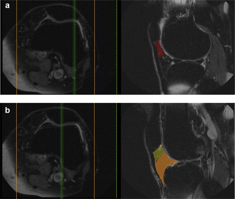
Purpose: To determine the relation of superolateral Hoffa's fat pad (SHFP) hyperintensity to cartilage damage and bone marrow lesions (BMLs) in the patellofemoral joint (PFJ) and tibiofemoral joint (TFJ).
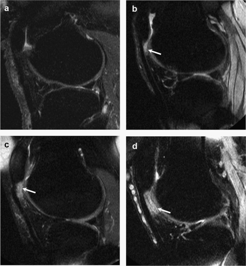
Methods: We used data from the 60 and 84-month study visits from the Multicenter Osteoarthritis (MOST) study. SHFP hyperintensity and Hoffa-synovitis were graded from 0 to 3. Cartilage damage and BMLs were scored in the PFJ and TFJ. Structural damage was defined as: any cartilage damage, full-thickness cartilage damage and any BML. Worsening structural damage was defined as any increase in cartilage and BML scores. Logistic regression was used to determine the relation of SHFP hyperintensity and Hoffa-synovitis (>0) to structural damage, adjusting for age, sex and body mass index (BMI).
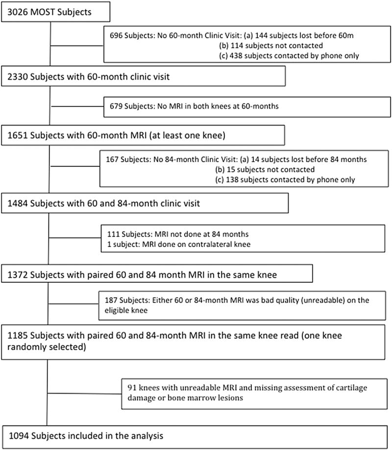
Results: 1,094 knees were included in the study. Compared to knees without SHFP hyperintensity, those with SHFP hyperintensity had 1.2 (95% Confidence Interval (CI), 1.1-1.4), 1.7 (1.3-2.3) and 1.6 (1.3-1.9) times the prevalence of any cartilage damage, full-thickness cartilage damage, and BMLs in the lateral PFJ respectively, and 1.1 (1.0-1.2), 1.3 (1.0-1.8), and 1.2 (1.0-1.4) times the prevalence of any cartilage damage, full-thickness cartilage damage, and BMLs in the medial PFJ. SHFP hyperintensity was associated with worsening BMLs in the medial PFJ (RR: 1.4 (1.0-1.9)). In general, there was no relation between SHFP hyperintensity and TFJ outcomes. Hoffa-synovitis was associated both cross-sectionally and longitudinally with structural damage, regardless of definition, in all compartments.
Conclusion: SHFP hyperintensity may be a local marker of PFJ structural damage.
Keywords: Bone marrow lesions; Cartilage; Hoffa's fat pad; Patellofemoral joint.
Copyright © 2017. Published by Elsevier Ltd.
Figures
Similar articles
-
Superolateral Hoffa's fat pad (SHFP) oedema and patellar cartilage volume loss: quantitative analysis using longitudinal data from the Foundation for the National Institute of Health (FNIH) Osteoarthritis Biomarkers Consortium.Eur Radiol. 2018 Oct;28(10):4134-4145. doi: 10.1007/s00330-018-5334-1. Epub 2018 Apr 12. Eur Radiol. 2018. PMID: 29651769
-
Association of markers of patellofemoral maltracking to cartilage damage and bone marrow lesions on MRI: Data from the 2016 Olympic Games of Rio De Janeiro.Eur J Radiol Open. 2021 Oct 4;8:100381. doi: 10.1016/j.ejro.2021.100381. eCollection 2021. Eur J Radiol Open. 2021. PMID: 34660850 Free PMC article.
-
Relationship of Trochlear Morphology and Patellofemoral Joint Alignment to Superolateral Hoffa Fat Pad Edema on MR Images in Individuals with or at Risk for Osteoarthritis of the Knee: The MOST Study.Radiology. 2017 Sep;284(3):806-814. doi: 10.1148/radiol.2017162342. Epub 2017 Apr 17. Radiology. 2017. PMID: 28418810 Free PMC article.
-
Superolateral Hoffa Fat Pad Edema and Patellofemoral Maltracking: Systematic Review and Meta-Analysis.AJR Am J Roentgenol. 2020 Sep;215(3):545-558. doi: 10.2214/AJR.19.22263. Epub 2020 Jun 6. AJR Am J Roentgenol. 2020. PMID: 32507017
-
Magnetic resonance imaging of Hoffa's fat pad and relevance for osteoarthritis research: a narrative review.Osteoarthritis Cartilage. 2016 Mar;24(3):383-97. doi: 10.1016/j.joca.2015.09.018. Epub 2015 Oct 9. Osteoarthritis Cartilage. 2016. PMID: 26455999 Review.
Cited by
-
Correlation analysis between the mDIXON-quant fat quantification parameters of the infrapatellar fat pad and the severity of knee osteoarthritis.J Orthop Surg Res. 2025 Mar 17;20(1):288. doi: 10.1186/s13018-025-05687-2. J Orthop Surg Res. 2025. PMID: 40091107 Free PMC article.
-
Quantitative Signal Intensity Alteration in Infrapatellar Fat Pad Predicts Incident Radiographic Osteoarthritis: The Osteoarthritis Initiative.Arthritis Care Res (Hoboken). 2019 Jan;71(1):30-38. doi: 10.1002/acr.23577. Arthritis Care Res (Hoboken). 2019. PMID: 29648688 Free PMC article.
-
Tibial tuberosity to trochlear groove distance and its association with patellofemoral osteoarthritis-related structural damage worsening: data from the osteoarthritis initiative.Eur Radiol. 2018 Nov;28(11):4669-4680. doi: 10.1007/s00330-018-5460-9. Epub 2018 Apr 30. Eur Radiol. 2018. PMID: 29713775
-
Magnetic resonance imaging of impingement and friction syndromes around the knee.Skeletal Radiol. 2020 Jun;49(6):823-836. doi: 10.1007/s00256-020-03379-y. Epub 2020 Jan 28. Skeletal Radiol. 2020. PMID: 31993687 Review.
-
Epidemiology of osteoarthritis: literature update.Curr Opin Rheumatol. 2018 Mar;30(2):160-167. doi: 10.1097/BOR.0000000000000479. Curr Opin Rheumatol. 2018. PMID: 29227353 Free PMC article. Review.
References
-
- Chung CB, Skaf A, Roger B, Campos J, Stump X, Resnick D. Patellar tendon-lateral femoral condyle friction syndrome: MR imaging in 42 patients. Skelet Radiol. 2001;30(12):694–7. http://dx.doi.org/10.1007/s002560100409. - DOI - PubMed
-
- Saddik D, McNally EG, Richardson M. MRI of Hoffa's fat pad. Skelet Radiol. 2004;33(8):433–44. http://dx.doi.org/10.1007/s00256-003-0724-z. - DOI - PubMed
-
- Chhabra A, Subhawong TK, Carrino JA. A systematised MRI approach to evaluating the patellofemoral joint. Skelet Radiol. 2011;40(4):375–87. http://dx.doi.org/10.1007/s00256-010-0909-1. - DOI - PMC - PubMed
-
- Campagna R, Pessis E, Biau DJ, Guerini H, Feydy A, Thevenin FS, et al. Is superolateral Hoffa fat pad edema a consequence of impingement between lateral femoral condyle and patellar ligament? Radiology. 2012;263(2):469–74. http://dx.doi.org/10.1148/radiol.12111066. - DOI - PubMed
-
- Subhawong TK, Thakkar RS, Padua A, Flammang A, Chhabra A, Carrino JA. Patellofemoral friction syndrome: magnetic resonance imaging correlation of morphologic and T2 cartilage imaging. J Comput Assist Tomogr. 2014;38(2):308–12. http://dx.doi.org/10.1097/RCT.0b013e3182aab187. - DOI - PMC - PubMed
Publication types
MeSH terms
Grants and funding
LinkOut - more resources
Full Text Sources
Other Literature Sources
