. 2017 Jul 3;216(7):2179-2199.
doi: 10.1083/jcb.201606065. Epub 2017 Jun 12.

Polarity of varicosity initiation in central neuron mechanosensation

Affiliations

Polarity of varicosity initiation in central neuron mechanosensation

Yuanzheng Gu et al. J Cell Biol. .

Abstract

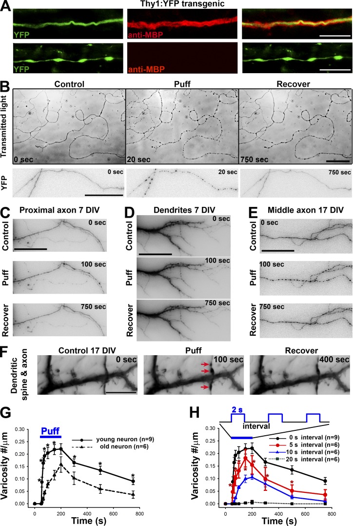
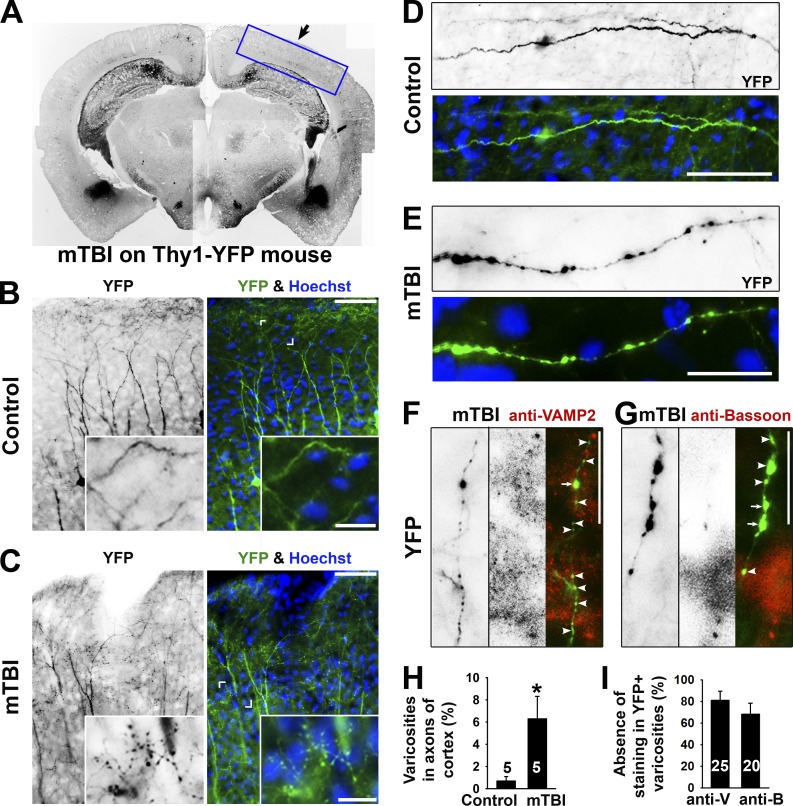
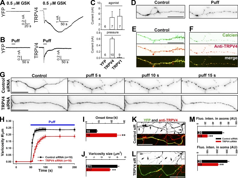
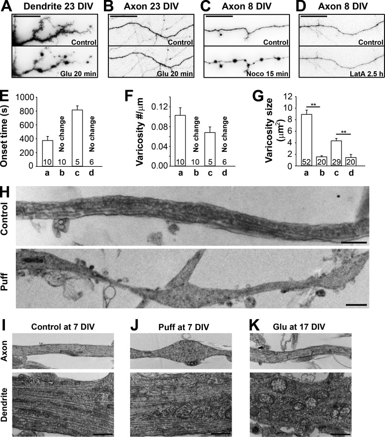
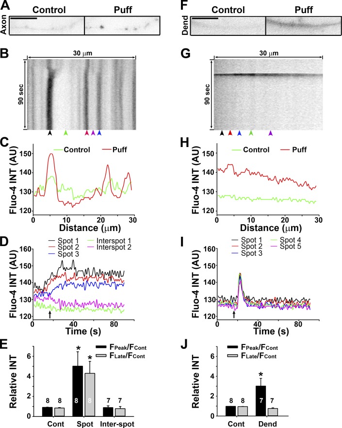
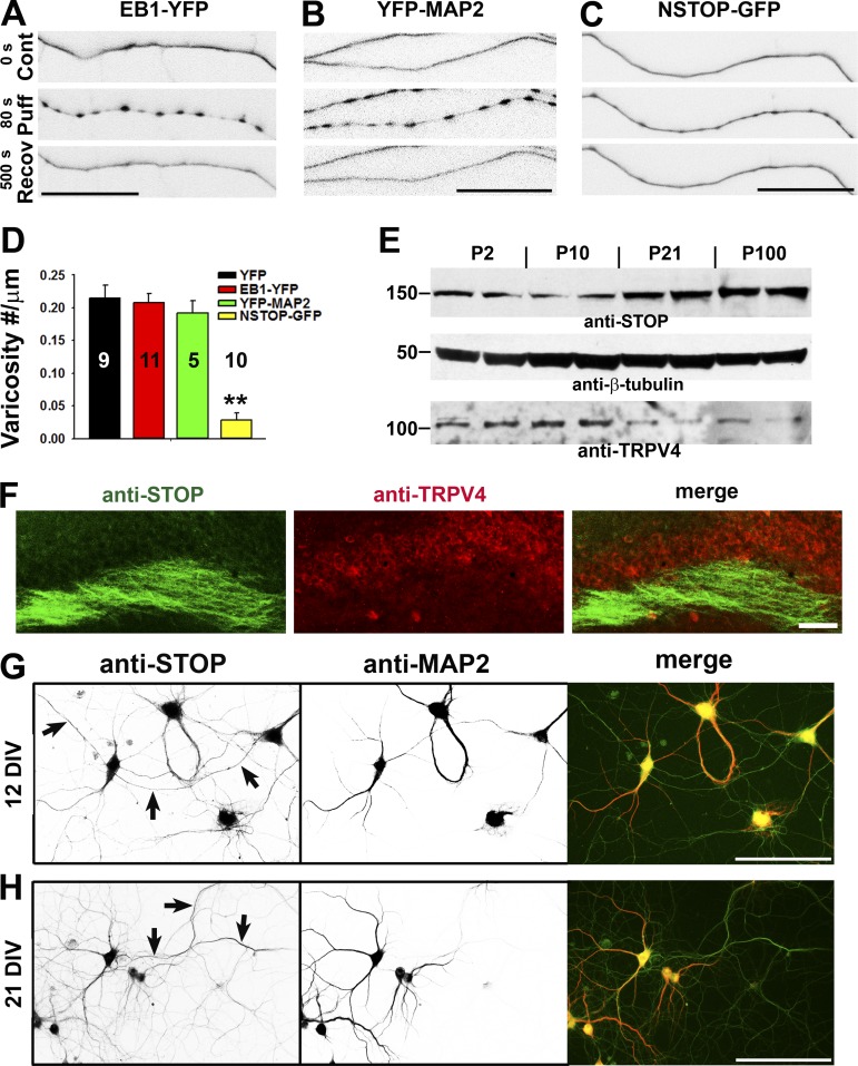
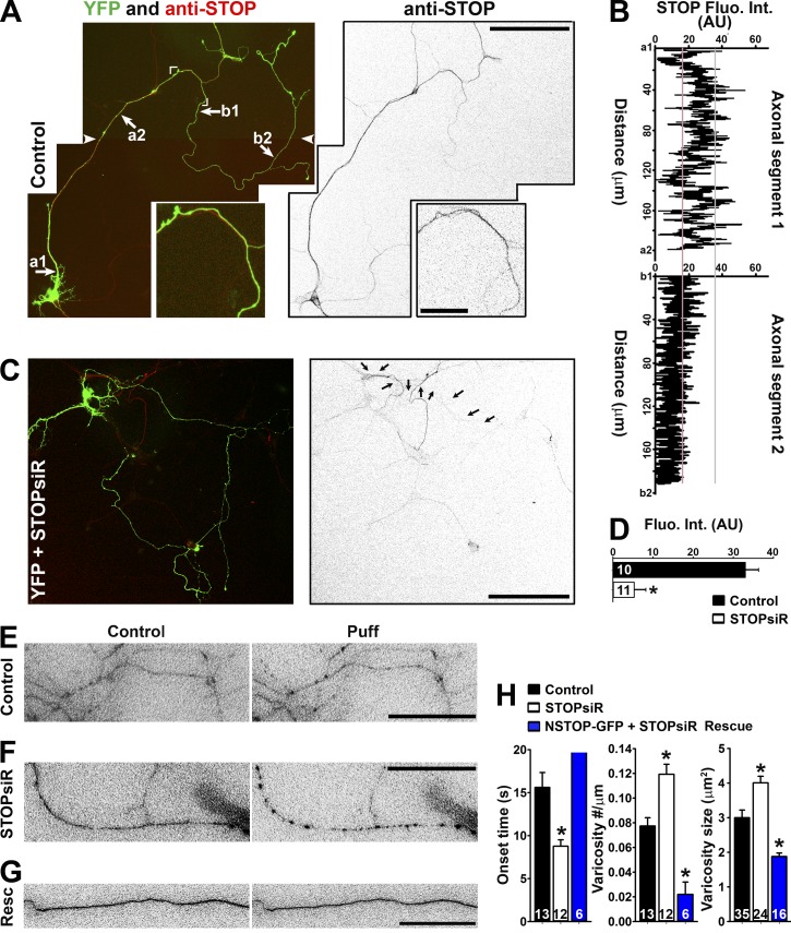
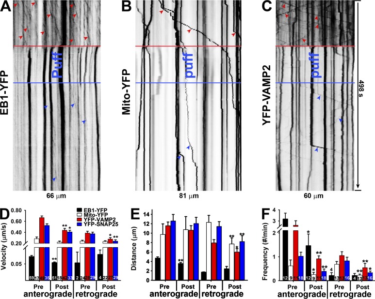
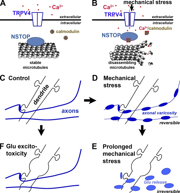
Little is known about mechanical regulation of morphological and functional polarity of central neurons. In this study, we report that mechanical stress specifically induces varicosities in the axons but not the dendrites of central neurons by activating TRPV4, a Ca2+/Na+-permeable mechanosensitive channel. This process is unexpectedly rapid and reversible, consistent with the formation of axonal varicosities in vivo induced by mechanical impact in a mouse model of mild traumatic brain injury. In contrast, prolonged stimulation of glutamate receptors induces varicosities in dendrites but not in axons. We further show that axonal varicosities are induced by persistent Ca2+ increase, disassembled microtubules (MTs), and subsequently reversible disruption of axonal transport, and are regulated by stable tubulin-only polypeptide, an MT-associated protein. Finally, axonal varicosity initiation can trigger action potentials to antidromically propagate to the soma in retrograde signaling. Therefore, our study demonstrates a new feature of neuronal polarity: axons and dendrites preferentially respond to physical and chemical stresses, respectively.


Figures

Figure 1.
Figure 1.
Mechanics-induced rapid and reversible initiation of axonal varicosities. (A) Axonal varicosities along unmyelinated (bottom) but not myelinated axons (top) in the hippocampus of Thy1-YFP transgenic mice. Axons are indicated by YFP fluorescence (green) and myelin segments by the anti-MBP staining (red). (B) Puffing induced rapid and reversible formation of axonal varicosities in cultured hippocampal neurons at 7 DIV, revealed by transmitted lights (top) and transiently expressed YFP (bottom). YFP signals are inverted. Transient or sustained mechanical pressures were delivered by puffing of Hank’s buffer via a glass pipette onto cultured hippocampal neurons as described in Fig. S1. The puffing time was 150 s. (C) Puffing did not induce varicosity formation in proximal axons (7 DIV). Neurons were transfected with YFP. The soma was on the left. (D) Puffing did not affect dendrite morphology (7 DIV). (E) Puffing induced varicosity formation in the middle axon of a neuron at 17 DIV. (F) Puffing induced varicosity formation along axons but not dendrites nor dendritic spines at 17 DIV. Red arrows indicate axonal varicosities. Related quantifications are included in Fig. S2. Bars: (A) 10 µm; (B) 50 µm; (C–F) 15 µm. (G) Older axons (21 DIV) were more resistant to puffing-induced varicosity formation than younger ones (7 DIV). (H) Frequency-dependent varicosity initiation in young axons receiving 2-s puffing pulses. Error bars show means ± SEM. Unpaired t test: *, P < 0.05. 5-s interval versus 20-s interval in H.
Figure 2.
Figure 2.
Axonal varicosity induction in a repetitive closed-skull mTBI mouse model. (A) Low-magnification image of a coronal section from the brain of a Thy1-YFP transgenic mouse that had received impact on the right side. YFP fluorescent signals are inverted. This is a compilation of multiple images. The boxed area indicates the brain region that this study focused on. The black arrow indicates head impact location. (B) YFP+ dendrites and axons in the cortex of a control mouse. The apical dendrites of layer V projection neurons in the cortex point in the upward direction. (C) YFP+ dendrites and axons in the cortex of an mTBI mouse. Cornered areas are provided in the insets to show normal axons (B) and axons with clear varicosities (C). (D) A normal axonal segment from a control mouse. (E) An axonal segment with many varicosities from an mTBI mouse. YFP is in green, and nuclear dye Hoechst is in blue in merged images. (F and G) Partial colocalization of axonal varicosities (left gray images; green in the merged images) with presynaptic markers (middle gray images; red in the merged images) in VAMP2 (F) and Bassoon (G) from mTBI mice. Arrows indicate axonal varicosities (indicated by YFP) containing a presynaptic marker (indicated by anti-VAMP2 or anti-Bassoon staining), and arrowheads indicate axonal varicosities without presynaptic markers. Bars: (B and C, main images) 100 µm; (B and C, insets) 30 µm; (D–G) 20 µm. (H) Percentages of axons with clear varicosities in control and mTBI mice. (I) Percentages of varicosities in axons from mTBI mice without costaining of the presynaptic markers. Error bars indicate means ± SEM. Unpaired t test: *, P < 0.05. anti-B, anti-Bassoon; anti-V, anti-VAMP2.
Figure 3.
Figure 3.
TRPV4 channel plays a major role in axonal varicosity initiation in hippocampal neuron mechanosensation. (A and B) When expressed in HEK293 cells, TRPV4 but not YFP was activated by TRPV4 agonist GSK101 (GSK; 0.5 µM; A) and puffing (B). (C) Summary of the effects of their respective agonist (top) or puffing (bottom) on HEK293 cells expressing YFP, TRPV4, or TRPV1. (D) An axonal segment loaded with calcien AM (inverted signals) before (left) and after (right) puffing. (E) A distal axonal segment loaded with calcien AM (green) without puffing and stained for endogenous TRPV4 (red). (F) Post hoc staining of the axon in D for endogenous TRPV4 (red). (G) Effects of puffing on axons transfected with control (top) or TRPV4 (bottom) siRNA (siR). (H) The onset and degree of varicosity formation along axons with TRPV4 siRNA markedly reduced. (I and J) Under the same puffing condition, axons with TRPV4 siRNA had significantly longer onset time (I) and smaller varicosities (J). (K) Young axons (7 DIV) expressing YFP (green) and control siRNA were stained for endogenous TRPV4 (red in merged image and inverted in grayscale image). (L) Young axons (7 DIV) expressing YFP (green) and TRPV4 siRNA were stained for endogenous TRPV4 (red in merged image and inverted in grayscale image). Arrows indicate coexpressing YFP and double-stranded small RNA. Bars, 20 µm. (M) TRPV4 siRNA significantly reduced TRPV4 staining intensity in both axons and soma of cultured neurons. Error bars indicate means ± SEM. Unpaired t test: **, P < 0.01.
Figure 4.
Figure 4.
MT disassembly in puffing-induced initiation of axonal varicosities. (A and B) Stimulating NMDA receptors (100 µM l-glutamate, 10 µM l-glycine, and 0.5 µM TTX) for 20 min induced varicosities in dendrites (A) but not axons (B) in mature neurons (23 DIV). (C and D) Depolymerizing MTs by Noco (10 µg/ml for 15 min; C) but not actin filaments by LatA (2.5 µM for 2.5 h; D) induced varicosities in axons. (E–G) The onset time (E), density (F), and size (G) of induced varicosities along dendrites or axons under the conditions in A–D. In the drug treatment experiments, the onset time was defined as the time needed to form clear varicosities. Error bars indicate means ± SEM. Unpaired t test: **, P < 0.05. (H) Long axonal segments under control conditions (top) or developing multiple varicosities induced by puffing (bottom) were imaged by TEM. TEM experiments showed ultrastructural changes of axons (top) and dendrites (bottom) under control (7 DIV; I), puffing (7 DIV; J), and Glu treatment (17 DIV; K) conditions. Bars: (A–D) 25 µm; (H–K) 1 µm.
Figure 5.
Figure 5.
Sustained Ca2+ increase in axonal varicosities induced by mechanical stress. (A) An axonal segment before (0 s; control) and after (50 s; Puff) puffing. Signals are inverted. (B) Kymograph of Fluo-4 fluorescence along the axonal segment in A. (C) Fluorescence profiles along the axonal segment at two time points in A. (D) Fluorescence intensities (INT) over time at five different locations indicated by arrowheads in B. (E) Summary of intensity changes of Fluo-4 fluorescence without (Cont) and with (Spot, in a varicosity; Inter-spot, between varicosities) puffing. (F) A dendrite before (0 s) and after (25 s) puffing. Bars, 10 µm. (F) Kymograph of Fluo-4 fluorescence along the dendrite in F. (H) Fluorescence profiles along the dendrite at two time points in F. (I) Fluorescence intensities over time at five different locations indicated by arrowheads in G. (J) Summary of intensity changes of Fluo-4 fluorescence without and with (Dend) puffing. Fcont, fluorescence intensity before puffing; FPeak, the maximal fluorescence intensity reached during puffing; FLate, fluorescence intensity at 80 s; arrows, the puffing onset. Background was not subtracted in C, D, H, or I. Background was subtracted in E and J. Error bars indicate means ± SEM. A one-way ANOVA followed by Dunnett's test was used in E and an unpaired t test was used in J. *, P < 0.05.
Figure 6.
Figure 6.
The level of MT-binding protein NSTOP correlates with the resistance of puffing-induced varicosity formation. (A and B) Axons expressing different MT-binding proteins were puffed, and expressing EB1-YFP (A) or YFP-MAP2 (B) in axons did not change puffing-induced varicosity formation. (C) The axon expressing NSTOP-GFP became more resistant to puffing. (D) Summary of the effects of three MT-binding proteins on axonal varicosity formation. Error bars indicate means ± SEM. One-way ANOVA followed by Dunnett’s test: **, P < 0.01. (E) Expression levels of STOP and TRPV4 in the brain during development. Western blotting for STOP and TRPV4 from mouse brain lysate was performed, and β-tubulin was used as a control. Molecular masses are indicated in kilodaltons. (F) Costaining for endogenous STOP (green) and TRPV4 (red) in the hippocampal slice of an adult mouse. (G and H) Costaining for endogenous STOP (green in merged) and MAP2 (red in merged) was performed in cultured hippocampal neurons at 12 DIV (G) and 21 DIV (H). Arrows indicate MAP2-negative axons containing anti-STOP staining signals. Bars: (A–C) 25 µm; (F–H) 100 µm.
Figure 7.
Figure 7.
Axonal distribution of STOP and its knockdown on varicosity formation. (A) Endogenous STOP (red in merged image on the left; inverted grayscale on the right) expression from neurons at 10 DIV. Cornered area, enlarged (2.5-fold) bottom panels. White arrowheads show the montage line of two overlapping images. (B) STOP fluorescence intensities along two labeled axonal segments in A. (C) Significant reduction of STOP in the neuron transfected with STOP siRNA (siR). Arrows indicate the axon of the transfected neuron. (D) Summary of the effect of STOP siRNA knockdown along middle axons. Mean fluorescence intensity was measured from a segment of middle axons from control (black bar) and STOPsiR (open bar) neurons. Unpaired t test: *, P < 0.05. (E) Middle axons transfected with YFP and control siRNA were puffed. YFP signals were inverted. (F) A middle axon transfected with YFP and STOP siRNA was puffed. (G) A middle axon transfected with NSTOP-GFP (rat) and STOP siRNA was puffed. Resc, Rescue. (H) Summary of the puffing results for the effects of STOP siRNA on onset (left), varicosity number (middle), and size (right). Mouse hippocampal neurons were cultured from mouse pups at P0–P2. Error bars indicate means ± SEM. One-way ANOVA followed by Dunnett's test: *, P < 0.05. Bars: (A [main images] and C) 100 µm; (A, inset) 20 µm; (E–G) 15 µm.
Figure 8.
Figure 8.
Mechanics-induced axonal varicosities are accompanied with transiently halted MT dynamics and axonal transport. (A) Kymograph of EB1-YFP plus end tracking before, during, and after puffing along an axonal segment. (B) Kymograph of Mito-YFP transport before, during, and after puffing along an axonal segment. (C) Kymograph of YFP-VAMP2 transport before, during, and after puffing along an axonal segment. Red arrowheads show movements before puffing, and blue arrowheads show movements after puffing. (D–F) Effects of puffing on moving velocity (D), distance (E), and frequency (F) of EB1-YFP plus end tracking, mitochondria transport, and presynaptic protein transport in anterograde and retrograde directions. Error bars indicate means ± SEM. Unpaired t test between pre- and post-puffing: *, P < 0.05; **, P < 0.01.
Figure 9.
Figure 9.
Axonal varicosity initiation can induce antidromic propagation of action potentials. (A) Experimental diagram of recording back-propagating action potentials initiated by puffing. The YFP-expressing neuron over the transmitted-light background was recorded by a patch pipette at the soma. Its axons received mechanical stimuli provided by the puffing pipette. The red arrow indicates the area containing YFP-positive axons shown before (Control; left) and after (Puff; right) puffing. (B) Simultaneous imaging of an axonal segment (top) and whole-cell current-clamp recording (bottom) of a young neuron (7 DIV) before (left) and after (right) puffing. (C) Simultaneous imaging of an axonal segment (top) and whole-cell current-clamp recording (bottom) of a more mature neuron (16 DIV) before (left) and after (right) puffing. Bars: (A) 30 µm; (B and C) 20 µm. (D) Summary of the time to induce action potentials for all neurons (left) and means ± SEM (right). Gray short lines in the left panel indicated the mean time for axonal varicosity initiation. AP, action potential. Error bars indicate means ± SEM.
Figure 10.
Figure 10.
A new model of polarized initiation of neuronal varicosities induced by mechanical stress. (A) Diagram of signaling molecules on both sides of axonal membrane under control condition. (B) Mechanical stress induces a signaling cascade across the axonal membrane to induce axonal varicosity formation. (C–E) A new model diagram of sequential axonal and dendritic swelling under pathological mechanical stress. (C) Dendrites, dendritic spines, and axons under normal conditions. (D) Under mechanical stress, axonal varicosities start to form while dendrites and dendritic spines remain unchanged. (E) Under prolonged or pathological mechanical stresses, axonal varicosities become irreversible and are eventually broken to release the neurotransmitter glutamate, which can overstimulate its receptors on dendrites to induce swelling and hence neuronal death. (F) Prolonged stimulation of Glu (particularly NMDA) receptors causes swelling in dendrites but not in axons.

Similar articles

Cited by

References

    1. Akhmanova A., and Steinmetz M.O.. 2008. Tracking the ends: a dynamic protein network controls the fate of microtubule tips. Nat. Rev. Mol. Cell Biol. 9:309–322. 10.1038/nrm2369 - DOI - PubMed
    1. Amack J.D., and Manning M.L.. 2012. Knowing the boundaries: Extending the differential adhesion hypothesis in embryonic cell sorting. Science. 338:212–215. 10.1126/science.1223953 - DOI - PubMed
    1. Andrieux A., Salin P.A., Vernet M., Kujala P., Baratier J., Gory-Fauré S., Bosc C., Pointu H., Proietto D., Schweitzer A., et al. . 2002. The suppression of brain cold-stable microtubules in mice induces synaptic defects associated with neuroleptic-sensitive behavioral disorders. Genes Dev. 16:2350–2364. 10.1101/gad.223302 - DOI - PMC - PubMed
    1. Árnadóttir J., and Chalfie M.. 2010. Eukaryotic mechanosensitive channels. Annu. Rev. Biophys. 39:111–137. 10.1146/annurev.biophys.37.032807.125836 - DOI - PubMed
    1. Baas P.W., Rao A.N., Matamoros A.J., and Leo L.. 2016. Stability properties of neuronal microtubules. Cytoskeleton (Hoboken). 73:442–460. - PMC - PubMed

Publication types

MeSH terms

Associated data