. 2017 Jul 15;6(7):1065-1073.
doi: 10.1242/bio.026203.

Cell lineage of timed cohorts of Tbx6-expressing cells in wild-type and Tbx6 mutant embryos

Affiliations

Cell lineage of timed cohorts of Tbx6-expressing cells in wild-type and Tbx6 mutant embryos

Daniel Concepcion et al. Biol Open. .

Abstract

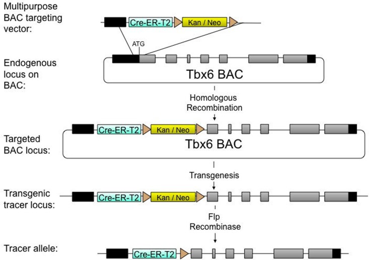
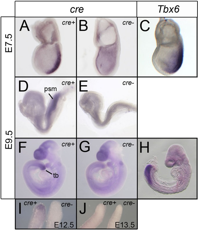
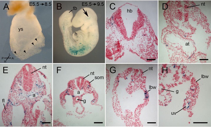
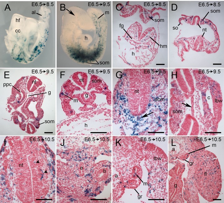
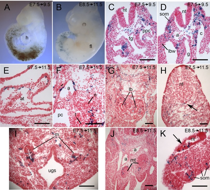
Tbx6 is a T-box transcription factor with multiple roles in embryonic development as evidenced by dramatic effects on mesoderm cell fate determination, left/right axis determination, and somite segmentation in mutant mice. The expression of Tbx6 is restricted to the primitive streak and presomitic mesoderm, but some of the phenotypic features of mutants are not easily explained by this expression pattern. We have used genetically-inducible fate mapping to trace the fate of Tbx6-expressing cells in wild-type and mutant embryos to explain some of the puzzling features of the mutant phenotype. We created an inducible Tbx6-creERT2 transgenic mouse in which cre expression closely recapitulates endogenous Tbx6 expression both temporally and spatially. Using a lacZ-based Cre reporter and timed tamoxifen injections, we followed temporally overlapping cohorts of cells that had expressed Tbx6 and found contributions to virtually all mesodermally-derived embryonic structures as well as the extraembryonic allantois. Contribution to the endothelium of major blood vessels may account for the embryonic death of homozygous mutant embryos. In mutant embryos, Tbx6-creERT2-traced cells contributed to the abnormally segmented anterior somites and formed the characteristic ectopic neural tubes. Retention of cells in the mutant tail bud indicates a deficiency in migratory behavior of the mutant cells and the presence of Tbx6-creERT2-traced cells in the notochord, a node derivative provides a possible explanation for the heterotaxia seen in mutant embryos.

Keywords: Cell fate; Lineage; Mouse; Mutant phenotype; T-box; Tbx6.


Conflict of interest statement

Competing interestsThe authors declare no competing or financial interests.

Figures

Fig. 1.
Fig. 1.
Construction of an inducible, cre lineage-tracing allele under the control of Tbx6 regulatory elements. A multipurpose targeting vector containing homology to the endogenous Tbx6 locus, an inducible cre gene, cre-ERT2, and a kanamycin/neomycin selection cassette surrounded by frt sites was targeted to a BAC containing the Tbx6 locus. Following homologous recombination, the targeted BAC was injected into the pronuclei of mouse zygotes to produce random-insertion transgenic mice. These were bred with mice containing a Flpe transgene to eliminate the selection cassette, resulting in a Tbx6 lineage-tracer allele inducible by tamoxifen.
Fig. 2.
Fig. 2.
Cre expression in Tg(Tbx6-creERT2) transgenic embryos recapitulates endogenous Tbx6 expression. (A-H) Whole mount ISH for Tbx6 (C,H) or cre (A,B,D-G,I,J) with transgene-negative (cre-) controls. At E7.5 cre expression is similar to Tbx6 expression in the primitive streak, excluding the node, although Tbx6 expression extends further into the lateral mesoderm. At E9.5, in embryos of different developmental stages, cre expression is limited to the presomitic mesoderm and tail bud although Tbx6 expression extends somewhat further into the presomitic mesoderm than cre expression. There is high background in the forebrain and otic vesicle in both transgenic and non-transgenic embryos at the advanced E9.5 stage. (I,J) cre expression is present in the tip of the tail of an E12.5 transgenic embryo (left) compared to a nontransgenic control, but expression is lost by E13.5 (J) in most embryos. psm, presomitic mesoderm; tb, tail bud.
Fig. 3.
Fig. 3.
Lineage tracing of a cohort of cells that expressed Tbx6-creERT2 between E6.5 and E7.0. Pregnant Cre-reporter females were injected with tamoxifen at E5.5 and embryos were dissected at E8.5 (A) or E9.5 (B-H) and stained for lacZ. (A) An E8.5 unturned, whole mount embryos with scattered lacZ-positive cells (arrowheads) in the headfolds, lateral mesoderm, primitive streak and base of the allantois. (B) A whole mount E9.5 embryo of approximately 15-20 somites with lacZ-positive cells in the head (arrow), heart, and lateral mesoderm including somites, with lower concentrations in the rostral and caudal somites. (C-H) Representative transverse sections of E9.5 embryos showing lacZ-positive cells in the head mesenchyme at the level of the hindbrain (C), in the atrium of the heart (D), in the endothelium of the dorsal aorta and umbilical vein (E,H), somites, lateral body wall, forelimb bud, intermediate mesoderm and splanchnopleure mesoderm. a, aorta; at, atrium; fl, forelimb bud; g, gut; h, heart; hb, hindbrain; hf, headfold; lbw, lateral body wall; nt, neural tube; som, somite; tb, tail bud; uv, umbilical vein; ys, yolk sac. Compass in A refers only to panel A. Scale bars: 100 μm.
Fig. 4.
Fig. 4.
Lineage tracing of a cohort of cells that expressed Tbx6-creERT2 between E6.75 and E8. Pregnant Cre-reporter females were injected with tamoxifen at E6.5 and embryos were dissected 2-4 days later as indicated on each panel and stained for lacZ. (A) Unturned whole-mount E8.5 embryo with lacZ-positive cells in the allantois, cardiac crescent and laterally along the length of the embryo. (B) A whole-mount E9.5 embryo of approximately 20-25 somites with lacZ-positive cells in the head (arrow), somites, with higher concentration in somites 4-15, lateral body wall, and in a posterior lateral stripe marking the mesonephros. (C-L) Transverse sections of embryos showing representative tissues with labeled cells, notably head mesenchyme (C), heart (C), differentiating somites (C-I), mesonephric ducts (F,K), mesenchyme surrounding the esophagus and bronchi (J), in the lateral body wall and limb (H,K,L), lining of the coelom and pericardio-peritoneal canal (E,H), and genital ridge (K,L). A minority of embryos had labeled cells in the neural tube (arrowheads) (I) and notochord (black arrows in G and H show labeled and unlabeled notochord, respectively). Dorsal is to the top of each panel. a, dorsal aorta; al, allantois; b, bronchus; c, coelom; cc, cardiac crescent; e, esophagus; fg, foregut; fl, forelimb; g, gut; gr, genital ridge; h, heart; hf, head folds; lbw, lateral body wall; m, mesonephric duct; nt, neural tube; ppc, pericardio-peritoneal canal; so, somatopleure; som, somite. Scale bars: 100 μm.
Fig. 5.
Fig. 5.
Lineage tracing of cohorts of cells that expressed Tbx6-creERT2 between E7.75 and E9 or between E8.75 and E10. Pregnant Cre-reporter females were injected with tamoxifen at E7.5 (A,C-I) or E8.5 (J,K) and embryos were dissected 2-4 days later as indicated on each panel and stained for lacZ. (A) A whole-mount embryo labeled at E7.5 with the bulk of the lacZ-positive cells in the somites and lateral mesoderm. (B) A whole-mount embryo labeled at E8.5 with a pronounced posterior shift in the distribution of labeled cells. (C-K) Transverse sections of embryos showing representative tissues with labeled cells, notably somites (C,D,K), lining of the coelom and pericardio-peritoneal canal (C,D), endothelium of the heart and aorta (D-F), mesenchyme surrounding the lung buds and trachea (F,G), mesonephric tubules and metanephric blastema (I,J), and tail mesenchyme (H,K). In a small number of embryos, labeled cells were seen in the posterior neural tube (arrow in H, in the floorplate of the tail neural tube; arrow in K, in tangential section of tail neural tube). a, dorsal aorta; at, atrium; c, coelom; e, esophagus; fg, foregut; fl, forelimb; g, gut; h, heart; lb, lung buds; lbw, lateral body wall; m, mesonephric duct; mt, metanephric blastemal; nt, neural tube; pc, pericardial cavity; ppc, pericardio-peritoneal canal; som, somite; t, trachea; ugc, urogenital sinus. Scale bars: 100 μm.
Fig. 6.
Fig. 6.
Lineage tracing in Tbx6 homozygous mutant embryos of a cohort of cells that expressed Tbx6-creERT2 between E6.5 and E7. Cre-reporter females heterozygous for a Tbx6 null allele were crossed with Tbx6 heterozygous males and were injected with tamoxifen at E6.5; embryos were recovered at E9.5 and stained for lacZ. (A-C) Whole-mount embryos of different developmental stages with lacZ-positive cells in the head region (arrows in B,C), in the irregular anterior somites, in the trunk corresponding to the ectopic neural tubes characteristic of the Tbx6 homozygous phenotype and in the expanded tail bud. The exaggerated twisting of the torso is a feature of the mutant phenotype. (D-H) Representative transverse sections showing lacZ-positive cells in the head mesenchyme (D,E), heart (E), ectopic neural tubes (F-H), notochord (1/7 embryos; arrow in F), lateral body wall (G), mesenchyme surrounding the gut (G) and throughout the mesenchyme of the expanded tail bud (H). ba, branchial arch; bp, branchial pouch; da, dorsal aorta; ec nt, ectopic neural tube; g, gut; h, heart; hg, hindgut; lbw, lateral body wall; nt, neural tube; p, pharynx; tb, tailbud. Scale bars: 100 μm.

Similar articles

Cited by

  • Tbx6 Induces Nascent Mesoderm from Pluripotent Stem Cells and Temporally Controls Cardiac versus Somite Lineage Diversification.
    Sadahiro T, Isomi M, Muraoka N, Kojima H, Haginiwa S, Kurotsu S, Tamura F, Tani H, Tohyama S, Fujita J, Miyoshi H, Kawamura Y, Goshima N, Iwasaki YW, Murano K, Saito K, Oda M, Andersen P, Kwon C, Uosaki H, Nishizono H, Fukuda K, Ieda M. Sadahiro T, et al. Cell Stem Cell. 2018 Sep 6;23(3):382-395.e5. doi: 10.1016/j.stem.2018.07.001. Epub 2018 Aug 9. Cell Stem Cell. 2018. PMID: 30100166 Free PMC article.
  • Rare genetic causes of complex kidney and urological diseases.
    Groopman EE, Povysil G, Goldstein DB, Gharavi AG. Groopman EE, et al. Nat Rev Nephrol. 2020 Nov;16(11):641-656. doi: 10.1038/s41581-020-0325-2. Epub 2020 Aug 17. Nat Rev Nephrol. 2020. PMID: 32807983 Free PMC article. Review.
  • The copy number variation landscape of congenital anomalies of the kidney and urinary tract.
    Verbitsky M, Westland R, Perez A, Kiryluk K, Liu Q, Krithivasan P, Mitrotti A, Fasel DA, Batourina E, Sampson MG, Bodria M, Werth M, Kao C, Martino J, Capone VP, Vivante A, Shril S, Kil BH, Marasà M, Zhang JY, Na YJ, Lim TY, Ahram D, Weng PL, Heinzen EL, Carrea A, Piaggio G, Gesualdo L, Manca V, Masnata G, Gigante M, Cusi D, Izzi C, Scolari F, van Wijk JAE, Saraga M, Santoro D, Conti G, Zamboli P, White H, Drozdz D, Zachwieja K, Miklaszewska M, Tkaczyk M, Tomczyk D, Krakowska A, Sikora P, Jarmoliński T, Borszewska-Kornacka MK, Pawluch R, Szczepanska M, Adamczyk P, Mizerska-Wasiak M, Krzemien G, Szmigielska A, Zaniew M, Dobson MG, Darlow JM, Puri P, Barton DE, Furth SL, Warady BA, Gucev Z, Lozanovski VJ, Tasic V, Pisani I, Allegri L, Rodas LM, Campistol JM, Jeanpierre C, Alam S, Casale P, Wong CS, Lin F, Miranda DM, Oliveira EA, Simões-E-Silva AC, Barasch JM, Levy B, Wu N, Hildebrandt F, Ghiggeri GM, Latos-Bielenska A, Materna-Kiryluk A, Zhang F, Hakonarson H, Papaioannou VE, Mendelsohn CL, Gharavi AG, Sanna-Cherchi S. Verbitsky M, et al. Nat Genet. 2019 Jan;51(1):117-127. doi: 10.1038/s41588-018-0281-y. Epub 2018 Dec 21. Nat Genet. 2019. PMID: 30578417 Free PMC article.
  • Human and mouse studies establish TBX6 in Mendelian CAKUT and as a potential driver of kidney defects associated with the 16p11.2 microdeletion syndrome.
    Yang N, Wu N, Dong S, Zhang L, Zhao Y, Chen W, Du R, Song C, Ren X, Liu J, Pehlivan D, Liu Z, Rao J, Wang C, Zhao S, Breman AM, Xue H, Sun H, Shen J, Zhang S, Posey JE, Xu H, Jin L, Zhang J, Liu P, Sanna-Cherchi S, Qiu G, Wu Z, Lupski JR, Zhang F. Yang N, et al. Kidney Int. 2020 Oct;98(4):1020-1030. doi: 10.1016/j.kint.2020.04.045. Epub 2020 May 22. Kidney Int. 2020. PMID: 32450157 Free PMC article.
  • Evolution of Somite Compartmentalization: A View From Xenopus.
    Della Gaspera B, Weill L, Chanoine C. Della Gaspera B, et al. Front Cell Dev Biol. 2022 Jan 17;9:790847. doi: 10.3389/fcell.2021.790847. eCollection 2021. Front Cell Dev Biol. 2022. PMID: 35111756 Free PMC article.

References

    1. Beddington R. S. P. (1994). Induction of a second neural axis by the mouse node. Development 120, 613-620. - PubMed
    1. Chapman D. L. and Papaioannou V. E. (1998). Three neural tubes in mouse embryos with mutations in the T-box gene Tbx6. Nature 391, 695-697. 10.1038/35624 - DOI - PubMed
    1. Chapman D. L., Agulnik I., Hancock S., Silver L. M. and Papaioannou V. E. (1996). Tbx6, a mouse T-box gene implicated in paraxial mesoderm formation at gastrulation. Dev. Biol. 180, 534-542. 10.1006/dbio.1996.0326 - DOI - PubMed
    1. Chapman D. L., Cooper-Morgan A., Harrelson Z. and Papaioannou V. E. (2003). Critical role for Tbx6 in mesoderm specification in the mouse embryo. Mech. Dev. 120, 837-847. 10.1016/S0925-4773(03)00066-2 - DOI - PubMed
    1. Farley F. W., Soriano P., Steffen L. S. and Dymecki S. M. (2000). Widespread recombinase expression using FLPeR (flipper) mice. Genesis 28, 106-110. 10.1002/1526-968X(200011/12)28:3/4<106::AID-GENE30>3.0.CO;2-T - DOI - PubMed

LinkOut - more resources