Review
. 2017 Sep;102(3):601-615.
doi: 10.1189/jlb.2RI0317-097R. Epub 2017 Jun 12.

Transcriptional and epigenetic regulation of T cell hyporesponsiveness

Affiliations
Review

Transcriptional and epigenetic regulation of T cell hyporesponsiveness

Renata M Pereira et al. J Leukoc Biol. 2017 Sep.

Abstract

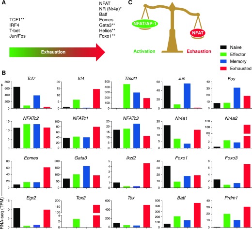
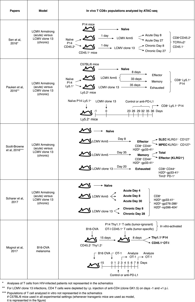
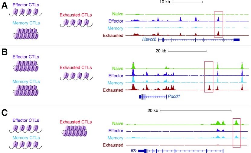
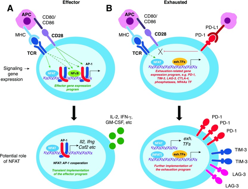
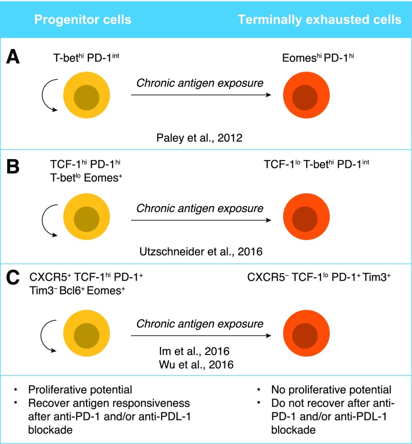
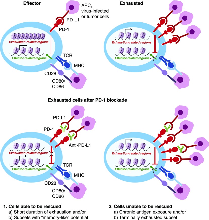
Naive CD8+ T cells differentiate into effector and memory cytolytic T cells (CTLs) during an acute infection. In contrast, in scenarios of persistent antigen stimulation, such as chronic infections and cancer, antigen-specific CTLs show a gradual decrease in effector function, a phenomenon that has been termed CD8+ T cell "exhaustion" or "dysfunction." Another hyporesponsive state, termed "anergy", is observed when T cells are activated in the absence of positive costimulatory signals. Among the many negative regulators induced in hyporesponsive T cells are inhibitory cell-surface receptors, such as PD-1, LAG-3, CTLA-4, and TIM-3; "checkpoint blockade" therapies that involve treatment of patients with cancer with blocking antibodies to those receptors show considerable promise in the clinic because the blocking antibodies can mitigate hyporesponsiveness and promote tumor rejection. In this review, we describe recent advances in our molecular understanding of these hyporesponsive states. We review evidence for the involvement of diverse transcription factors, metabolic programs, and chromatin accessibility changes in hyporesponsive T cells, and we discuss how checkpoint blockade therapies affect the molecular program of CD8+ T cell exhaustion.

Keywords: epigenetic; exhaustion; transcription factors.


Figures

Figure 1.
Figure 1.. Transcriptional regulators of T cell exhaustion.
(A) Relative importance of the indicated transcription factors in exhaustion. Factors shown toward the right are reported to have a more-prominent role in T cell exhaustion. *Transcription factors whose motifs have been identified by ATAC-seq as enriched in accessible-chromatin regions in exhausted cells. **Transcription factors identified by transcriptomic analyses or genetic approaches, but whose motifs have not been identified by ATAC-seq as enriched in accessible-chromatin regions in one population or the other. (B) Expression of indicated transcription factors measured by RNA-seq and displayed as transcripts per million (TPM) in naive (black bars), effector (green bars), memory (blue bars) or exhausted (red bars) cells. Data from Scott-Browne et al. [19]. (C) Schematic diagram showing how an initiating transcription factor NFAT governs effector and exhaustion programs in T cells in the presence or absence of AP-1 cooperation, respectively.
Figure 2.
Figure 2.. Experimental designs used in studies of chromatin accessibility in hyporesponsive CD8 T cells.
Schematic representations of the experimental approaches of 5 research articles that have recently used ATAC-seq to identify accessible-chromatin regions in CD8+ T cell populations in vivo.
Figure 3.
Figure 3.. Genome browser views of representative loci showing differential chromatin accessibility when comparing naive, effector, memory, and exhausted Ag-specific cells in acute and chronic LCMV infection.
(A) The Havcr2 locus, which codes for TIM-3, contains several regions in which the chromatin is similarly accessible in effector and exhausted cells but is less accessible in naive or memory T cells. A representative region is highlighted by the red rectangle. (B) The Pdcd1 locus, which codes for PD-1, contains genomic regions at the promoter and at the distal 5′ enhancer, in which the chromatin is more accessible in exhausted cells compared with naive, effector, or memory cells. (C) The Il7r locus contains a genomic region in which the chromatin is more accessible in memory and naive cells compared with exhausted cells. Representative differentially accessible regions are indicated by red rectangles.
Figure 4.
Figure 4.. In the absence of AP-1 cooperation, NFAT transcription factors drive expression of genes associated with a hyporesponsive state.
(A) Effector CTLs show activated NFAT1, AP-1, and NF-κB transcription factors among others, which together drive the expression of effector-related genes. Many of those genes (e.g., genes encoding cytokines, such as IL-2, IFN-γ, GM-CSF, etc.; see bottom panel) are dependent on NFAT:AP-1 cooperation. (B) Hyporesponsive cells lack or have turned off signals that lead to the induction or activation of AP-1 (Fos-Jun family) factors but sustain calcium signaling at a low level sufficient for NFAT activation. NFAT binds alone, or more likely in cooperation with yet-to-be-determined transcription factors, to NFAT-responsive elements, leading to the induction of genes associated with a hyporesponsive state (e.g., inhibitory receptors and transcription factors as Nr4a members, see bottom panel). In turn, those newly induced transcription factors bind—perhaps with other factors including NFAT itself—to regions that are accessible in hyporesponsive cells, potentially contributing to the maintenance of the hyporesponsive state. Exh. TF, exhaustion-related transcription factors.
Figure 5.
Figure 5.. Proposed models for progenitor and terminally differentiated exhausted T cells.
(A, Left) T-bethi PD-1int cells retain some proliferation potential as well as the ability to produce cytokines. (A, Right) Eomeshi PD-1hi cells have lower proliferation potential as well as lower cytokine production. T-bethi PD-1int cells can give rise to the Eomeshi PD-1hi population upon persistent Ag exposure, thus, are suggested to be “progenitor” and “terminally differentiated” cells, respectively [36, 37]. (B) Progenitor cells have a distinct phenotype characterized by TCF-1hi PD-1hi Eomes+ T-betlo, and that population can give rise to TCF-1lo PD-1int T-bethi [75]. (C) Cells expressing the surface markers CXCR5 and PD-1, as well as transcription factors TCF-1, Bcl6, and Eomes, have a greater proliferation potential and act as progenitors, giving rise to more terminally differentiated, exhausted T cells, which show a reduction in CXCR5 and TCF-1 and an increase in Blimp-1 and TIM-3 expression [78, 79].
Figure 6.
Figure 6.. Anti–PD-1/PD-L1 blockade affects a distinct population of exhausted cells.
Anti–PD-1 or anti–PD-L1 therapies have been recently shown to affect “progenitor” cells (see Fig. 5) and/or cells that have been exposed to Ags for shorter periods [24] and result in only a few chromatin-accessibility changes [27, 39]. Cells able to be reverted temporarily from the exhaustion phenotype are depicted in the left panel, whereas “terminally differentiated” cells that are unable to be rescued from checkpoint blockade are depicted in the right panel.

Similar articles

Cited by

References

    1. Kaech S. M., Cui W. (2012) Transcriptional control of effector and memory CD8+ T cell differentiation. Nat. Rev. Immunol. 12, 749–761. - PMC - PubMed
    1. Weng N. P., Araki Y., Subedi K. (2012) The molecular basis of the memory T cell response: differential gene expression and its epigenetic regulation. Nat. Rev. Immunol. 12, 306–315. - PMC - PubMed
    1. Zediak V. P., Johnnidis J. B., Wherry E. J., Berger S. L. (2011) Cutting edge: persistently open chromatin at effector gene loci in resting memory CD8+ T cells independent of transcriptional status. J. Immunol. 186, 2705–2709. - PMC - PubMed
    1. Zajac A. J., Blattman J. N., Murali-Krishna K., Sourdive D. J., Suresh M., Altman J. D., Ahmed R. (1998) Viral immune evasion due to persistence of activated T cells without effector function. J. Exp. Med. 188, 2205–2213. - PMC - PubMed
    1. Wherry E. J. (2011) T cell exhaustion. Nat. Immunol. 12, 492–499. - PubMed

Publication types

MeSH terms