. 2017 Jul 3;214(7):1913-1923.
doi: 10.1084/jem.20170355. Epub 2017 Jun 12.

The fate and lifespan of human monocyte subsets in steady state and systemic inflammation

Affiliations

The fate and lifespan of human monocyte subsets in steady state and systemic inflammation

Amit A Patel et al. J Exp Med. .

Abstract

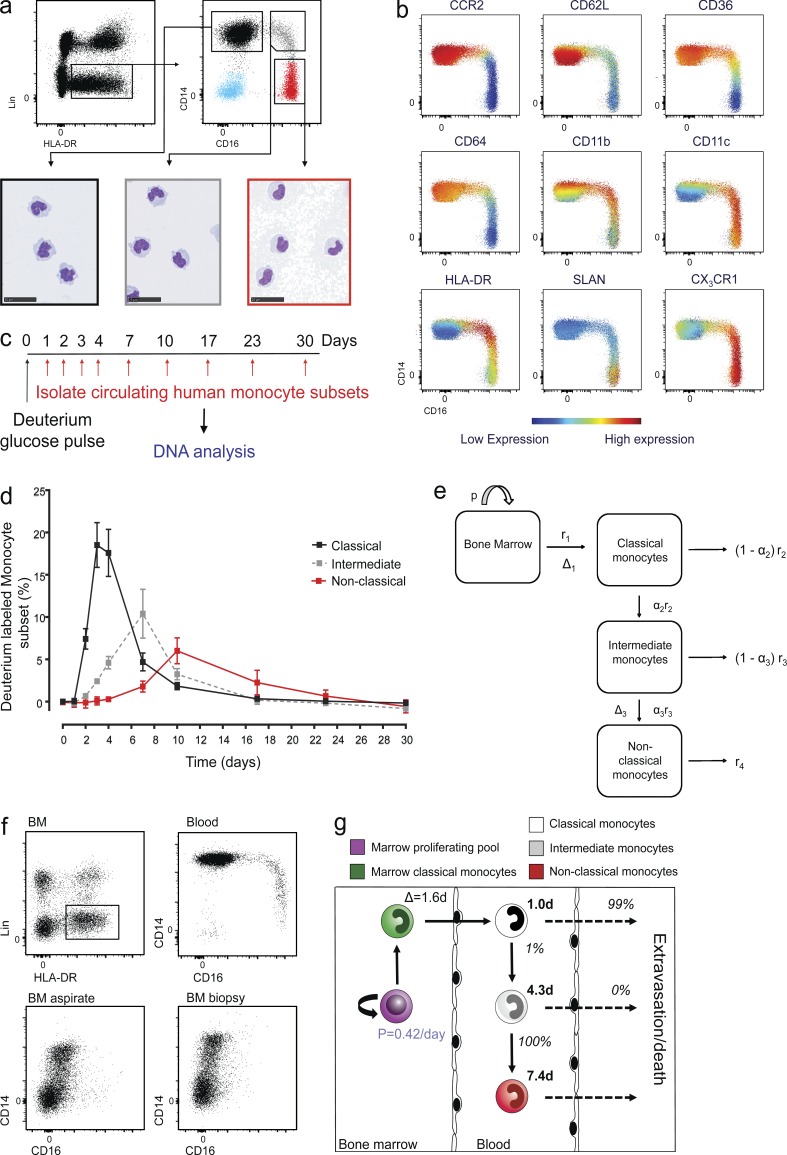
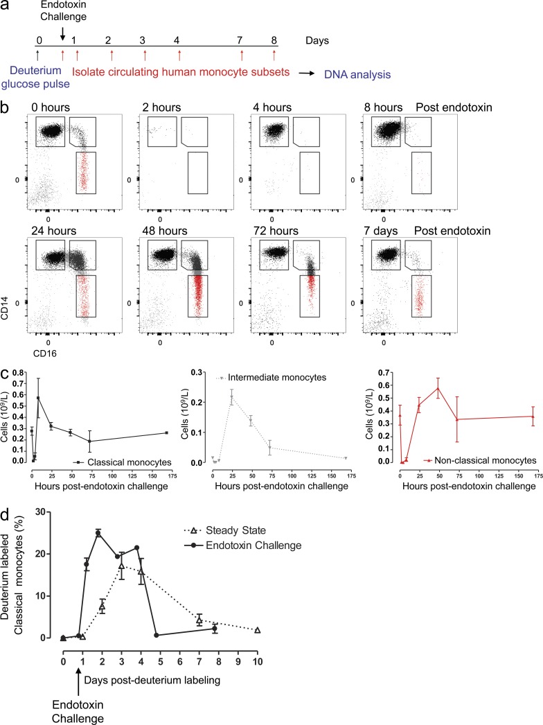
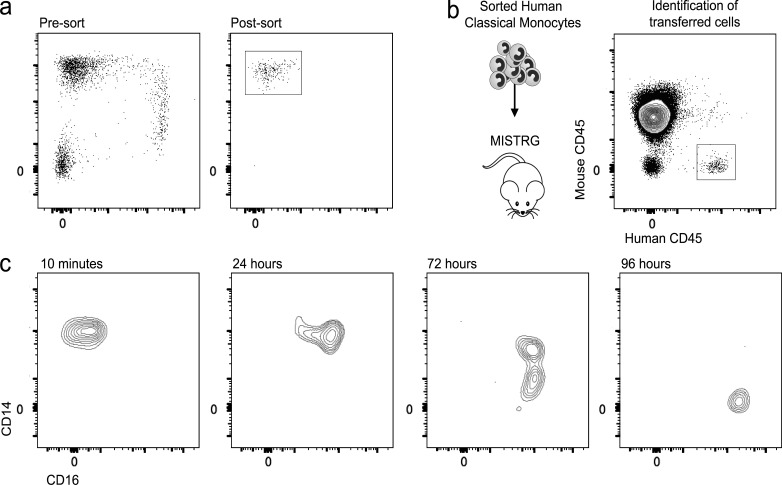
In humans, the monocyte pool comprises three subsets (classical, intermediate, and nonclassical) that circulate in dynamic equilibrium. The kinetics underlying their generation, differentiation, and disappearance are critical to understanding both steady-state homeostasis and inflammatory responses. Here, using human in vivo deuterium labeling, we demonstrate that classical monocytes emerge first from marrow, after a postmitotic interval of 1.6 d, and circulate for a day. Subsequent labeling of intermediate and nonclassical monocytes is consistent with a model of sequential transition. Intermediate and nonclassical monocytes have longer circulating lifespans (∼4 and ∼7 d, respectively). In a human experimental endotoxemia model, a transient but profound monocytopenia was observed; restoration of circulating monocytes was achieved by the early release of classical monocytes from bone marrow. The sequence of repopulation recapitulated the order of maturation in healthy homeostasis. This developmental relationship between monocyte subsets was verified by fate mapping grafted human classical monocytes into humanized mice, which were able to differentiate sequentially into intermediate and nonclassical cells.


Figures

Figure 1.
Figure 1.
In vivo labeling and a methodological approach of modeling human monocyte subset kinetics at steady state. (a) Polychromatic flow cytometry gating strategy for blood monocyte subsets. Peripheral blood mononuclear phagocyte cells were identified as Lin (CD3, CD19, CD20, CD56, CD66b) HLA-DR+ cells. This population comprises classical monocytes (CD14+ CD16: black gated population), intermediate monocytes (CD14+ CD16+: gray gated population), and nonclassical monocytes (CD14lo CD16+: red gated population) representative of >10 subjects. Representative cytospin images from 10 healthy volunteers stained with hematoxylin and eosin (bottom). Bar, 25 µm. (b) Flow cytometry viSNE analysis of monocyte subsets illustrating membrane expression for CCR2, CD62L, CD36, CD64, CD11b, CD11c, HLA-DR, SLAN, and CX3CR1, representative of eight healthy volunteers. (c) Schematic of protocol for labeling newly divided cells. Healthy volunteers received 20 g deuterium-labeled glucose over 3 h. Monocytes subsets were then sorted from whole blood over a 30-d period, DNA was extracted to quantify the deuterium enrichment in each monocyte subset by gas chromatography mass spectrometry. (d) Percentage of deuterium label in peripheral blood classical (black), intermediate (gray), and nonclassical (red) monocytes following oral admission of deuterated glucose in four healthy volunteers; values shown are mean ± SEM. (e) Model of circulating monocyte kinetics. Cartoon depicts the sequential model for the fate of circulating monocyte subsets. Monocytes mature in the bone marrow, where their precursors proliferate at a rate p. Classical monocytes leave the bone marrow at rate r1, after a delay of Δ1 days between the last proliferation and release into the circulation. In the blood, classical monocytes either mature into intermediate monocytes at rate α2 r2, where α = proportion of the subset, or they disappear from the blood (either by death or by moving to other organs) at rate (1–α2)r2. The total disappearance rate is thus r2. Likewise, a proportion α3 of the intermediate monocyte subset develop into nonclassical monocytes, the remainder disappearing from blood. A parameter Δ3 has been included to allow for a potential delay in the differentiation of intermediate monocyte to nonclassical monocytes. (f) Polychromatic flow cytometry comparing BM with circulating monocyte subsets. Human BM was initially gated as LinHLA-DR+. Human BM obtained as either an aspirate or femoral head excavated biopsy was examined by flow cytometry to identify resident monocyte subsets. Only classical monocytes could be detected in the biopsy specimen. These data are representative of three donors for each procedure. (g) Summary of the steady-state kinetics for monocyte subsets. Figures in black bold text denote lifespans in each compartment figures in italics denote the relative probability of each cell undergoing the respective fate (death/disappearance versus phenotype transition). Progenitor cells in the bone marrow proliferate at rate of 0.42/d (blue), where the postmitotic cells remain within the bone marrow for 1.6 d before being released into the circulation as classical monocytes. Classical monocytes contribute 87% to the total monocyte pool, whereas intermediate and nonclassical monocytes make up 5% and 8%, respectively. 99% of classical monocytes leave the circulation, and 1% go onto become intermediate monocytes. 100% of intermediate monocytes mature in the circulation to become nonclassical monocytes under steady state.
Figure 2.
Figure 2.
Sequential reappearance of monocytes subsets after endotoxin challenge. (a) Schematic protocol for administrating deuterium-labeled glucose 20 h before i.v. endotoxin 2 ng/kg in healthy volunteers. Classical monocytes were then sorted from whole blood, DNA extracted, and deuterium enrichment quantified by gas chromatography mass spectrometry over the ensuing 8 d. (b) Flow cytometry analysis of human monocyte subsets at 0, 2, 4, 8, 24, 48, and 72 h and 7 d after i.v. administration of endotoxin, representative of 10 individuals. (c) Time course of absolute monocyte numbers at selected time points following endotoxin challenge for classical, intermediate, and nonclassical monocytes (mean ± SEM × 109/L of three individual subjects; note the different scale for each subset). (d) Comparison of deuterium-labeled classical monocyte egression from the BM under normal physiological conditions (triangles, dashed line, four subjects) and after endotoxin challenge (circles, solid line, three subjects). Values represent mean ± SEM.
Figure 3.
Figure 3.
Development of intermediate and nonclassical human monocytes from classical monocytes. (a) Classical human monocyte LIN HLA-DR+ CD14+ CD16 cells were sorted from healthy blood by FACS. (b) 1.5 × 10⁶ sorted classical monocytes were grafted i.v. into the humanized MISTRG mouse. Grafted cells could be readily identified by expression of the human isoform of CD45 compared with recipient leukocytes expressing mouse CD45. (c) Flow cytometry analysis identified human CD45+ circulating monocytes from MISTRG recipients following adoptive transfer of human CD14+CD16 classical monocytes at 10 min and 24, 72, and 96 h after infusion. Results are representative of three analyzed mice per time point.

Similar articles

Cited by

References

    1. Arnold L., Henry A., Poron F., Baba-Amer Y., van Rooijen N., Plonquet A., Gherardi R.K., and Chazaud B.. 2007. Inflammatory monocytes recruited after skeletal muscle injury switch into antiinflammatory macrophages to support myogenesis. J. Exp. Med. 204:1057–1069. 10.1084/jem.20070075 - DOI - PMC - PubMed
    1. Auffray C., Fogg D., Garfa M., Elain G., Join-Lambert O., Kayal S., Sarnacki S., Cumano A., Lauvau G., and Geissmann F.. 2007. Monitoring of blood vessels and tissues by a population of monocytes with patrolling behavior. Science. 317:666–670. 10.1126/science.1142883 - DOI - PubMed
    1. Avraham-Davidi I., Yona S., Grunewald M., Landsman L., Cochain C., Silvestre J.S., Mizrahi H., Faroja M., Strauss-Ayali D., Mack M., et al. . 2013. On-site education of VEGF-recruited monocytes improves their performance as angiogenic and arteriogenic accessory cells. J. Exp. Med. 210:2611–2625. 10.1084/jem.20120690 - DOI - PMC - PubMed
    1. Bain C.C., Bravo-Blas A., Scott C.L., Gomez Perdiguero E., Geissmann F., Henri S., Malissen B., Osborne L.C., Artis D., and Mowat A.M.. 2014. Constant replenishment from circulating monocytes maintains the macrophage pool in the intestine of adult mice. Nat. Immunol. 15:929–937. 10.1038/ni.2967 - DOI - PMC - PubMed
    1. Bigley V., Haniffa M., Doulatov S., Wang X.N., Dickinson R., McGovern N., Jardine L., Pagan S., Dimmick I., Chua I., et al. . 2011. The human syndrome of dendritic cell, monocyte, B and NK lymphoid deficiency. J. Exp. Med. 208:227–234. 10.1084/jem.20101459 - DOI - PMC - PubMed