Case Reports
. 2017 Jul 3;214(7):1949-1972.
doi: 10.1084/jem.20161759. Epub 2017 Jun 12.

Recurrent rhinovirus infections in a child with inherited MDA5 deficiency

Affiliations
Case Reports

Recurrent rhinovirus infections in a child with inherited MDA5 deficiency

Ian T Lamborn et al. J Exp Med. .

Abstract

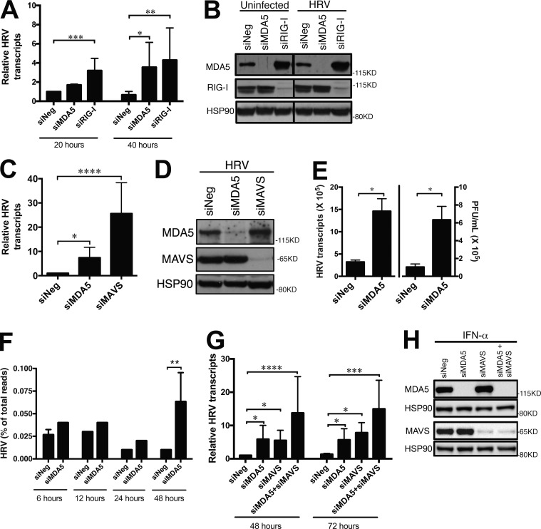
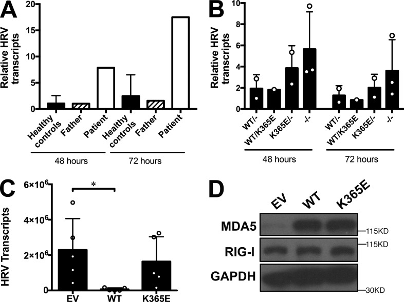
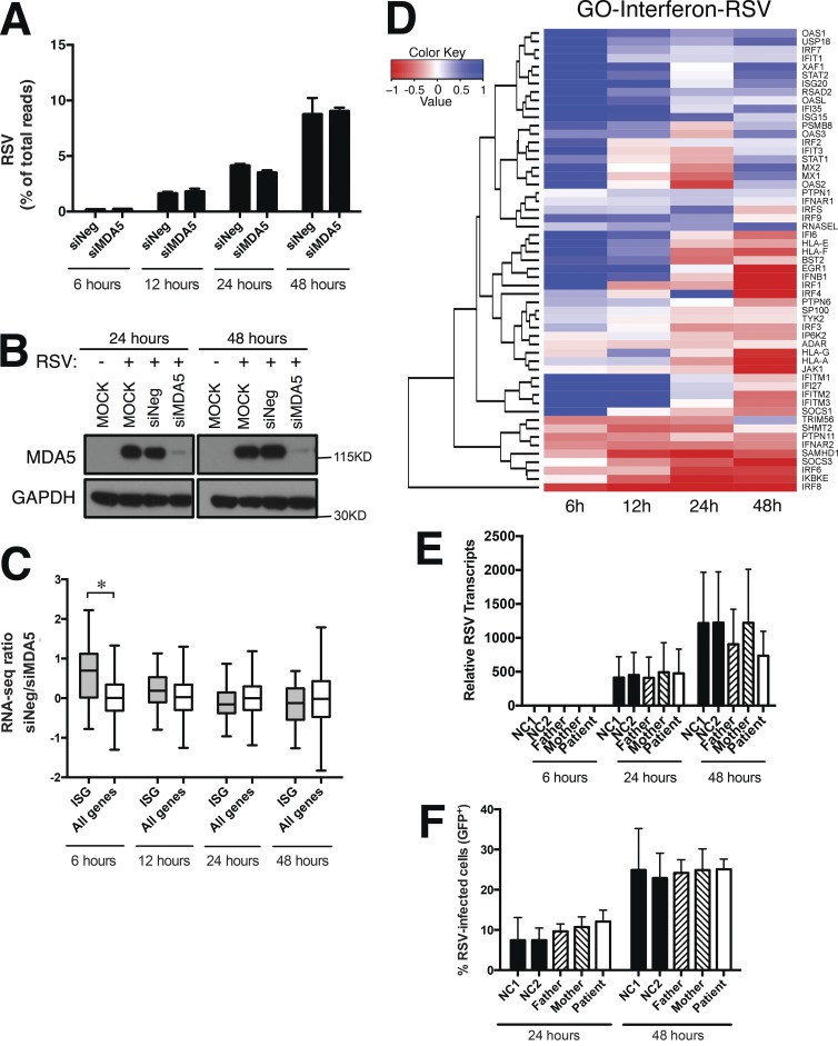
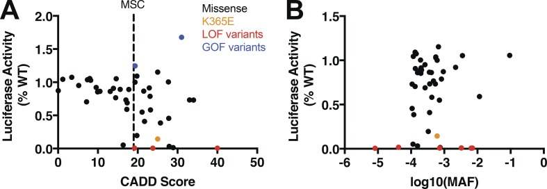
MDA5 is a cytosolic sensor of double-stranded RNA (ds)RNA including viral byproducts and intermediates. We studied a child with life-threatening, recurrent respiratory tract infections, caused by viruses including human rhinovirus (HRV), influenza virus, and respiratory syncytial virus (RSV). We identified in her a homozygous missense mutation in IFIH1 that encodes MDA5. Mutant MDA5 was expressed but did not recognize the synthetic MDA5 agonist/(ds)RNA mimic polyinosinic-polycytidylic acid. When overexpressed, mutant MDA5 failed to drive luciferase activity from the IFNB1 promoter or promoters containing ISRE or NF-κB sequence motifs. In respiratory epithelial cells or fibroblasts, wild-type but not knockdown of MDA5 restricted HRV infection while increasing IFN-stimulated gene expression and IFN-β/λ. However, wild-type MDA5 did not restrict influenza virus or RSV replication. Moreover, nasal epithelial cells from the patient, or fibroblasts gene-edited to express mutant MDA5, showed increased replication of HRV but not influenza or RSV. Thus, human MDA5 deficiency is a novel inborn error of innate and/or intrinsic immunity that causes impaired (ds)RNA sensing, reduced IFN induction, and susceptibility to the common cold virus.


Figures

Figure 1.
Figure 1.
Infection history in a human with recurrent respiratory tract infections. (A) Timeline of pathogens recovered from the respiratory tract. These were classified as positive single-stranded RNA virus, negative single-stranded RNA virus, double-stranded DNA virus, or bacteria as indicated in the adjacent symbol key. (B) RT-PCR molecular typing of RNA isolated from nasopharyngeal samples, using primer sets that preferentially amplify the indicated HRV species. Patient’s samples a to h, as indicated in A, were collected between 2 and 4 yr of age. Sample a was negative for respiratory pathogens and h positive for influenza A. Purified HRV-B14 and -A16 virus stocks, and a sample from a different subject having respiratory symptoms but negative for HRV/enterovirus, were also tested. Results are representative of three repeats. (C) Phylogenetic tree based on nucleotide sequences of 5′UTR of HRV isolates showing evolutionary relationship of the patient’s samples to closest serotypes and representative HRV species.
Figure 2.
Figure 2.
Autosomal recessive, homozygous IFIH1 mutation in the proband. (A) Pedigree indicating genotypes. (B) Confirmatory Sanger sequencing. Schematic below showing mutation location relative to MDA5 protein domains. (C) Ribbon diagram of the MDA5 structure with close-up showing lysine 365 interaction with ribose in RNA, and effects of glutamic acid substitution on distances (Å) between charged groups (yellow sphere, positive; red spheres, negative). (D) Immunoblot of MDA5, RIG-I, and MAVS proteins, relative to β-actin, in cycling T cells, either untreated or treated for 20 h with IFN-α. NC, healthy normal control. Pt, patient. (E) Immunoblot of overexpressed MDA5 (WT or K365E) after affinity precipitation with biotinylated-poly(I:C). D and E are representative of four repeats.
Figure 3.
Figure 3.
Loss-of-function IFIH1 mutation. (A, C, and D) Relative increase in normalized luciferase activity driven by the IFNΒ1 promoter (A), ISRE (C), or NF-κB (D) reporter constructs. Cells were cotransfected with WT or mutant MDA5, and with (filled) or without (open) transfected poly(I:C) as indicated. R337G and R779H are gain-of-function mutants. (B) Immunoblot for MDA5 proteins after transient transfection of 20 ng WT or mutant K365E MDA5, both under CMV promoters. (E) Similar to A, except that cells were cotransfected with 20 ng WT MDA5 (under CMV promoter) and either K365E MDA5 or empty vector (EV; under MSCV promoter). (F) Immunoblot for MDA5 proteins in lysates from E. Data show means ± SD from four (A and E), three (C), and five experiments (D). Data in B and F are examples from experiments shown in A and C–E, respectively. 293T cells lack endogenous MDA5 expression (not depicted). Equivalent lysates from ∼30,000 cells were run across lanes. **, P < 0.01; ***, P < 0.001; ****, P < 0.0001, by one-way ANOVA.
Figure 4.
Figure 4.
Loss of MDA5 function results in increased replication of HRV in respiratory epithelial cells. (A) HRV transcripts, normalized to nonspecific siRNA negative (siNeg) control at 20 h. HRV-B14–infected (MOI: 1) A549 cells were previously transfected with the indicated siRNA. (B) Immunoblot showing efficiencies of MDA5 and RIG-I knockdown in A. Transfected cells were left uninfected or infected with HRV-B14 for 48 h. 120 µg of lysates were run per lane. (C) Similar to A, at 48 h after infection and two rounds of transient transfection with the indicated siRNA. (D) Immunoblot showing efficiencies of MAVS and MDA5 knockdown in C. (E) HRV transcripts and HRV simultaneously quantitated by infectious plaque assay, at 60 h after HRV-B14 infection. (F) Number of HRV reads in RNA-seq data during HRV-B14 infection. A549 cells were previously transfected with MDA5 or nonspecific negative control siRNA (siNeg). Mean of triplicate expression ratios from triplicate infections were shown for each time point. (G) HRV transcripts, normalized to nonspecific siRNA negative (siNeg) control at 48 h, HRV-A16–infected (MOI: 10). Human primary fibroblasts were previously transfected with the indicated siRNA. (H) Immunoblot showing efficiencies of MDA5 and MAVS knockdown in G, except that cells were treated overnight with IFN-α. Data show means ± SD from six (A); eight (C), four (E), and seven experiments (G). Representative experiments are shown in B, D, and H. *, P < 0.05; **, P < 0.01; ***, P < 0.001; ****, P < 0.0001, by Kruskal-Wallis test (A, C, and G); Mann-Whitney U test in (E); and Kolmogorov-Smirnov test (F); other comparisons in A and F were nonsignificant.
Figure 5.
Figure 5.
MDA5 effects on IFN response during HRV infection. (A) “Response to type I interferon” genes (GO term GO0034340) versus all genes. (B) Heatmap representation of RNA-seq data showing HRV-induced IFN-regulated gene transcripts (RPKM) over the course of infection with HRV-B14 (MOI: 1). Data in A show box-and-whisker plots from triplicate infections. A549 cells were previously transfected with MDA5 or nonspecific negative control siRNA (siNeg) and correspond to Fig. 4 F. Mean of triplicate expression ratios from triplicate infections were shown for each time point in B. *, P < 0.05, by Kolmogorov-Smirnov test in A and B; other comparisons were nonsignificant. (C) IFNB1 transcripts were first normalized to β-actin expression and then shown relative to uninfected cells for each siRNA. HRV-A16-infected (MOI: 10) primary human fibroblasts were previously transfected with the indicated siRNA. (D) Similar to C, except for IFNL3 (IL-28) transcripts. (E) Immunoblot showing efficiencies of MAVS and MDA5 knockdown in C and D, except that cells were treated overnight with IFN-α. Data show means ± SD from four independent experiments in C and D, and a representative experiment in E. *, P < 0.05, by Kruskal-Wallis test comparing areas under the curves in C and D.
Figure 6.
Figure 6.
Loss of MDA5 function results in increased replication of HRV in respiratory epithelial cells and fibroblasts. (A) HRV transcripts, normalized to father control at 48 h. Primary nasal epithelial cells from the patient, parents, and six normal healthy controls were infected with HRV-B14 (MOI: 1) as indicated. (B) HRV transcripts in maternal fibroblasts, gene-edited to have IFIH1 genotypes WT/- (2 clones), WT/K365E (1 clone), K365E/- (2 clones), −/− (3 clones), after infection with HRV-B14 (MOI: 1). (C) HRV transcripts in A549 cells previously transduced with empty vector (EV), WT, or K365E MDA5, at 72 h after infection with HRV-B14 (MOI:1). (D) Immunoblot showing MDA5 overexpression and comparable RIG-Iexpression in C. 10.5 µg of lysates were run per lane. Data show means ± SD from two (A), six (B), and five experiments (C). *, P < 0.05; ***, P < 0.001 by Kruskal-Wallis test; comparisons in B and C were nonsignificant.
Figure 7.
Figure 7.
Loss of MDA5 function does not affect replication of influenza virus or production of proinflammatory cytokines in respiratory epithelial cells. (A) Influenza (MOI: 0.1) transcripts, normalized to siNeg control at 24 h. Influenza-infected A549 cells were previously transfected with the indicated siRNA. (B) Immunoblotting showing efficiencies of MDA5, RIG-I, and MAVS protein expression, relative to HSP90 loading control, after transient transfection with the indicated siRNA into A549 cells. Transfected cells were left uninfected or infected with influenza strain A/Victoria/361/2011 (MOI: 0.5) for 24 h or treated with IFN-β (10 IU/ml) for 24 h. 20 µg of lysates were run per lane. Shown is a representative experiment corresponding to A. (C–F) Proinflammatory gene transcripts quantitated by qRT-PCR from A. Levels of IL-1α (C), IL-6 (D), IL-8 (E), and TNF (F) were normalized to β-actin and are shown relative to normalized levels at 8 h after infection. Data show means ± SD from six to seven experiments (A) and three independent experiments in (C–F). *, P < 0.05, by Kruskal-Wallis test (A); all other comparisons were nonsignificant.
Figure 8.
Figure 8.
Loss of MDA5 function does not affect influenza virus replication, or influenza-induced IFN production or cytotoxicity, in respiratory epithelial cells or fibroblasts. (A) Influenza transcripts, normalized to father control at 24 h. Primary nasal epithelial cells from the patient, parents, and two normal healthy controls were infected with influenza (MOI: 0.02). (B) Influenza virus quantitated by infectious plaque assay, after infection (MOI: 1) of SV40-transformed fibroblasts having the indicated genotypes: empty squares, IFIH1−/−; empty inverted triangles, IFIH1K365E/-; solid triangles, IFIH1K365E/WT; empty circles, STAT1−/−; solid circles or solid squares, healthy controls. (C) IFN-β released into supernatants as measured by ELISA after infection with influenza strain A/Puerto Rico/8/1934 (MOI: 0.37–54). Decreasing MOI of influenza virus correspond to the bars proceeding from left to right for each cell line. Sendai Virus (SeV) was included as positive control of IFN induction as the right-most bar for each cell line. Genotypes of SV40-transformed fibroblasts are as indicated, with each cell line separated by vertical dotted lines. (D) LDH released into samples from C. Data show means ± SD from four experiments (A) and three independent experiments (B) that are representative of 11 experiments with varying MOIs (0.1–30) and three different influenza strains (A/Netherlands/602/2009, A/California/4/2009, and A/Puerto Rico/8/1934), and three independent experiments (C and D) that are representative of 6 and 7 experiments, respectively, in which the MOI were varied.
Figure 9.
Figure 9.
Loss of MDA5 function does not affect RSV replication, whereas affecting RSV-induced IFN-regulated transcripts. (A) Number of RSV reads in RNA-seq data during RSV infection. RSV-infected (MOI: 1) A549 cells were previously transfected with MDA5 or nonspecific negative control siRNA (siNeg). (B) Immunoblotting showing efficiencies of MDA5 protein expression, after transient transfection of A549 cells with the indicated siRNA, or without transfection (MOCK). Cells were either left uninfected or infected with RSV, as indicated. 20 µg of lysates were run per lane. Shown is a representative experiment corresponding to A. (C) “Response to type I interferon” genes (GO term GO0034340) versus all genes, from A. (D) Heatmap representation of RNA-seq data showing the expression change between MDA5 siRNA and nonspecific siRNA control of IFN-regulated gene over the course of RSV infection. Mean of triplicate expression ratios from triplicate infections were shown for each time point in A, C, and D. (E) RSV transcripts in primary nasal epithelial cells from the patient (open bar), parents (hatched bars), and two normal healthy controls (solid bars), normalized to father at 6 h, after RSV-GFP infection (MOI: 0.2). (F) Percent GFP+ of gated live RSV-infected cells from (E). Data show means ± SD from four experiments (E and F). *, P < 0.05, by Kruskal-Wallis test (C and D); other comparisons were nonsignificant.
Figure 10.
Figure 10.
Functional activity of population-level missense variants in IFIH1. (A) IFNB1-driven luciferase activity of overexpressed IFIH1 variants, measured as means % of the wild-type from at least three independent experiments, plotted against CADD score (A) or MAF (B). Black, missense variants. Orange, patient K365E variant. Red, loss-of-function (nonsense, splice acceptor/donor, frameshift) variants. Blue, gain-of-function variants. MSC, mutation significance cutoff. Variant information with corresponding plotted values (100 ng of construct) are presented in Table S3.

References

    1. Arora R., Kaplan M., and Nelson M.. 2011. Enterovirus-specific IgG in intravenous immunoglobulin preparations. Ann. Allergy Asthma Immunol. 106:544–545. 10.1016/j.anai.2011.03.007 - DOI - PubMed
    1. Balish A.L., Katz J.M., and Klimov A.I.. 2013. Influenza: propagation, quantification, and storage. Curr Protoc Microbiol Chapter 15:Unit 15G 11. - PubMed
    1. Baños-Lara M.R., Ghosh A., and Guerrero-Plata A.. 2013. Critical role of MDA5 in the interferon response induced by human metapneumovirus infection in dendritic cells and in vivo. J. Virol. 87:1242–1251. 10.1128/JVI.01213-12 - DOI - PMC - PubMed
    1. Benitez A.A., Panis M., Xue J., Varble A., Shim J.V., Frick A.L., López C.B., Sachs D., and tenOever B.R.. 2015. In Vivo RNAi Screening Identifies MDA5 as a Significant Contributor to the Cellular Defense against Influenza A Virus. Cell Reports. 11:1714–1726. 10.1016/j.celrep.2015.05.032 - DOI - PMC - PubMed
    1. Bochkov Y.A., Grindle K., Vang F., Evans M.D., and Gern J.E.. 2014. Improved molecular typing assay for rhinovirus species A, B, and C. J. Clin. Microbiol. 52:2461–2471. 10.1128/JCM.00075-14 - DOI - PMC - PubMed

Publication types

MeSH terms

LinkOut - more resources