. 2017 Jun 27;114(26):6782-6787.
doi: 10.1073/pnas.1700868114. Epub 2017 Jun 12.

Ca2+-induced movement of tropomyosin on native cardiac thin filaments revealed by cryoelectron microscopy

Affiliations

Ca2+-induced movement of tropomyosin on native cardiac thin filaments revealed by cryoelectron microscopy

Cristina Risi et al. Proc Natl Acad Sci U S A. .

Abstract

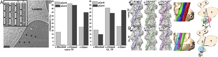
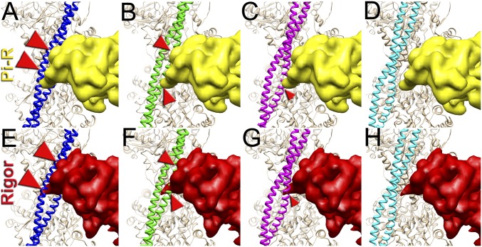
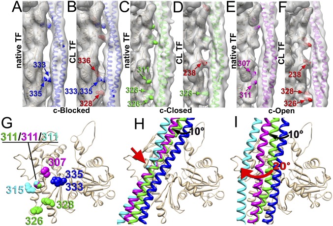
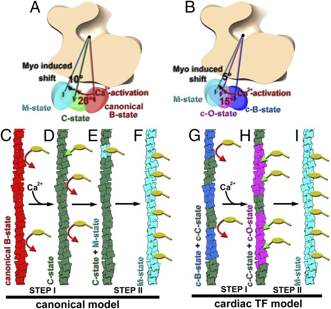
Muscle contraction relies on the interaction of myosin motors with F-actin, which is regulated through a translocation of tropomyosin by the troponin complex in response to Ca2+ The current model of muscle regulation holds that at relaxing (low-Ca2+) conditions tropomyosin blocks myosin binding sites on F-actin, whereas at activating (high-Ca2+) conditions tropomyosin translocation only partially exposes myosin binding sites on F-actin so that binding of rigor myosin is required to fully activate the thin filament (TF). Here we used a single-particle approach to helical reconstruction of frozen hydrated native cardiac TFs under relaxing and activating conditions to reveal the azimuthal movement of the tropomyosin on the surface of the native cardiac TF upon Ca2+ activation. We demonstrate that at either relaxing or activating conditions tropomyosin is not constrained in one structural state, but rather is distributed between three structural positions on the surface of the TF. We show that two of these tropomyosin positions restrain actomyosin interactions, whereas in the third position, which is significantly enhanced at high Ca2+, tropomyosin does not block myosin binding sites on F-actin. Our data provide a structural framework for the enhanced activation of the cardiac TF over the skeletal TF by Ca2+ and lead to a mechanistic model for the regulation of the cardiac TF.

Keywords: cardiac muscle regulation; cryoelectron microscopy; thin filament.


Conflict of interest statement

The authors declare no conflict of interest.

Figures

Fig. 1.
Fig. 1.
Three-dimensional reconstruction of frozen hydrated native and cross-linked cardiac TFs. (A) Electron micrograph of frozen hydrated cardiac TFs (pCa = 4) shows that some filaments possess well-defined Tn densities (black arrows), whereas others do not (white arrow). (Inset) Examples of segments of native and cross-linked (CL) TFs at low (pCa > 8) and high (pCa = 4) Ca2+ used for image analysis. Tn complexes are marked with black arrowheads. (B) Pseudoatomic models of the canonical-blocked (14), apo (15), and myosin (17) structural states of Tpm were used as reference structures in cross-correlation sorting of native and CL TFs at low (pCa > 8) and high (pCa = 4) Ca2+. (C and D) Three-dimensional reconstructions of native (C) and CL (D) cardiac TFs possessing Tpm in the c-blocked (blue ribbons), c-closed (green ribbons), or c-open (magenta ribbons) structural states. Density maps are shown as transparent gray surfaces, whereas actin subunits are tan ribbons. (E) Position of Tpm in the c-blocked state (blue surface) is compared with the previously observed canonical-blocked (14) (red surface) and apo (15) (green surface) positions of Tpm on F-actin. The top view shows an ∼10° swing of Tpm in the c-blocked state from its apo state. (F) Tpm position in the c-open state (magenta surface) is compared with the previously defined apo (15) (green surface) and myosin (17) (cyan surface) positions on F-actin. The top view shows that the c-open state is located between the apo and the myosin positions of Tpm on F-actin.
Fig. 2.
Fig. 2.
Superimposition of the c-blocked, c-closed, and c-open structural states with the Pi-release (Pi-R) (AD) and rigor (EH) states of myosin. (AD) Tpm in either c-blocked (A) or c-closed (B) positions has a severe sterical clash with myosin in the Pi-R state (large and medium red arrowheads, respectively), whereas in the c-open state only a minor sterical hindrance is present (C, small red arrowhead). In the c-myosin state myosin and Tpm have no clashes (D). (EH) In the c-blocked and c-closed structural states Tpm yielded an overwhelming sterical clash with the rigor-bound myosin head (E and F, large and medium red arrowheads, respectively). In the c-open state only a small portion of loop-4 of the myosin head clashes with Tpm (G, small red arrowhead). (H) Atomic model of rigor myosin–Tpm–F-actin complex (17).
Fig. 3.
Fig. 3.
Contacts of Tpm with F-actin in the c-blocked (A and B), c-closed (C and D), and c-open (E and F) states of the native (A, C, and E) and CL (B, D, and F) cardiac TFs. Residues of F-actin involved in the interaction with Tpm in native TF are marked in blue (A), green (D), and magenta (E), whereas actin residues presumably CL to Tpm by glutaraldehyde (B, D, and F) are marked in red. (G) In the native cardiac TF the Tpm interface on actin comprises residues located in SD3 of actin: 333/335 for the c-blocked state (blue spheres), 326/328/311 for the closed state (green spheres), and 307/311 for the c-open state (magenta spheres). (H) The transition of the inner Tpm strand, which interacts with F-actin, between the c-blocked (blue ribbons) and c-closed (green ribbons) state requires a 10° azimuthal movement of Tpm (black arrow), whereas the transition between the c-closed (green ribbons), c-open (magenta ribbons), and c-myosin (cyan ribbons) states involves rocking movement around a common anchor point (red arrow). (I) The outer Tpm strand, which is distal from F-actin, makes an ∼20° swing upon transition from the c-closed (green ribbons) to the c-myosin (cyan ribbons) structural state (red arrow). Actual density maps are shown as transparent gray surfaces, whereas actin molecules are tan ribbons.
Fig. 4.
Fig. 4.
Model for the Ca2+-dependent activation of the TF. (A) In the canonical model derived from the negatively stained TFs the swing of the Tpm cable from the canonical-blocked to the closed structural state upon Ca2+-induced activation of the TF is ∼25° (red arrow), whereas rigor-bound myosin adds an additional ∼10° azimuthal rotation of Tpm (black arrow) (5). (B) CryoEM-based model for the activation of the cardiac TF shows an ∼15° swing of Tpm upon Ca2+-induced activation of the cardiac TF (red arrow), whereas rigor-bound myosin adds an additional ∼5° azimuthal rotation of the Tpm cable toward subdomain 4 of actin (black arrow). (CI) Comparison of the canonical model for the TF activation (CF) with the model for cardiac TF activation (GI). (C) At low Ca2+ Tpm in the canonical-blocked state (red) blocks myosin heads (yellow) binding to F-actin (red arrows). (D) Upon Ca2+-induced activation of the TF, Tpm moves to the closed structural state (green) partially exposing myosin binding sites so that a few myosin molecules (yellow) can bind to the TF (green arrow). (E) Strongly bound myosin heads locally activate the TF by switching Tpm from its closed (green) to myosin (cyan) state. (F) The activated myosin Tpm state cooperatively propagates along the TF. (G) At low Ca2+ Tpm in the cardiac TF is in either c-blocked (blue) or c-closed (green) inhibitive states which block myosin (yellow) binding to F-actin (red arrows). (H) Upon Ca2+-induced activation of the cardiac TF Tpm occupies either c-closed (green) or c-open structural state (magenta). In c-open structural state myosin binding sites are almost fully exposed and myosin molecules (yellow) freely bind to the TF (green arrows). (I) Strongly bound myosin heads shift Tpm further from either c-closed or c-open into the myosin (cyan) structural state.

References

    1. Brown JH, Cohen C. Regulation of muscle contraction by tropomyosin and troponin: How structure illuminates function. Adv Protein Chem. 2005;71:121–159. - PubMed
    1. Spudich JA, Huxley HE, Finch JT. Regulation of skeletal muscle contraction. II. Structural studies of the interaction of the tropomyosin-troponin complex with actin. J Mol Biol. 1972;72:619–632. - PubMed
    1. Vibert P, Craig R, Lehman W. Steric-model for activation of muscle thin filaments. J Mol Biol. 1997;266:8–14. - PubMed
    1. McKillop DFA, Geeves MA. Regulation of the interaction between actin and myosin subfragment 1: Evidence for three states of the thin filament. Biophys J. 1993;65:693–701. - PMC - PubMed
    1. Pirani A, et al. Single particle analysis of relaxed and activated muscle thin filaments. J Mol Biol. 2005;346:761–772. - PubMed

Publication types

LinkOut - more resources