. 2017 Jun 27;114(26):E5216-E5225.
doi: 10.1073/pnas.1705795114. Epub 2017 Jun 12.

Systems-guided forward genetic screen reveals a critical role of the replication stress response protein ETAA1 in T cell clonal expansion

Affiliations

Systems-guided forward genetic screen reveals a critical role of the replication stress response protein ETAA1 in T cell clonal expansion

Lisa A Miosge et al. Proc Natl Acad Sci U S A. .

Abstract

T-cell immunity requires extremely rapid clonal proliferation of rare, antigen-specific T lymphocytes to form effector cells. Here we identify a critical role for ETAA1 in this process by surveying random germ line mutations in mice using exome sequencing and bioinformatic annotation to prioritize mutations in genes of unknown function with potential effects on the immune system, followed by breeding to homozygosity and testing for immune system phenotypes. Effector CD8+ and CD4+ T-cell formation following immunization, lymphocytic choriomeningitis virus (LCMV) infection, or herpes simplex virus 1 (HSV1) infection was profoundly decreased despite normal immune cell development in adult mice homozygous for two different Etaa1 mutations: an exon 2 skipping allele that deletes Gly78-Leu119, and a Cys166Stop truncating allele that eliminates most of the 877-aa protein. ETAA1 deficiency decreased clonal expansion cell autonomously within the responding T cells, causing no decrease in their division rate but increasing TP53-induced mRNAs and phosphorylation of H2AX, a marker of DNA replication stress induced by the ATM and ATR kinases. Homozygous ETAA1-deficient adult mice were otherwise normal, healthy, and fertile, although slightly smaller, and homozygotes were born at lower frequency than expected, consistent with partial lethality after embryonic day 12. Taken together with recently reported evidence in human cancer cell lines that ETAA1 activates ATR kinase through an exon 2-encoded domain, these findings reveal a surprisingly specific requirement for this ATR activator in adult mice restricted to rapidly dividing effector T cells. This specific requirement may provide new ways to suppress pathological T-cell responses in transplantation or autoimmunity.

Keywords: DNA damage; Etaa1; T cell; immunity; replication stress.


Conflict of interest statement

The authors declare no conflict of interest.

Figures

Fig. 1.
Fig. 1.
Etaa1 is required for effector T-cell expansion after immunization and LCMV infection. (A) Schematic of the Etaa1 gene, with exons numbered, coding sequence in blue, and location of the ΔEx2 splice and C166X mutations. (B–D) Representative and compiled data showing KLRG1hi effector T-cell percentage (B and C) and CGG antibody titer (D) in blood at day 14 after immunization of Etaa1+/+ or Etaa1ΔEx2/ΔEx2 mice with CGG and inactivated B. pertussis. (E) Agarose gel electrophoresis of PCR-amplified cDNA using primers in Etaa1 exons 1 and 5 from Etaa1+/+ or Etaa1ΔEx2/ΔEx2 spleen cells. The mRNA structure of the indicated bands deduced by Sanger sequencing is shown on the right. (F) Wild-type or ΔEx2 mutant Etaa1 cDNA sequences fused to GFP were transiently transfected into HEK293T cells. Western blot analysis was conducted on total cell lysates prepared at 48 h after transfection to assess GFP-fused ETAA1 or control β-actin protein expression. (G) Etaa1+/+, Etaa1C166X/C166X, or Etaa1ΔEx2/ΔEx2 mice were infected with LCMV-Arm, and the numbers of splenic CD8+GP33–41, CD8+NP396–404, and CD4+GP66–77 tetramer-positive cells were assessed at day 8 postinfection. The dotted line in each graph indicates the limit of detection based on staining of uninfected animals. ***P < 0.001, t test (C and D) or one-way ANOVA (G).
Fig. 2.
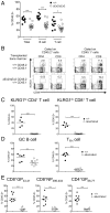
Fig. 2.
Etaa1 control of effector T-cell expansion is cell intrinsic. Rag1−/− mice were lethally irradiated and reconstituted with a 50:50 mixture of CD45.1+ wild-type bone marrow and CD45.2+ Etaa1+/+ or Etaa1ΔEx2/ΔEx2 bone marrow. (A) Contributions of CD45.2+ Etaa1+/+ and Etaa1ΔEx2/ΔEx2 cells to total lymphocytes, B cells, and T cells in competitive mixed bone marrow chimeras, measured in blood before immunization or infection. (B and C) Wild-type and mutant mixed bone marrow chimeras were immunized with CGG and inactivated B. pertussis, and blood was analyzed at day 28 postimmunization. (B) Representative plots showing the percentage of CD45.2+ and CD45.1+ cells falling within the KLRG1hi effector T-cell gate. (C) %CD45.2+ cells among all KLRG1hi effector T cells in individual wild-type or mutant mixed bone marrow chimeras. To correct for individual differences in overall reconstitution by CD45.2+ marrow in A, each postimmunization value was normalized to the %CD45.2+ cells among all CD4 or CD8 cells in the same chimeric mouse before immunization. (D) %CD45.2+ cells within germinal center B cells (GC; B220+GL-7+CD95+) and TFH cells (CD4+PD-1hiCXCR5hi) in the spleen on day 7 postimmunization with SRBCs, normalized to the %CD45.2+ cells in all B cells or to the CD4 T cells in the same chimera before immunization. (E) %CD45.2+ splenic cells within the CD8+GP33–41, CD8+NP396–404, and CD4+GP66–77 tetramer-positive cells at day 8 postinfection with LCMV-Arm in either wild-type or mutant mixed bone marrow chimeras, normalized to the %CD45.2+ cells among all CD8 cells in the same chimera before infection. ***P < 0.001, **P < 0.01, t test.
Fig. 3.
Fig. 3.
Etaa1-deficient mice display little or no steady-state phenotype. (A) Fecundity of male Etaa1ΔEx2/ΔEx2 mice vs. male Etaa1ΔEx2/+ controls, expressed as the number of pups sired per male over a 6-mo period. (B) Length, weight, and BMI of young (8–12 wk) or old (1 y) Etaa1+/+ or Etaa1ΔEx2/ΔEx2 mice. (C) Survival curves of Etaa1+/+, Etaa1ΔEx2/+, and Etaa1ΔEx2/ΔEx2 mice. (D) Numbers of thymic (Left) and splenic (Right) T-cell subsets, defined as follows: double-negative (DN), B220CD4CD8; double-positive (DP), B220CD4+CD8+; CD4 single-positive (SP), B220CD4+CD8; CD8 SP, B220CD4CD8+; naïve, CD4+ or CD8+ and CD62LhiCD44lo; Tcm, CD4+ or CD8+ and CD62LhiCD44hi; Tem (or effector), CD4+ or CD8+ and CD62LloCD44hi; and Treg, CD4+Foxp3+CD25int-hi. **P < 0.01; *P < 0.05; ns, not significant by t test.
Fig. 4.
Fig. 4.
Etaa1-deficient mice exhibit defective T-cell responses to HSV1 infection. Etaa1+/+ or Etaa1ΔEx2/ΔEx2 mice were infected on the flank with HSV1. (A and B) At day 19 postinfection, splenic CD8+gB498–505+ dextramer cells, or IFN-γ+ CD8+ cells identified after stimulation with the listed peptides, were enumerated. (C) Mean ± SEM size of the flank lesion over the course of infection (n = 6). ***P < 0.001, **P < 0.01, t test.
Fig. 5.
Fig. 5.
Etaa1-deficient T cells differentiate into functional effector cells. Etaa1+/+ and Etaa1ΔEx2/ΔEx2 mice were infected with LCMV-Arm, and at day 8 postinfection either CD8+GP33–41 (A–D) or CD4+GP66–77 (E) tetramer-positive cells were assessed for short-lived effector (CD127loKLRG1hi) and memory precursor (CD127hiKLRG1lo and potentially CD62Lint-hi) markers (A and B), effector function (GzmB expression and IFN-γ, TNFα, and IL-2 production) (C and D), and TH1 (PSGL1hiLy6Chi), CD4 memory precursor (PSGL1hiLy6Clo) and TFH (PSGL1lo) differentiation (E). ***P < 0.001; **P < 0.01; ns, not significant, t test.
Fig. 6.
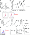
Fig. 6.
Etaa1-deficient T cells exhibit normal proliferation and tissue migration. (A and B) CD45.2+ C57BL/6 mice were injected i.v. with 5 × 10⁴ CD45.1+ Etaa1+/+ or Etaa1ΔEx2/ΔEx2 P14 cells before infection with LCMV-Arm. (A) Splenic P14 cells enumerated at days 5 and 8 postinfection showing mean and SD (n = 10–14/group). (B) P14 numbers in the indicated tissues on day 8 postinfection (C) CD45.1+ wild-type and CD45.2+ Etaa1ΔEx2/ΔEx2 splenocytes were mixed in a 50:50 ratio, labeled with CTV, and stimulated with 1 µg/mL anti-CD3. Then 3–4 d later, CTV dilution in CD4+ and CD8+ T cells from wild-type (CD45.1+) and mutant (CD45.2+) cells was compared. (D) Etaa1+/+ and Etaa1ΔEx2/ΔEx2 P14 cells were stimulated in vitro with GP33–41 peptide and IL-2, live P14 cells were enumerated (expressed as fold change over starting cell number), and Ki67 expression and viability (% 7AAD Live/Dead dye) were measured by flow cytometry at each timepoint. (E) CD45.2+ C57BL/6 mice were injected i.v. with 8 × 10⁵ CTV-labeled CD45.1+ Etaa1+/+ or Etaa1ΔEx2/ΔEx2 P14 cells before infection with LCMV-Arm. At day 2.5 postinfection, CTV dilution in the transferred P14 cells was measured by flow cytometry, with the CTV MFI in divided P14 cells from each mouse normalized to the average MFI in wild-type P14 cells in the same experiment. Data are pooled from two experiments. (F) The percentage of Ki67+ P14 cells at day 5 postinfection in the experiment outlined in A and B. ***P < 0.001; ns, not significant, two-way ANOVA (A) or t test (B, E, and F).
Fig. 7.
Fig. 7.
Etaa1-deficient T cells have an enhanced DNA damage response. CD45.2+ C57BL/6 mice were injected i.v. with 5 × 10⁴ CD45.1+ Etaa1+/+ or Etaa1ΔEx2/ΔEx2 P14 cells before infection with LCMV-Arm. (A) The Etaa1+/+ and Etaa1ΔEx2/ΔEx2 P14 cells were sorted at day 5 postinfection, and gene expression was analyzed by RNA-seq. The plot shows GSEA demonstrating a significant up-regulation of the p53 (TP53)-induced genes within the ranked RNA-seq data (ordered from most up-regulated to most down-regulated in mutant vs. wild-type cells). NES, normalized enrichment score; FDR q, false discovery rate q score. (B and C) Representative and compiled data of γH2AX staining within Etaa1+/+ and Etaa1ΔEx2/ΔEx2 P14 cells at day 5 (B and C) and day 8 (C) postinfection ***P < 0.001, t test.

References

    1. Masopust D, Murali-Krishna K, Ahmed R. Quantitating the magnitude of the lymphocytic choriomeningitis virus-specific CD8 T-cell response: It is even bigger than we thought. J Virol. 2007;81:2002–2011. - PMC - PubMed
    1. Zhang N, Bevan MJ. CD8(+) T cells: Foot soldiers of the immune system. Immunity. 2011;35:161–168. - PMC - PubMed
    1. Yoon H, Kim TS, Braciale TJ. The cell cycle time of CD8+ T cells responding in vivo is controlled by the type of antigenic stimulus. PLoS One. 2010;5:e15423. - PMC - PubMed
    1. O’Sullivan D, Pearce EL. Targeting T cell metabolism for therapy. Trends Immunol. 2015;36:71–80. - PMC - PubMed
    1. Johnson TS, et al. Etoposide selectively ablates activated T cells to control the immunoregulatory disorder hemophagocytic lymphohistiocytosis. J Immunol. 2014;192:84–91. - PMC - PubMed

Publication types

MeSH terms