. 2017 Jun 27;114(26):6842-6847.
doi: 10.1073/pnas.1703228114. Epub 2017 Jun 12.

Gel-forming mucins form distinct morphologic structures in airways

Affiliations

Gel-forming mucins form distinct morphologic structures in airways

Lynda S Ostedgaard et al. Proc Natl Acad Sci U S A. .

Abstract

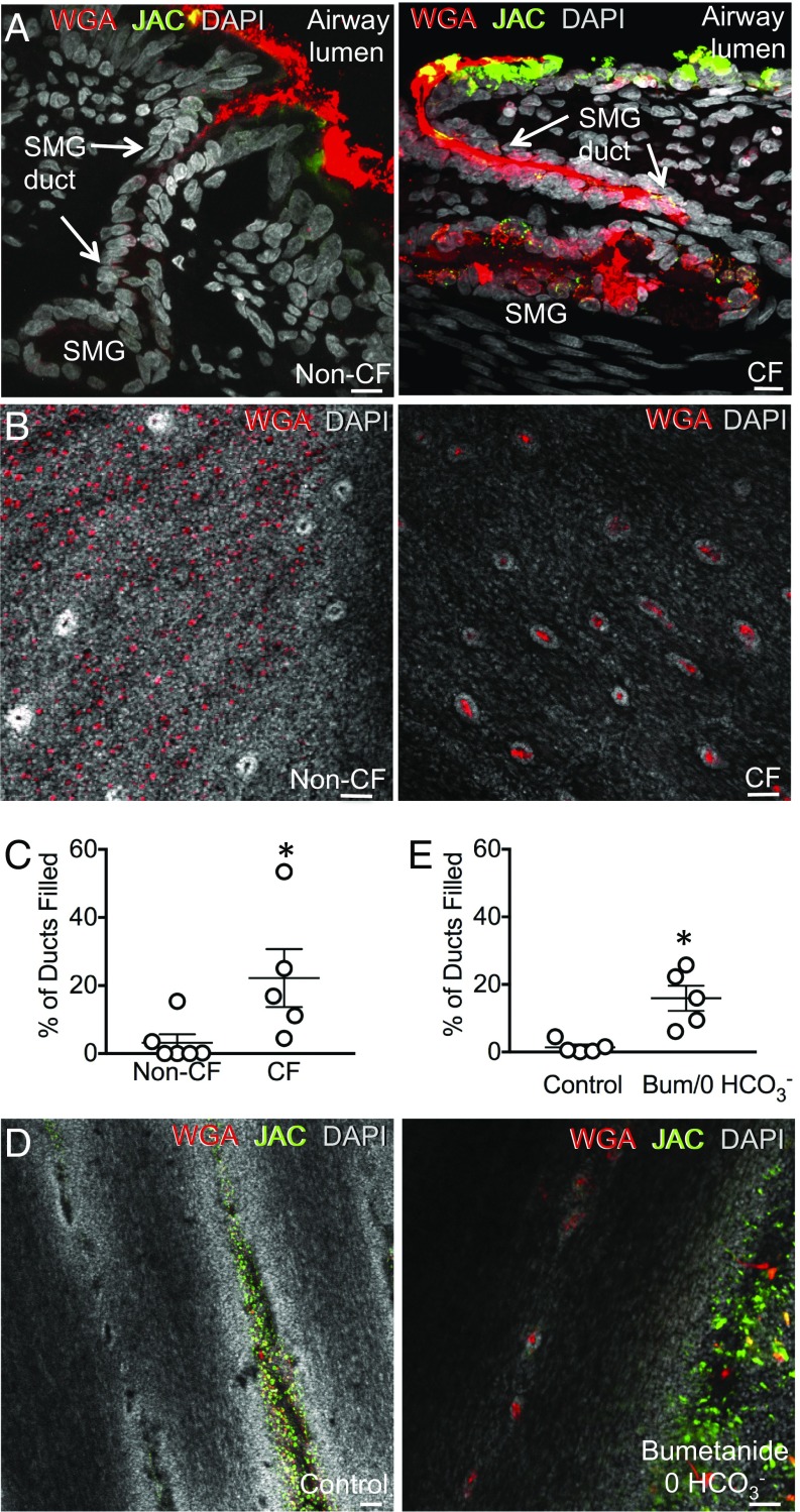
Gel-forming mucins, the primary macromolecular components of airway mucus, facilitate airway clearance by mucociliary transport. In cystic fibrosis (CF) altered mucus properties impair mucociliary transport. Airways primarily secrete two closely related gel-forming mucins, MUC5B and MUC5AC. However, their morphologic structures and associations in airways that contain abundant submucosal glands and goblet cells are uncertain. Moreover, there is limited knowledge about mucins in airways not affected by inflammation, infection, or remodeling or in CF airways. Therefore, we examined airways freshly excised from newborn non-CF pigs and CF pigs before secondary manifestations develop. We found that porcine submucosal glands produce MUC5B, whereas goblet cells produce predominantly MUC5AC plus some MUC5B. We found that MUC5B emerged from submucosal gland ducts in the form of strands composed of multiple MUC5B filaments. In contrast, MUC5AC emerged from goblet cells as wispy threads and sometimes formed mucin sheets. In addition, MUC5AC often partially coated the MUC5B strands. Compared with non-CF, MUC5B more often filled CF submucosal gland ducts. MUC5AC sheets also accumulated in CF airways overlying MUC5B strands. These results reveal distinct morphology and interactions for MUC5B and MUC5AC and suggest that the two mucins make distinct contributions to mucociliary transport. Thus, they provide a framework for understanding abnormalities in disease.

Keywords: COPD; asthma; cystic fibrosis; lung; mucus.


Conflict of interest statement

Conflict of interest statement: The University of Iowa has licensed CF pigs to Exemplar Genetics, and M.J.W. receives royalties from the license.

Figures

Fig. 1.
Fig. 1.
WGA and JAC lectins preferentially label MUC5B and MUC5AC. Figure shows labeling of trachea from newborn pigs by fluorophore-linked lectins and anti-mucin antibodies. (A) Images are tracheal sections showing submucosal glands labeled with anti-MUC5B antibody (white) and WGA (red) (Top) or anti-MUC5AC antibody (white) and JAC (green) (Bottom). Also shown are actin labeling (phalloidin, yellow) and nuclei labeling (DAPI, blue). (Scale bar, 10 μm.) (B) Images are airway surface epithelium showing goblet cells labeled with anti-MUC5B antibody (white) and WGA (red) (Top) or with anti-MUC5AC antibody (white) and JAC (green) (Bottom). Also shown are actin labeling (phalloidin, yellow) and nuclei labeling (DAPI, blue). (Scale bar, 10 μm.)
Fig. 2.
Fig. 2.
WGA and JAC lectins label surface goblet cells. (A) En face image of excised airway surface epithelium labeled with WGA (red), JAC (green), and nuclei (DAPI, gray). (Scale bar, 50 μm.) In subsequent en face images, red and green mucin staining in goblet cells, with proportions varying in individual fields, can be seen below secreted mucin. (B) Percentage of goblet cells in airway surface epithelium labeled by JAC (MUC5AC), WGA (MUC5B), or both. Each symbol represents average of experiments on epithelia from one animal, and error bars indicate SD.
Fig. S1.
Fig. S1.
Adjacent sections of the same airway were treated identically in A and B. (A) En face image of excised airway surface epithelium showing goblet cells labeled with anti-MUC5AC antibody (red), JAC (green), and merged image (yellow). Nuclei—DAPI, gray. (Scale bar, 50 μm.) Mucus in submucosal gland ducts was not labeled by anti-MUC5AC antibody or JAC. (B) En face image of excised trachea showing colocalization of mucus filling submucosal gland ducts with anti-MUC5B antibody (green) and WGA (red). Nuclei—DAPI, gray. (Scale bar, 50 μm.)
Fig. S2.
Fig. S2.
(A) Images are tracheal sections showing submucosal glands and mucus in duct labeled with anti-MUC5B antibody (red). Surface goblet cells, but not submucosal glands, are detected by JAC (green). Also shown are actin labeling (phalloidin, gray) and nuclei labeling (DAPI, blue). (Scale bar, 10 μm.) (B) Images are tracheal sections showing submucosal glands detected by WGA (red) and surface goblet cells labeled by anti-Muc5AC antibody (green). Also shown are β-catenin (gray) and nuclei labeling (DAPI, blue). (Scale bar, 10 μm.) (C) WGA (red) detects glycocalyx at plasma membrane of trachea. β-catenin, gray; nuclei, DAPI, blue. JAC (green) detects glycocalyx at the plasma membrane of trachea. Actin, phalloidin, gray; nuclei, DAPI, blue. (Scale bar, 10 μm.)
Fig. 3.
Fig. 3.
Mucus emerging from submucosal gland ducts labels with WGA lectin. Images in A and B are z stacks and in C are single confocal images of the excised non-CF tracheal surface. WGA is red, JAC is green, and DAPI (nuclei) is gray. (A) Airway surface with mucus strands emerging from submucosal gland (SMG) ducts. (Scale bar, 50 μm.) (See also Fig. S6.) (B) Image shows that mucin strands are comprised of WGA-labeled filaments. JAC-labeled mucus lies on the surface of the WGA-labeled strands. (Scale bar, 50 μm.) (C) Successive single-plane confocal images from the epithelial surface (Bottom) to just above the surface (Top), as indicated by blue dashed lines in Inset. (Scale bar, 50 μm.)
Fig. S6.
Fig. S6.
En face image of unfixed, nonpermeabilized, freshly excised trachea labeled with WGA (red) and JAC (green). DAPI is gray. (Scale bar, 50 μm.) Arrows point to submucosal gland ducts.
Fig. S3.
Fig. S3.
En face image of excised airway surface epithelium showing mucus strands labeled with anti-MUC5B antibody (green), WGA (red), and merged image (yellow). Nuclei, DAPI, gray. (Scale bar, 50 μm.)
Fig. S4.
Fig. S4.
Scanning electron microscopic image of mucus strand emerging from submucosal gland duct. Trachea was stimulated with methacholine in the presence of bumetanide and the absence of HCO3/CO2. (Scale bar, 10 μm.)
Fig. 4.
Fig. 4.
Mucus from goblet cells forms threads and sheets. Images are z stacks of confocal images of excised trachea of non-CF pigs. (A) Left shows threads of mucin detected by MUC5AC antibody (green), a small sheet of mucus (indicated by an arrow), and position of submucosal gland ducts (indicated by white arrowheads). Right shows nuclei (DAPI, gray) to identify the position of submucosal gland ducts (indicated by white arrowheads). A small sheet of mucus is indicated by arrow. (Scale bar, 50 μm.) (B) Image of JAC-labeled mucus threads (green) from goblet cells with rare WGA (red) thread. Goblet cells are labeled by JAC and WGA beneath the threads. (Scale bar, 50 μm.)
Fig. 5.
Fig. 5.
CF airways showed entangled mucus strands and increased mucus sheets. (A) Methacholine-stimulated airways from newborn non-CF (Left) and CF pigs (Right). WGA is red, and JAC is green. (Scale bar, 50 μm.) (B) Large MUC5AC sheet (JAC, green) floating on MUC5B (WGA, red) strands in methacholine-stimulated CF trachea. (Scale bar, 50 μm.)
Fig. 6.
Fig. 6.
CF submucosal gland ducts are filled with mucus. (A) Images are from pigs treated in vivo with methacholine. WGA (MUC5B) is red, JAC (MUC5AC) is green, and DAPI (nuclei) is gray. Shown are vertical sections of airway excised from non-CF (Left) and CF (Right) pigs. (Scale bar, 10 μm.) (B) En face image of excised trachea from methacholine-stimulated newborn non-CF (Left) and CF (Right) pigs. (Scale bar, 50 μm.) (C) Percentage of submucosal gland ducts filled with mucin in excised trachea from non-CF and CF pigs treated in vivo with methacholine. Each data point is from a different pig. Bars and whiskers indicate mean ± SEM. *P < 0.05. (D) Data are z stacks of confocal images at the level of the apical membrane. Excised tracheas from non-CF pigs incubated with methacholine in HCO3/CO2-buffered saline (control) or Hepes-buffered saline containing bumetanide. WGA, red; JAC, green; DAPI, gray. (Scale bar, 50 μm.) (E) Percentage of submucosal gland ducts filled with mucus. Pigs received methacholine in vivo. Each data point is from a different pig. n = 5 pigs for each condition. Average number of ducts counted per condition = 340 ± 53. Bars and whiskers indicate mean ± SEM. *P < 0.05. (See also Fig. S7.)
Fig. S5.
Fig. S5.
ASL pH 30 min after basolateral incubation of tracheal rings. Control, Krebs bicarbonate buffer, pH 7.4, with methacholine incubated at 37 °C in a humidified incubator with 5% CO2. Bumet, 0 HCO3, 10 mM Hepes, pH 7.4, no HCO3, with bumetanide and methacholine incubated at 37 °C in a humidified incubator without CO2. Each symbol represents a single pH measurement. Results are from 3–4 measurements per condition in two pigs.
Fig. 7.
Fig. 7.
Model of mucin secretion in pig airway.
Fig. S7.
Fig. S7.
Percentage of submucosal gland ducts that were filled with mucus. Control, Krebs bicarbonate buffer, pH 7.4, with methacholine incubated at 37 °C in a humidified incubator with 5% CO2. Bumet & 0 HCO3, 10 mM Hepes, pH 7.4, no HCO3, with bumetanide and methacholine incubated at 37 °C in a humidified incubator without CO2. n = 5 paired trachea samples each from a different pig. Each symbol represents data from one pig. Each panel shows percent filled ducts from a different reader. Bars and whiskers indicate mean ± SEM.

References

    1. Widdicombe JH, Wine JJ. Airway gland structure and function. Physiol Rev. 2015;95:1241–1319. - PubMed
    1. Wanner A, Salathé M, O’Riordan TG. Mucociliary clearance in the airways. Am J Respir Crit Care Med. 1996;154:1868–1902. - PubMed
    1. Robinson M, Bye PT. Mucociliary clearance in cystic fibrosis. Pediatr Pulmonol. 2002;33:293–306. - PubMed
    1. Thornton DJ, Rousseau K, McGuckin MA. Structure and function of the polymeric mucins in airways mucus. Annu Rev Physiol. 2008;70:459–486. - PubMed
    1. Fahy JV, Dickey BF. Airway mucus function and dysfunction. N Engl J Med. 2010;363:2233–2247. - PMC - PubMed

Publication types

LinkOut - more resources