. 2017 Aug;206(4):1895-1907.
doi: 10.1534/genetics.117.201632. Epub 2017 Jun 12.

Epigenetic Transcriptional Memory of GAL Genes Depends on Growth in Glucose and the Tup1 Transcription Factor in Saccharomyces cerevisiae

Affiliations

Epigenetic Transcriptional Memory of GAL Genes Depends on Growth in Glucose and the Tup1 Transcription Factor in Saccharomyces cerevisiae

Varun Sood et al. Genetics. 2017 Aug.

Abstract

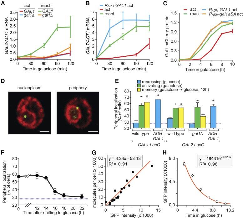
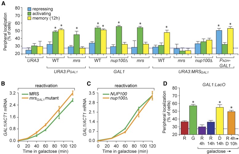
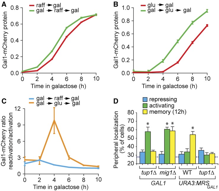
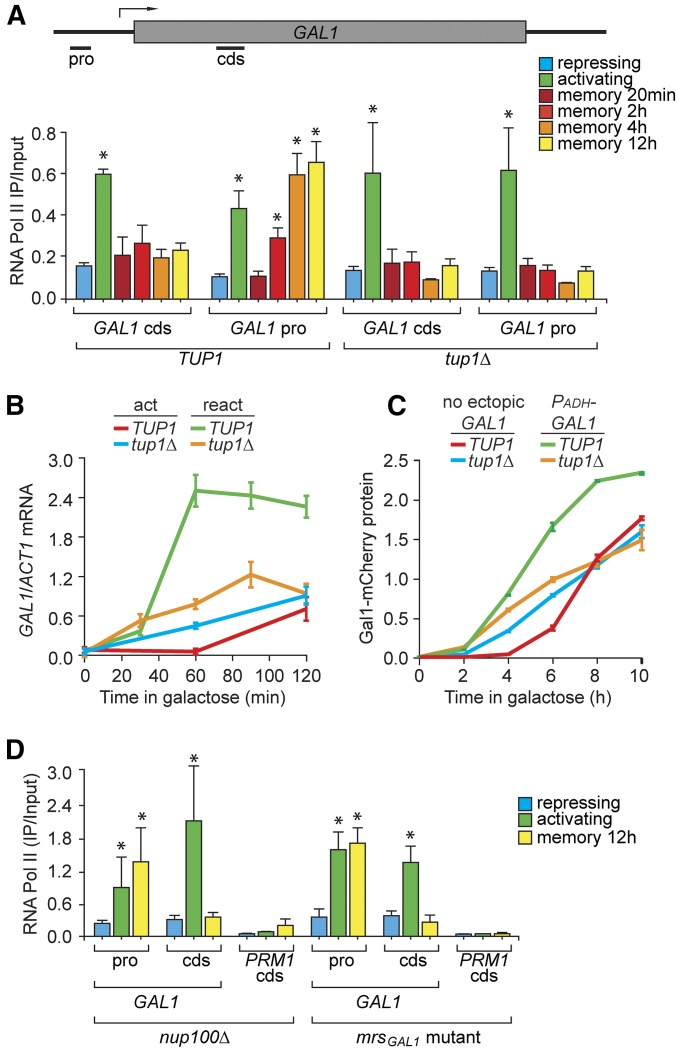
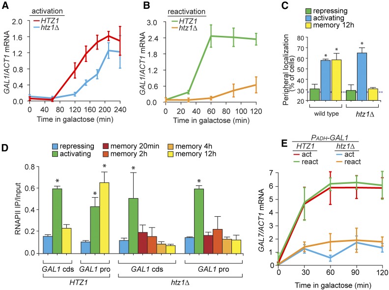
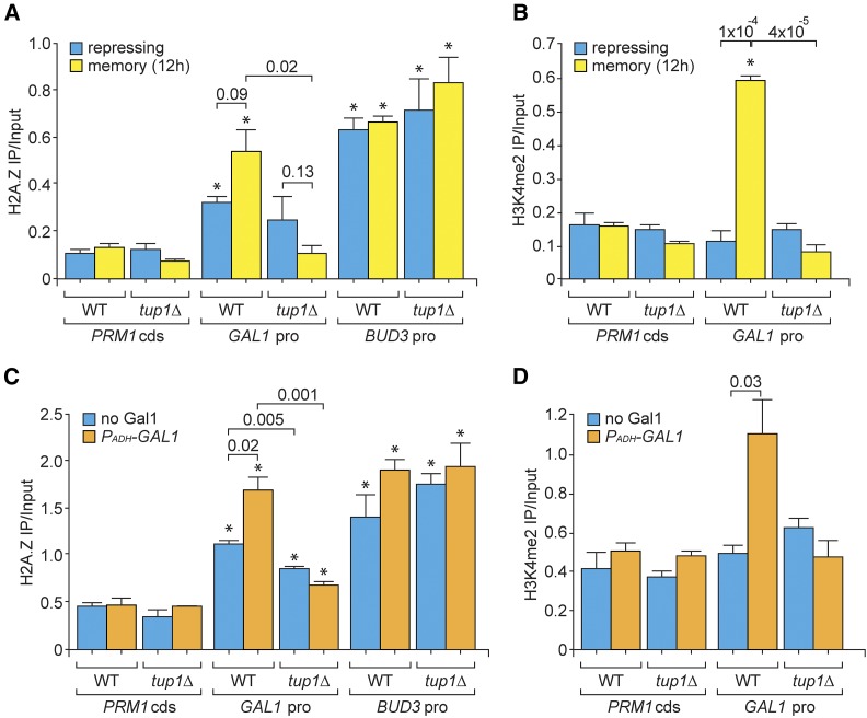
Previously expressed inducible genes can remain poised for faster reactivation for multiple cell divisions, a conserved phenomenon called epigenetic transcriptional memory. The GAL genes in Saccharomyces cerevisiae show faster reactivation for up to seven generations after being repressed. During memory, previously produced Gal1 protein enhances the rate of reactivation of GAL1, GAL10, GAL2, and GAL7 These genes also interact with the nuclear pore complex (NPC) and localize to the nuclear periphery both when active and during memory. Peripheral localization of GAL1 during memory requires the Gal1 protein, a memory-specific cis-acting element in the promoter, and the NPC protein Nup100 However, unlike other examples of transcriptional memory, the interaction with NPC is not required for faster GAL gene reactivation. Rather, downstream of Gal1, the Tup1 transcription factor and growth in glucose promote GAL transcriptional memory. Cells only show signs of memory and only benefit from memory when growing in glucose. Tup1 promotes memory-specific chromatin changes at the GAL1 promoter: incorporation of histone variant H2A.Z and dimethylation of histone H3, lysine 4. Tup1 and H2A.Z function downstream of Gal1 to promote binding of a preinitiation form of RNA Polymerase II at the GAL1 promoter, poising the gene for faster reactivation. This mechanism allows cells to integrate a previous experience (growth in galactose, reflected by Gal1 levels) with current conditions (growth in glucose, potentially through Tup1 function) to overcome repression and to poise critical GAL genes for future reactivation.

Keywords: GAL genes; RNA polymerase II; chromatin; epigenetic; gene positioning; nuclear pore complex; transcriptional memory.


Figures

Figure 1
Figure 1
Gal1 promotes GAL gene localization at the nuclear periphery during memory. (A–C) Cells were shifted from glucose to galactose (act; activation) or grown overnight in galactose, shifted to glucose for 12 hr and then shifted to galactose (react; reactivation). Cells were harvested at the indicated times, RNA was prepared and mRNA levels were quantified relative to ACT1 by reverse-transcriptase quantitative PCR (RT qPCR) (A and B) or fluorescence was quantified using flow cytometry (C). (A) GAL2 activation and reactivation in wild-type and gal1∆ cells. (B) GAL7 activation and reactivation or activation with PADH-GAL1. (C) Gal1-mCherry levels, normalized to constitutively expressed cyan fluorescent protein (CFP) (PTDH-CFP) during activation, reactivation, and activation in cells with ectopically expressed wild-type GAL1 (PADH-GAL1) or catalytically inactive mutant (PADH-gal1-∆SA). (D) Immunofluorescence images of cells having the LacO array integrated downstream of the GAL1 gene, stained for GFP-LacI (green) and Sec-63myc (red) and scored as either nucleoplasmic or peripheral. Bar, 1 µm. (E) Peripheral localization of GAL1 and GAL2 under repressing (glucose), activating (galactose), and memory (galactose → glucose, 12 hr) conditions in wild-type or gal1∆ cells and in the presence of PADH-GAL1. (F) Cells with the LacO array downstream of GAL1 were shifted from galactose to glucose media for the indicated length of times and the percentage of cells in which GAL1 colocalized with the nuclear envelope was plotted. The hatched blue line in (E and F) represents the baseline colocalization predicted by chance (Brickner and Walter 2004). (G) Plot of the fluorescence intensities of 20 GFP-tagged proteins (Ghaemmaghami et al. 2003; Huh et al. 2003), measured by flow cytometry, against protein copy number per cell (Newman et al. 2006). (H) Gal1-GFP fluorescence decay after shifting from galactose to glucose. Note: to avoid potential effects of continued translation and maturation of GFP, the initial point for curve fitting was 2 hr after repression. Error bars represent SEM for ≥ three biological replicates. Each replicate for localization (E and F) consisted of 30–50 cells and for fluorescence estimation using flow cytometer (C, G, and H) consisted of ≥ 5000 cells, respectively. * P ≤ 0.05 (Student’s t-test) relative to the repressing condition.
Figure 2
Figure 2
Memory Recruitment Sequence (MRS)GAL1-dependent peripheral localization of GAL1 during memory requires growth in glucose and Tup1. (A) Peripheral localization of URA3, GAL1, URA3:PGAL1, or URA3:MRSGAL1 was quantified under repressing (glucose), activating (galactose), and memory (galactose → glucose, 12 hr) conditions in wild-type (WT) or nup100∆ cells using immunofluorescence or live cell microscopy. The full-length GAL1 promoter (PGAL1, 667 bp) or the 63-bp MRSGAL1 were inserted at URA3 along with a LacO array as described (Egecioglu et al. 2014). The mrs mutation is shown in Figure S2B in File S1. (B and C) Cells were grown in galactose overnight, shifted to glucose for 12 hr, and then shifted to galactose (reactivation) to assay GAL1 expression using RT-quantitative PCR in WT, mrsGAL1 (B), and nup100∆ (C) mutant cells. (D) Peripheral localization of GAL1 in cells grown in raffinose (R), galactose (G), and upon shift from galactose to: raffinose for 4 hr (R 4 hr), raffinose for 14 hr (R 14 hr), glucose for 14 hr (D 14 hr), or raffinose 4 hr followed by glucose 10 hr (R 4 hr → D 10 hr). The hatched line represents the level of colocalization with the nuclear envelope predicted by chance (A and D). Error bars represent SEM for at least three biological replicates. * P ≤ 0.05 (Student’s t-test) relative to the repressing condition.
Figure 3
Figure 3
The adaptive value of memory in cells grown in nonrepressing and repressing carbon sources. (A and B) Gal1-mCherry expression during activation and reactivation, measured by flow cytometry. Activation: cells were shifted to galactose (gal) from either a nonrepressing carbon source, raffinose (raff) (A), or a repressing carbon source, glucose (glu) (B). Reactivation: cells were shifted from gal to either raff (A) or glu (B) for around seven cell divisions and then reactivated in galactose. (C) Gal1-mCherry reactivation:activation ratio at the indicated time points after shifting cells from raffinose to galactose or glucose to galactose. (D) Peripheral localization of GAL1 or URA3:MRSGAL1 in tup1∆ and mig1∆ mutant strains. The hatched line represents the level of colocalization with the nuclear envelope predicted by chance. WT, wild type. * P ≤ 0.05 (Student’s t-test) relative to the repressing condition. Error bars represent SEM for at least three biological replicates.
Figure 4
Figure 4
Tup1 functions downstream of Gal1 to promote binding of RNAPII to the promoter and faster reactivation of GAL1 during memory. (A) RNAPII ChIP from wild-type and tup1∆ cells under repressing (glucose), activating (galactose), and at different times during memory (galactose → glucose, 20 min to 12 hr) conditions. Recovery of the GAL1 promoter and cds was quantified relative to input by qPCR. (B) Time course of RT-qPCR for GAL1 expression relative to ACT1 during activation (act; glucose → galactose) and reactivation (react; galactose → glucose 12 hr → galactose) in WT and tup1Δ cells. (C) Gal1-mCherry expression during activation in wild-type and tup1Δ cells with or without PADH-GAL1 integrated at the TRP1 locus. (D) RNAPII ChIP under repressing (glucose), activating (galactose), and memory (galactose → glucose, 12 hr) conditions for mrsGAL1 and nup100∆ mutant. Error bars represent SEM for at least three biological replicates. * P ≤ 0.05 (Student’s t-test) relative to the repressing condition. cds, coding sequence; ChIP, chromatin immunoprecipitation; pro, promoter; qPCR, quantitative PCR; RNAPII, RNA polymerase II; WT, wild type.
Figure 5
Figure 5
H2A.Z functions downstream of Gal1 to promote GAL transcriptional memory. (A and B) GAL1 expression, relative to ACT1, measured by RT-qPCR over time in wild-type and htz1∆ cells during activation (A) and reactivation after 12 hr of repression (B). (C) Peripheral localization of GAL1 under repressing (glucose), activating (galactose), and memory (galactose → glucose, 12 hr) conditions in wild-type and htz1∆ cells. The hatched line represents the level of colocalization with the nuclear envelope predicted by chance. (D) RNAPII ChIP from wild-type and htz1∆ cells under repressing, activating, and at different times during memory (galactose → glucose, 20 min to 12 hr) conditions. (E) GAL7 expression, relative to ACT1, measured by RT-qPCR during activation or reactivation in wild-type and htz1∆ cells transformed with PADH-GAL1. Error bars represent SEM from at least three independent replicates. * P ≤ 0.05 (Student’s t-test) relative to the repressing condition. cds, coding sequence; ChIP, chromatin immunoprecipitation; pro, promoter; qPCR, quantitative PCR; RNAPII, RNA polymerase II.
Figure 6
Figure 6
Tup1 promotes H2A.Z incorporation and H3K4me2 modification during GAL memory. (A and C) H2A.Z ChIP in WT and tup1∆ cells under repressing (glucose) and memory (galactose → glucose, 12 hr) conditions (A) or under repressing conditions with PADH-GAL1 (C). The recovered DNA fragments in IP were analyzed for sequences arising from the GAL1 promoter, PRM1 coding sequence (negative control), and BUD3 promoter (positive control) and plotted relative to input fraction. (B and D) H3K4me2 ChIP in WT and tup1∆ cells, performed as described in (A and C). Error bars represent SEM from at least three independent replicates. * P ≤ 0.05 (Student’s t-test) relative to the repressing condition. cds, coding sequence; ChIP, chromatin immunoprecipitation; H3K4me2, histone 3 dimethyl lysine 4; IP, immunoprecipitation; pro, promoter; qPCR, quantitative PCR; WT, wild type.

Similar articles

Cited by

References

    1. Ahmed S., Brickner D. G., Light W. H., Cajigas I., McDonough M., et al. , 2010. DNA zip codes control an ancient mechanism for gene targeting to the nuclear periphery. Nat. Cell Biol. 12: 111–118. - PMC - PubMed
    1. Bhat P. J., Hopper J. E., 1992. Overproduction of the GAL1 or GAL3 protein causes galactose-independent activation of the GAL4 protein: evidence for a new model of induction for the yeast GAL/MEL regulon. Mol. Cell. Biol. 12: 2701–2707. - PMC - PubMed
    1. Biggar S. R., Crabtree G. R., 2001. Cell signaling can direct either binary or graded transcriptional responses. EMBO J. 20: 3167–3176. - PMC - PubMed
    1. Brickner D. G., Cajigas I., Fondufe-Mittendorf Y., Ahmed S., Lee P. C., et al. , 2007. H2A.Z-mediated localization of genes at the nuclear periphery confers epigenetic memory of previous transcriptional state. PLoS Biol. 5: e81. - PMC - PubMed
    1. Brickner D. G., Light W., Brickner J. H., 2010. Quantitative localization of chromosomal loci by immunofluorescence. Methods Enzymol. 470: 569–580. - PubMed

MeSH terms