. 2017 Jun 12;7(1):3315.
doi: 10.1038/s41598-017-03628-6.

Development of a root canal treatment model in the rat

Affiliations

Development of a root canal treatment model in the rat

Naomichi Yoneda et al. Sci Rep. .

Abstract

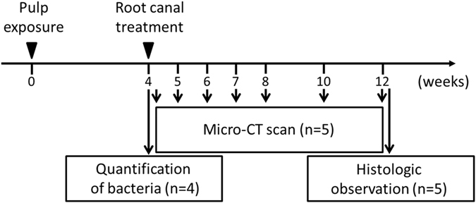
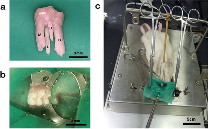
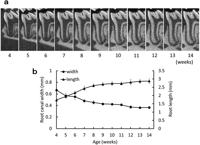
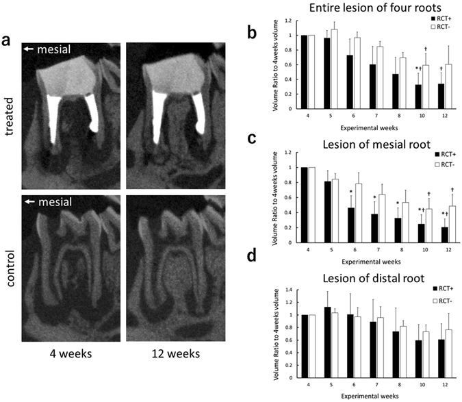
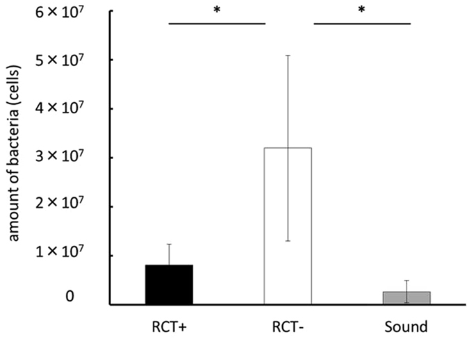
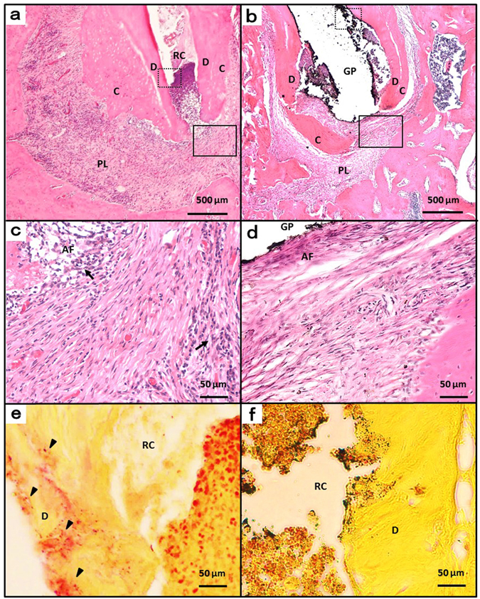
Root canal treatment is performed to treat apical periodontitis, and various procedures and techniques are currently used. Although animal models have been used in the developmental research of root canal treatment, little of this research has used small animals such as rats, because of their small size. In this study, root canal treatment was performed on the rat mandibular first molar, which had four root canals, using a microscope, and the therapeutic effect was evaluated bacteriologically, radiologically and histopathologically. By performing root canal treatment, the level of bacteria in the mesial root of the treated teeth was reduced by 75% compared with the control. Additionally, the volume of the periapical lesions of the treated teeth as measured by micro-computed tomography decreased significantly 2 weeks after the root canal treatment when compared with the control. Histological evidence of healing was observed in the treatment group 8 weeks after root canal treatment. These results suggest that a root canal treatment model using rats can be used in developmental research for novel methods of root canal treatment.


Conflict of interest statement

The authors declare that they have no competing interests.

Figures

Figure 1
Figure 1
Experimental protocol. Root canal treatment was performed 4 weeks after pulp exposure and was evaluated by micro-CT scanning, quantification of bacteria, and histological observation.
Figure 2
Figure 2
Rubber-dam isolation of the rat mandibular molar. (a) Rat mandibular first molar with four roots and a crown with a mesiodistal diameter of approximately 3 mm. (b,c) Isolation of the tooth with a custom-made rubber-dam clamp.
Figure 3
Figure 3
Age-related change in the mesial root of the mandibular first molar. (a) Representative micro-CT images of the mesial root of the mandibular first molar at each week of age. (b) Age-related change in root length and root canal width. Root length was measured from the pulpal floor to the root apex; the root canal width was measured at 1 mm apically from the pulpal floor. Data represent the means of four sample measurements; error bars indicate standard deviations.
Figure 4
Figure 4
Micro-CT analysis of periapical lesions after root canal treatment in rat. (a) Representative micro-CT images of experimental teeth in the treatment group, and the control group, at the 4-week and 12-week time points. (bd) Comparison of changes in the volume of periapical lesions (*P < 0.05 indicates significant differences compared with the control group at the same time point, Welch’s t-test; P < 0.05 indicates significant differences compared with the 4-week data of the same group, Kruskal-Wallis test). Data represent the means of five sample measurements; error bars indicate standard deviations. The volume of the periapical lesions of the mesial root for the treatment group were significantly lower than that of the control group at each time point after 6 weeks following pulp exposure.
Figure 5
Figure 5
Quantification of bacteria derived from the mesial root immediately after root canal treatment using real-time PCR (*P < 0.05 indicates significant differences between the two groups, Steel-Dwass test). Data represent the means of four sample measurements; error bars indicate standard deviations.
Figure 6
Figure 6
Histologic images at week 12. (a) Periapical areas of the control group stained with haematoxylin and eosin. (b) Periapical areas of the treatment group stained with haematoxylin and eosin. (c and d) High-magnification views of the solid inset in panels a and b, respectively. (e and f) High-magnification views of the dotted insets in panels b and e, respectively, stained with a modified Brown and Brenn method. AF, apical foramen; GP, gutta-percha point; RC, root canal; C, cementum; D, dentin; PL, periapical lesion; arrow head, bacteria; arrow, inflammatory cells.

References

    1. Kakehashi S, Stanley HR, Fitzgerald RJ. The effects of surgical exposures of dental pulps in germ-free and conventional laboratory rats. Oral Surg. Oral Med. Oral Pathol. 1965;20:340–349. doi: 10.1016/0030-4220(65)90166-0. - DOI - PubMed
    1. Ng YL, Mann V, Rahbaran S, Lewsey J, Gulabivala K. Outcome of primary root canal treatment: systematic review of the literature - Part 2. Influence of clinical factors. Int. Endod. J. 2008;41:6–31. doi: 10.1111/j.1365-2591.2008.01484.x. - DOI - PubMed
    1. Tronstad L, Barnett F, Riso K, Slots J. Extraradicular endodontic infections. Endod. Dent. Traumatol. 1987;3:86–90. doi: 10.1111/j.1600-9657.1987.tb00549.x. - DOI - PubMed
    1. Tronstad L, Kreshtool D, Barnett F. Microbiological monitoring and results of treatment of extraradicular endodontic infection. Endod. Dent. Traumatol. 1990;6:129–136. doi: 10.1111/j.1600-9657.1990.tb00407.x. - DOI - PubMed
    1. Sunde PT, Olsen I, Lind PO, Tronstad L. Extraradicular infection: a methodological study. Endod. Dent. Traumatol. 2000;16:84–90. doi: 10.1034/j.1600-9657.2000.016002084.x. - DOI - PubMed

Publication types