. 2017 Jun 12;7(1):3323.
doi: 10.1038/s41598-017-02532-3.

A Novel Strategy to Engineer Pre-Vascularized Full-Length Dental Pulp-like Tissue Constructs

Affiliations

A Novel Strategy to Engineer Pre-Vascularized Full-Length Dental Pulp-like Tissue Constructs

Avathamsa Athirasala et al. Sci Rep. .

Abstract

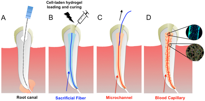
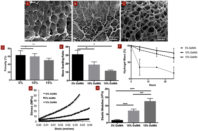
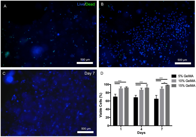
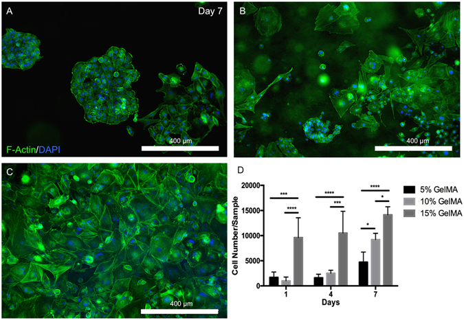
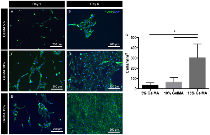
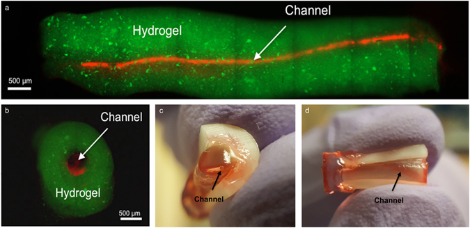
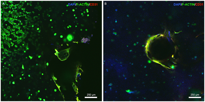
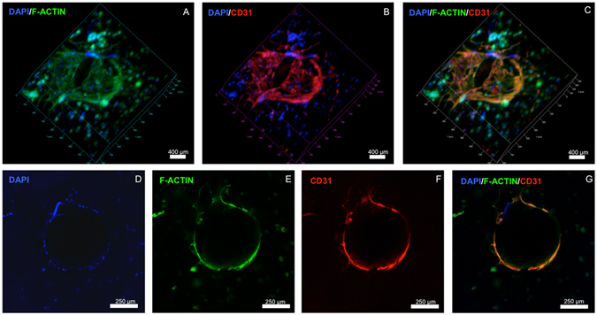
The requirement for immediate vascularization of engineered dental pulp poses a major hurdle towards successful implementation of pulp regeneration as an effective therapeutic strategy for root canal therapy, especially in adult teeth. Here, we demonstrate a novel strategy to engineer pre-vascularized, cell-laden hydrogel pulp-like tissue constructs in full-length root canals for dental pulp regeneration. We utilized gelatin methacryloyl (GelMA) hydrogels with tunable physical and mechanical properties to determine the microenvironmental conditions (microstructure, degradation, swelling and elastic modulus) that enhanced viability, spreading and proliferation of encapsulated odontoblast-like cells (OD21), and the formation of endothelial monolayers by endothelial colony forming cells (ECFCs). GelMA hydrogels with higher polymer concentration (15% w/v) and stiffness enhanced OD21 cell viability, spreading and proliferation, as well as endothelial cell spreading and monolayer formation. We then fabricated pre-vascularized, full-length, dental pulp-like tissue constructs by dispensing OD21 cell-laden GelMA hydrogel prepolymer in root canals of extracted teeth and fabricating 500 µm channels throughout the root canals. ECFCs seeded into the microchannels successfully formed monolayers and underwent angiogenic sprouting within 7 days in culture. In summary, the proposed approach is a simple and effective strategy for engineering of pre-vascularized dental pulp constructs offering potentially beneficial translational outcomes.


Conflict of interest statement

The authors declare that they have no competing interests.

Figures

Figure 1
Figure 1
Schematic diagram illustrating the basic steps of the proposed strategy to engineer pre-vascularized full-length dental pulp-like tissue constructs. (A) The root canal is prepared following common endodontic procedure using endodontic files. (B) A pre-made sacrificial fiber is positioned in the root canal. A cell-laden hydrogel is loaded into the canal and photopolymerized. (C) After the hydrogel photopolymerization, the sacrificial fiber is removed, creating a hollow microchannel that traverses the entire length of the canal, from the apex through the pulp chamber. (D) Endothelial cells are seeded in the fabricated microchannel to engineer the core vascular capillary in the dental pulp, thus resulting in a pre-vascularized full-length dental pulp-like tissue construct. Details are provided in Supplementary Figure S1.
Figure 2
Figure 2
Physical and mechanical properties of GelMA hydrogels. SEM images of (A) 5%, (B) 10% and (C) 15% (w/v) GelMA hydrogels show a decrease in apparent pore sizes and (d) relative percentage of porosity with increasing hydrogel concentration, especially comparing 5% to 10 or 15% hydrogels. Mass swelling ratios and (E) degradation profiles of 5, 10 and 15% GelMA hydrogels in DPBS, and in 2.5 U ml–1 collagenase, respectively showing a marked decrease in swelling properties and degradability with increasing polymer volume fraction. (F) Stress-strain curves and (G) elastic modulus for 5, 10 and 15% GelMA hydrogels in unconfined compression, respectively demonstrate enhanced mechanical properties in more densely crosslinked hydrogels (15%). For degradation data, p < 0.0001 between 5% and 10%, and between 5% and 15% for time points 15 and 25 h. Statistical significance is represented by *for p < 0.05, **for p < 0.01, ***for p < 0.001 and ****for p < 0.0001.
Figure 3
Figure 3
Viability of OD21 cells in GelMA hydrogel scaffolds. Representative images of OD21 cells encapsulated in (A) 5%, (B) 10% and (C) 15% GelMA hydrogels stained for live (blue) and dead (green) cells on day 7. (D) Percentage of live cells in the hydrogels after 1, 4 and 7 days showed consistently enhanced OD21 cell survival in 10 and 15% GelMA hydrogels compared to the softer 5% GelMA hydrogels at all time points, with a statistically significant increase in cell viability in 15% GelMA hydrogels over 10% GelMA hydrogels by day 7. Statistical significance is represented by **for p < 0.01 and ****for p < 0.0001.
Figure 4
Figure 4
Spreading and proliferation of OD21 cells in 3D GelMA hydrogels. Representative images of OD21 cells encapsulated in (A) 5%, (B) 10% and (C) 15% GelMA hydrogels stained for actin (green) and DAPI (blue) shows increased cell spreading in stiffer over softer hydrogels. (D) Quantification of the number of cells per gel after 1, 4 and 7 days in culture indicates a significantly higher rate of proliferation of OD21 cells in scaffolds of higher polymer concentration. Statistical significance is represented by *for p < 0.05, ***for p < 0.001 and ****for p < 0.0001.
Figure 5
Figure 5
Cell spreading and monolayer formation by ECFCs on 2D GelMA hydrogels. Representative images of ECFCs on (A,B) 5%, (C,D) 10% and (E,F) 15% GelMA hydrogels after 1 and 6 days in culture, stained for actin (green) and DAPI (blue) showed increased cell spreading and cell density on stiffer substrates at both early and late time points. (G) Quantification of the number of cells per unit surface area on day 6, representative of cell coverage on the hydrogels and the propensity for endothelial monolayer formation suggests that stiffer GelMA hydrogel substrates support ECFC proliferation and monolayer formation. Statistical significance is represented by *for p < 0.05.
Figure 6
Figure 6
Representative images of pre-vascularized pulp-like tissue construct. (A) Longitudinal and (B) cross-sectional views of GelMA hydrogels loaded with green fluorescent microparticles showing the fabricated microchannel after being perfused with a red fluorescent microparticle solution. The channels cross the entire length of the root. (C,D) Photographs of GelMA hydrogels from longitudinal and occlusal perspectives inside a full-length root fragment. Root fragments were stabilized prior to hydrogel loading and microchannel fabrication, and were separated to retrieve the constructs and illustrate the position of the hydrogel inside the tooth. Microchannels were perfused with red food dye.
Figure 7
Figure 7
Confocal images of OD21 and ECFCs cultured in GelMA hydrogels in the full-length dental pulp-like tissue constructs. (A) OD21 cells had visibly higher spreading near the dentin walls (upper left corner of (A)) than in areas around the fabricated microchannels (B). Cells were stained for actin (green), DAPI (blue) and CD31 (red) on day 7. Z-stack movie for 3D rendering of this image is available as Supplementary Video 1.
Figure 8
Figure 8
Confocal images of endothelialized microchannels in an OD21-laden GelMA hydrogel cultured in a full-length dental pulp-like tissue constructs. (AC) 3D rendering and (DG) cross-sectional slices of confocal images showing ECFC monolayer formation and angiogenic sprouts in the engineered microchannel in the dental pulp-like tissue constructs on day 7. Cells were stained for actin (green), DAPI (blue) and CD31 (red). Z-stack movie for 3D rendering of this image is available as Supplementary Video 2.

References

    1. Pashley, D. H., Walton, R. E. & Slavkin, H. C. Endodontics. 5 edn, 25 (PMPH-USA, 2002).
    1. Yu C, Abbott PV. An overview of the dental pulp: its functions and responses to injury. Aust Dent J. 2007;52:S4–16. doi: 10.1111/j.1834-7819.2007.tb00525.x. - DOI - PubMed
    1. I., A. In The Dental Pulp Vol. 1 Ch. 5, 61–74 (Springer-Verlag Berlin Heidelberg, 2014).
    1. Goldberg, M. The Dental Pulp - Biology, Pathology, and Regenerative Therapies. Vol. 1 (Springer-Verlag Berlin Heidelberg, 2014).
    1. Smith AJ, Lumley PJ, Tomson PL, Cooper PR. Dental regeneration and materials: a partnership. Clin Oral Investig. 2008;12:103–108. doi: 10.1007/s00784-008-0189-5. - DOI - PubMed

Publication types