. 2017 Jun 12;7(1):3301.
doi: 10.1038/s41598-017-03280-0.

Neuropilin-1 Associated Molecules in the Blood Distinguish Poor Prognosis Breast Cancer: A Cross-Sectional Study

Affiliations

Neuropilin-1 Associated Molecules in the Blood Distinguish Poor Prognosis Breast Cancer: A Cross-Sectional Study

Adviti Naik et al. Sci Rep. .

Abstract

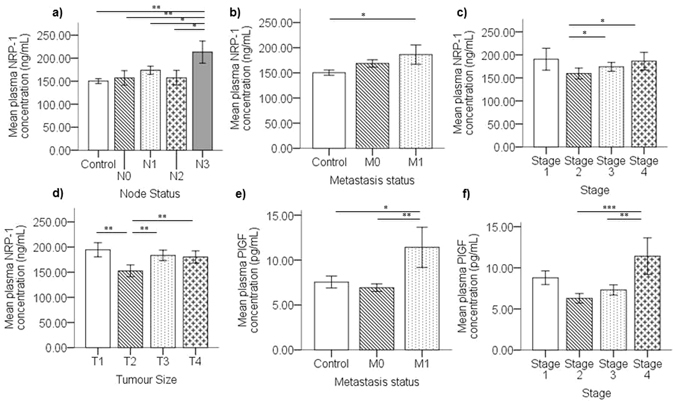
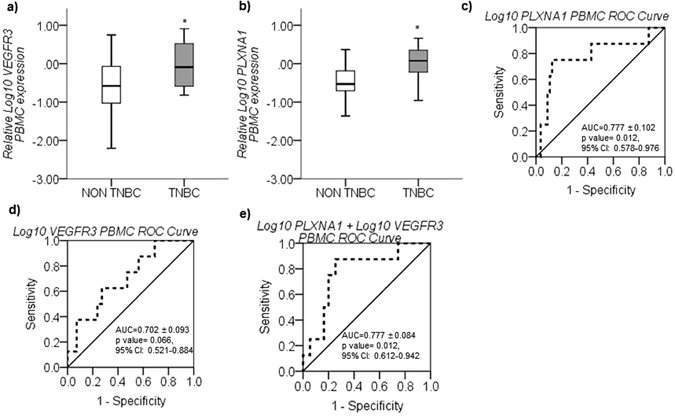
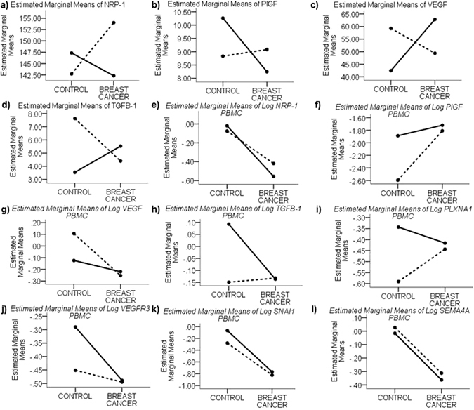
Circulating plasma and peripheral blood mononuclear (PBMCs) cells provide an informative snapshot of the systemic physiological state. Moreover, they provide a non-invasively accessible compartment to identify biomarkers for personalized medicine in advanced breast cancer. The role of Neuropilin-1 (NRP-1) and its interacting molecules in breast tumor tissue was correlated with cancer progression; however, the clinical impact of their systemic levels was not extensively evaluated. In this cross-sectional study, we found that circulating and tumor tissue expression of NRP-1 and circulating placental growth factor (PlGF) increase in advanced nodal and metastatic breast cancer compared with locally advanced disease. Tumor tissue expression of NRP-1 and PlGF is also upregulated in triple negative breast cancer (TNBC) compared to other subtypes. Conversely, in PBMCs, NRP-1 and its interacting molecules SEMA4A and SNAI1 are significantly downregulated in breast cancer patients compared to healthy controls, indicating a protective role. Moreover, we report differential PBMC expression profiles that correlate inversely with disease stage (SEMA4A, SNAI1, PLXNA1 and VEGFR3) and can differentiate between the TNBC and non-TNBC tumor subtypes (VEGFR3 and PLXNA1). This work supports the importance of NRP-1-associated molecules in circulation to characterize poor prognosis breast cancer and emphasizes on their role as favorable drug targets.


Conflict of interest statement

The authors declare that they have no competing interests.

Figures

Figure 1
Figure 1
Elevated plasma NRP-1 and PlGF associates with advanced breast cancer. The graphs represent mean concentration of plasma NRP-1 ± SEM measured by ELISA in breast cancer patients which indicates significant upregulation in (a) advanced nodal disease and (b) metastasis, (c) advanced disease stage and (d) tumor size. (e) Plasma PlGF mean concentration ± SEM shows significantly upregulated levels in metastatic breast cancer cases compared to non-metastatic and healthy controls and (f) upregulation in Stage 4 cases compared to stage 2 and 3. p < 0.05 considered to indicate statistical significance. *p < 0.05, **p < 0.01, ***p < 0.001, Multivariate ANOVA followed by Fisher’s LSD post hoc test.
Figure 2
Figure 2
NRP-1 and PlGF tumor tissue expression associated with poor prognosis breast cancer. Representative immunohistochemical staining images for NRP-1 tumor tissue expression according to patient subgroups based on (a,b) nodal status, (c,d) metastatic status; and (e–g) NRP-1 and PlGF tumor expression in breast cancer patients tissues grouped according to their tumor molecular subtype. Graphs represent calculated mean immuno-reactive score (IRS) of NRP-1 and/or PlGF expression ± SEM in breast tumor tissue grouped according to the respective subtypes. NRP-1 tumor tissue expression was significantly higher in cases with metastasis and advanced nodal disease. Moreover, NRP-1 was significantly upregulated in TNBC cases compared to luminal B and luminal B-like; however, PlGF was significantly higher in TNBC cases compared to luminal B subtype only. p < 0.05 considered to indicate statistical significance. Scale bar = 50 µm, *p < 0.05, **p < 0.01, Multivariate ANOVA followed by Fisher’s LSD post hoc test.
Figure 3
Figure 3
Differential PBMC gene expression between healthy controls vs breast cancer patients. Graphs (ac) represent the mean relative log10 transformed gene expression (±SEM) as quantified using quantitative real time qRT-PCR. The relative expression of the following genes is significantly downregulated in PBMCs isolated from breast cancer patients compared to healthy controls: (a) Log10 NRP-1, (b) Log10 SNAI1 and (c) Log10 SEMA4A. Target gene expression levels are normalized against GUSB gene expression and scaled to expression levels in control cases. P < 0.05 considered to indicate statistical significance. ***p < 0.001, Independent samples T-test.
Figure 4
Figure 4
Decreased SNAI1, SEMA4A, VEGFR3 and PLXNA1 PBMC gene expression in patients with large tumor size and advanced disease stage. (a) The mean relative Log10 SNAI1 expression ( ± SEM), as quantified by real time PCR, is significantly downregulated in PBMCs from breast cancer cases with tumor size ≥ 2 cm (T2, T3 and T4) compared to <2 cm (T1) (median value is represented by black line inside boxplot). Similarly, (b) Log10 SEMA4A, (c) Log10 VEGFR3 and (d) Log10 PLXNA1 mean relative gene expression decreases as the tumor size increases. (e) Mean relative Log10 SNAI1 expression and (f) Log10 SEMA4A (±SEM) is significantly downregulated in advanced disease stages 2, 3 and 4 compared to Stage 1. (g) ROC analysis confirms that the expression of Log10 SNAI1 in PBMCs differentiates between Stage 1 cases and all other advanced breast cancer stages (Stage 2–4) (AUC = 0.870 ± 0.056, p = 0.003, 95% CI: 0.760–0.979). Similarly, mean relative gene expression of (h) Log10 PLXNA1 and (i) Log10 VEGFR3 decreases in advancing disease with lowest expression in Stage 4 compared to the other stages. Target gene expression levels normalized against GUSB gene expression and scaled to expression levels in healthy controls. p < 0.05 considered to indicate statistical significance. *p < 0.05, **p < 0.01, ***p < 0.001, Multivariate ANOVA followed by Fisher’s LSD post hoc test.
Figure 5
Figure 5
VEGFR3 and PLXNA1 PBMC gene expression associated with TNBC. (a) Log10 VEGFR3 and (b) Log10 PLXNA1 mean relative gene expression (±SEM), quantified by real-time PCR, were significantly upregulated in PBMCs from TNBC compared to non-TNBC cases. (c) ROC analysis for TNBC cases against other breast cancer molecular subtypes based on PBMC expression of Log10 PLXNA1 (AUC = 0.777 ± 0.102, p = 0.012), (d) Log10 VEGFR3 (AUC = 0.702 ± 0.093, p = 0.066) and (e) a combination of Log10 PLXNA1 + Log10 VEGFR3 (AUC = 0.777 ± 0.084, p = 0.012). Combining Log10 VEGFR3 and Log10 PLXNA1 expression improved the specificity of the test to 74.5% from 57.1% while maintaining the high sensitivity (85.7%) to differentiate TNBC cases from other molecular subtypes. Target gene expression levels normalized against GUSB gene expression and scaled to expression levels in healthy controls. p < 0.05 considered to indicate statistical significance. *p < 0.05, Independent samples t-test (a and b).
Figure 6
Figure 6
Age-dependent differential plasma proteins and PBMC expression profiles between breast cancer patients and healthy controls. Interaction plots of the estimated marginal means for soluble proteins (a) NRP-1, (b) PlGF, (c) VEGF and (d) TGFB-1; and estimated marginal means of relative log10 transformed gene expression in PBMCs of (e) NRP-1, (f) PlGF, (g) VEGF, (h) TGFβ-1, (i) PLXNA1, (j) VEGFR3, (k) SNAI1 and (l)SEMA4A compared between early onset breast cancer patients (18–35 years) (solid black line) and older patients (>35 years) (dashed black line) vs age-matched healthy controls.

References

    1. Siegel RL, Miller KD, Jemal A. Cancer statistics, 2016. CA Cancer J. Clin. 2016;66:7–30. doi: 10.3322/caac.21332. - DOI - PubMed
    1. Sledge GW, et al. Past, Present, and Future Challenges in Breast Cancer Treatment. J. Clin. Oncol. 2014;32:1979–1986. doi: 10.1200/JCO.2014.55.4139. - DOI - PMC - PubMed
    1. Anderson NL. The Human Plasma Proteome: History, Character, and Diagnostic Prospects. Mol. Cell. Proteomics. 2002;1:845–867. doi: 10.1074/mcp.R200007-MCP200. - DOI - PubMed
    1. Grivennikov SI, Greten FR, Karin M. Immunity, Inflammation, and Cancer. Cell. 2010;140:883–899. doi: 10.1016/j.cell.2010.01.025. - DOI - PMC - PubMed
    1. Baine, M. J., Mallya, K. & Batra, S. K. Quantitative Real-Time PCR Expression Analysis of Peripheral Blood Mononuclear Cells in Pancreatic Cancer Patients. Methods Mol. Biol., 157–173, doi:10.1007/978-1-62703-287-2_8 (2012). - PMC - PubMed

Publication types

MeSH terms