. 2017 Jun 12;7(1):3277.
doi: 10.1038/s41598-017-02917-4.

NMR structure of the Bacillus cereus hemolysin II C-terminal domain reveals a novel fold

Affiliations

NMR structure of the Bacillus cereus hemolysin II C-terminal domain reveals a novel fold

Anne R Kaplan et al. Sci Rep. .

Abstract

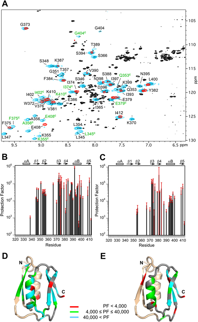
In addition to multiple virulence factors, Bacillus cereus a pathogen that causes food poisoning and life-threatening wound infections, secretes the pore-forming toxin hemolysin II (HlyII). The HlyII toxin has a unique 94 amino acid C-terminal domain (HlyIIC). HlyIIC exhibits splitting of NMR resonances due to cis/trans isomerization of a single proline near the C-terminus. To overcome heterogeneity, we solved the structure of P405M-HlyIIC, a mutant that exclusively stabilizes the trans state. The NMR structure of HlyIIC reveals a novel fold, consisting of two subdomains αA-β1-β2 and β3-β4-αB-β5, that come together in a barrel-like structure. The barrel core is fastened by three layers of hydrophobic residues. The barrel end opposite the HlyIIC-core has a positively charged surface, that by binding negatively charged moieties on cellular membranes, may play a role in target-cell surface recognition or stabilization of the heptameric pore complex. In the WT domain, dynamic flexibility occurs at the N-terminus and the first α-helix that connects the HlyIIC domain to the HlyII-core structure. In the destabilizing P405M mutant, increased flexibility is evident throughout the first subdomain, suggesting that the HlyIIC structure may have arisen through gene fusion.


Conflict of interest statement

The authors declare that they have no competing interests.

Figures

Figure 1
Figure 1
NMR structure of HlyIIC. (A) Best-fit superposition for the backbone structures of residues 330–412 from the ensemble of 25 lowest energy NMR structures. The first 11 residues at the disordered N-terminus are not shown. The N- and C-terminal halves of the molecule are colored blue and magenta, respectively, to illustrate the better precision for the 370–412 segment in subdomain 2 (magenta). (B) Stereo diagram (‘wall-eyed’ view) of the NMR structure closest to the ensemble average. The structure is colored on a gradient running from blue (N-terminus) to red (C-terminus). The view is the same as in (A). (C) Diagram summarizing the folding topology of the structure. Cylinders depict α-helices and arrows indicate β-strands. Secondary structure elements are labeled according to their position in the sequence, together with their start and end residues. The coloring scheme is the same as in (B). Additional elements of secondary structure not shown in the figure, are a 310 helix between residues E386-T389 which is present in most of the NMR structures, and a 310 helix between N320-L324 that occurs at the disordered N-terminus of only some of the structures in the NMR ensemble.
Figure 2
Figure 2
Structural properties of the HlyIIC domain. (A) Cartoon of the HlyIIC NMR structure closest to the ensemble average, illustrating the 3-layer hydrophobic core of HlyIIC. (B) The relationship between P405, which is subject to cis/trans isomerization in WT-HlyIIC and a patch of positively charged lysine residues at the bottom of the structure. (C) Electrostatic surface of HlyIIC calculated using the APBS method, showing the positively charged patch formed by basic lysine residues. (D) Structural mapping of chemical shift differences between the conformational states of WT-HlyIIC related by cis/trans isomerization of P405. The composite (|HN| + 0.1|N|) chemical shift index data are colored on a gradient running from cyan for the smallest differences to red for the largest. Residues that do not show splitting of 1H-15N resonances due to cis/trans isomerization are in gray.
Figure 3
Figure 3
CD spectroscopy of HlyIIC. (A) Wavelength scans for folded WT-HlyIIC (solid line) and P405M-HlyIIC (dotted line) at 20 °C, together with thermally unfolded WT-HlyIIC (short dash) and P405M-HlyIIC (long dash) at 90 °C. (B) Thermal unfolding of WT-HlyIIC (filled circles, solid line) and P405M-HlyIIC (gray squares, dotted line).
Figure 4
Figure 4
Backbone dynamics of the HlyIIC domain. (A) S 2 order parameters describing the amplitudes of 1H-15N bond motions on the ps-ns timescale. S 2 values smaller than 0.8 are shown in green and those smaller than 0.5 in red. (B) R2ex line-broadening contributions to 15N R2 relaxation due to conformational averaging on the µs-ms timescale. R2ex values greater than 0 Hz are shown in green and those greater than 5 Hz in red. The secondary structure of HlyIIC is shown at the top of panels. Error bars are shown for all values but in some cases are smaller than the symbols used to depict the data. The insets show the S 2 and R2ex values mapped on the HlyIIC structure, with the first half of the molecule colored peach and the second half gray.
Figure 5
Figure 5
Hydrogen exchange of HlyIIC. (A) Superposition of the 1H-15N HSQC spectrum of the WT-HlyIIC domain in H2O (black) with those of WT-HlyIIC (cyan) and the P405M-HlyIIC mutant (red) after 2 h of incubation in D2O. Amide protons protected after 2 h of exchange in D2O are labeled (the full assignments for WT-HlyIIC and P405M-HlyIIC are published). Protected amide protons from the cis form of WT-HlyIIC are labeled with green letters, these signals are missing in the P405M mutant, which eliminates the cis conformational form. E408 has a large chemical shift difference between the WT and the mutant because it is close to the site of the P405M mutation. Note that compared to WT-HlyIIC, only amides from the second half of the domain, residues 372–410, persist for 2 h in D2O for the P405M-HlyIIC mutant. Protection factors for (B) WT-HlyIIC and (C) P405M-HlyIIC. Protection factors are only given for amide protons from the trans form of the WT protein. Additional protection is seen for the side-chain Nε1 proton of W372, which exchanges with rates of 0.0029 ± 0.0006 min−1 and 0.106 ± 0.008 min−1 in WT-HlyIIC and P405M-HlyIIC, corresponding to protection factors of about 17,000 and 500 at pH 6. Protection factors for (D) WT-HlyIIC and (C) P405M-HlyIIC, mapped on the NMR structure of P405M-HlyIIC with the indicated scale.
Figure 6
Figure 6
Modeling of full-length HlyII. (A) Model of the HlyII monomer based on the X-ray structure of monomeric S. aureus α-hemolysin (PDB code 4IDJ). The stem loop, which forms the trans-membrane β-barrel in the heptameric pore is colored gray, the core structure of HlyII in orange, and the HlyIIC domain in rainbow colors. (B) Model of the HlyII pore based on the X-ray structure of heptameric S. aureus α-hemolysin (PDB code 7AHL). The view is down the axis of the pore from the extracellular side, perpendicular to the plane of the membrane lipid bilayer. (C) View of the HlyII pore parallel to the plane of the lipid bilayer. The pore has a mushroom like structure, with the membrane-traversing β-barrel formed from the stem loops of seven monomers. The mushroom cap consists of the core domains. A protomer of the heptamer is colored in orange. In the model of the HlyII pore, the HlyIIC domain (shown in rainbow colors for one of the protomers) extends from the mushroom cap towards the membrane. (D) Expansion of the view in (C). The lysines that make up the positively charged patch of the HlyIIC domain are shown as blue sticks. The G404-P405 dipeptide, in a loop adjacent to the N-terminal connector, is depicted by gray spheres. Exposed aromatic residues that may dock the HlyIIC domain to the HlyII-core are labeled in the expansion.

Similar articles

Cited by

References

    1. Bottone EJ. Bacillus cereus, a volatile human pathogen. Clin. Microbiol. Rev. 2010;23:382–398. doi: 10.1128/CMR.00073-09. - DOI - PMC - PubMed
    1. Drobniewski FA. Bacillus cereus and related species. Clin. Microbiol. Rev. 1993;6:324–338. doi: 10.1128/CMR.6.4.324. - DOI - PMC - PubMed
    1. Logan NA. Bacillus and relatives in foodborne illness. J. Appl. Microbiol. 2012;112:417–429. doi: 10.1111/j.1365-2672.2011.05204.x. - DOI - PubMed
    1. Stenfors Arnesen LP, Fagerlund A, Granum PE. From soil to gut: Bacillus cereus and its food poisoning toxins. FEMS Microbiol. Rev. 2008;32:579–606. doi: 10.1111/j.1574-6976.2008.00112.x. - DOI - PubMed
    1. Ramarao N, Sanchis V. The pore-forming haemolysins of bacillus cereus: a review. Toxins (Basel) 2013;5:1119–1139. doi: 10.3390/toxins5061119. - DOI - PMC - PubMed

Publication types

LinkOut - more resources