

Diagnostic capability of Pulsar perimetry in pre-perimetric and early glaucoma
- PMID: 28607414
- PMCID: PMC5468287
- DOI: 10.1038/s41598-017-03550-x
Diagnostic capability of Pulsar perimetry in pre-perimetric and early glaucoma
Abstract
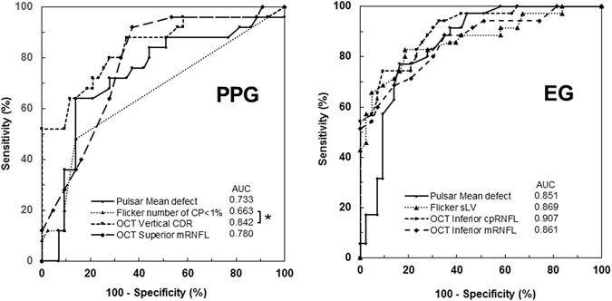
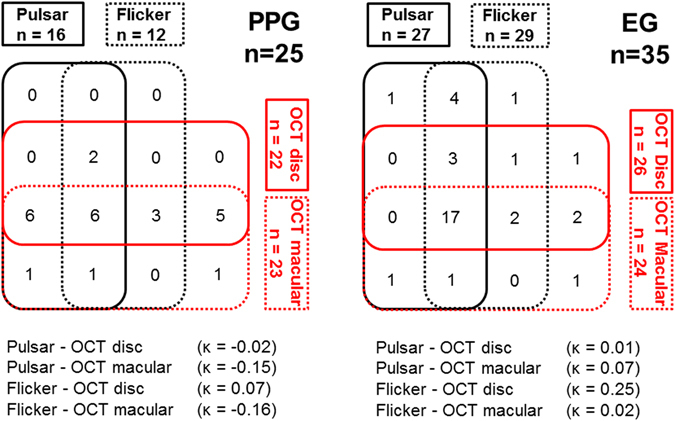
This study aimed to compare the diagnostic capability of Pulsar perimetry (Pulsar) in pre-perimetric glaucoma (PPG) and early glaucoma (EG) with that of Flicker perimetry (Flicker) and spectral-domain optical conference tomography (SD-OCT). This prospective cross-sectional study included 25 eyes of 25 PPG patients, 35 eyes of 35 EG patients, and 42 eyes of 42 healthy participants. The diagnostic capability using the area under the curve (AUC) of the best parameter and agreement of detectability between structural and functional measurements were compared. For PPG patients, the AUC of Pulsar, Flicker, OCT-disc, and OCT-macular was 0.733, 0.663, 0.842, and 0.780, respectively. The AUC of Flicker was significantly lower than that of OCT-disc (p = 0.016). For EG patients, the AUC of Pulsar, Flicker, OCT-disc, and OCT-macular were 0.851, 0.869, 0.907, and 0.861, respectively. There was no significant difference in AUC among these methods. The agreement between structural and functional measurements expressed by kappa value ranged from -0.16 to 0.07 for PPG and from 0.01 to 0.25 for EG. Although the diagnostic capability of Pulsar in the PPG and EG groups was equal to that of Flicker and SD-OCT, the agreements between structural and functional measurements for both PPG and EG were poor.
Conflict of interest statement
K.H. and N.S. received financial support from RE Medical (Osaka, Japan).
Figures
Similar articles
-
The Diagnostic Value of Pulsar Perimetry, Optical Coherence Tomography, and Optical Coherence Tomography Angiography in Pre-Perimetric and Perimetric Glaucoma.J Clin Med. 2021 Dec 13;10(24):5825. doi: 10.3390/jcm10245825. J Clin Med. 2021. PMID: 34945121 Free PMC article.
-
Diagnostic Ability of Wide-field Retinal Nerve Fiber Layer Maps Using Swept-Source Optical Coherence Tomography for Detection of Preperimetric and Early Perimetric Glaucoma.J Glaucoma. 2017 Jun;26(6):577-585. doi: 10.1097/IJG.0000000000000662. J Glaucoma. 2017. PMID: 28368998
-
Detectability of glaucomatous changes using SAP, FDT, flicker perimetry, and OCT.J Glaucoma. 2009 Feb;18(2):165-71. doi: 10.1097/IJG.0b013e318179f7ca. J Glaucoma. 2009. PMID: 19225357
-
Evaluation of spectral domain optical coherence tomography parameters in ocular hypertension, preperimetric, and early glaucoma.Indian J Ophthalmol. 2017 Nov;65(11):1143-1150. doi: 10.4103/ijo.IJO_157_17. Indian J Ophthalmol. 2017. PMID: 29133640 Free PMC article.
-
[Pulsar perimetry. A review and new results].Ophthalmologe. 2013 Feb;110(2):107-15. doi: 10.1007/s00347-012-2690-0. Ophthalmologe. 2013. PMID: 23392836 Review. German.
Cited by
-
Degree of loss in the tissue thickness, microvascular density, specific perimetry and standard perimetry in early glaucoma.BMJ Open Ophthalmol. 2023 Apr;8(1):e001256. doi: 10.1136/bmjophth-2023-001256. BMJ Open Ophthalmol. 2023. PMID: 37278436 Free PMC article. Clinical Trial.
-
The Diagnostic Value of Pulsar Perimetry, Optical Coherence Tomography, and Optical Coherence Tomography Angiography in Pre-Perimetric and Perimetric Glaucoma.J Clin Med. 2021 Dec 13;10(24):5825. doi: 10.3390/jcm10245825. J Clin Med. 2021. PMID: 34945121 Free PMC article.
-
Glaucoma Clinic Monitoring Over 6 Months Using Online Circular Contrast Perimetry in Comparison with Standard Automatic Perimetry: The Developing-World Setting.Clin Ophthalmol. 2024 Dec 14;18:3767-3780. doi: 10.2147/OPTH.S496728. eCollection 2024. Clin Ophthalmol. 2024. PMID: 39697638 Free PMC article.
-
A hybrid multi model artificial intelligence approach for glaucoma screening using fundus images.NPJ Digit Med. 2025 Feb 27;8(1):130. doi: 10.1038/s41746-025-01473-w. NPJ Digit Med. 2025. PMID: 40016437 Free PMC article.
-
Evaluation of RETICs Glaucoma Diagnostic Calculators in Preperimetric Glaucoma.Transl Vis Sci Technol. 2018 Nov 30;7(6):13. doi: 10.1167/tvst.7.6.13. eCollection 2018 Nov. Transl Vis Sci Technol. 2018. PMID: 30519498 Free PMC article.
References
-
- Chauhan BC, McCormick TA, Nicolela MT, LeBlanc RP. Optic disc and visual field changes in a prospective longitudinal study of patients with glaucoma: comparison of scanning laser tomography with conventional perimetry and optic disc photography. Arch Ophthalmol. 2001;119:1492–1499. doi: 10.1001/archopht.119.10.1492. - DOI - PubMed
Publication types
MeSH terms
LinkOut - more resources
Full Text Sources
Other Literature Sources
Medical
