. 2017:2017:1960965.
doi: 10.1155/2017/1960965. Epub 2017 May 10.

Characterization of Mesenchymal Stem Cell-Like Cells Derived From Human iPSCs via Neural Crest Development and Their Application for Osteochondral Repair

Affiliations

Characterization of Mesenchymal Stem Cell-Like Cells Derived From Human iPSCs via Neural Crest Development and Their Application for Osteochondral Repair

Ryota Chijimatsu et al. Stem Cells Int. 2017.

Abstract

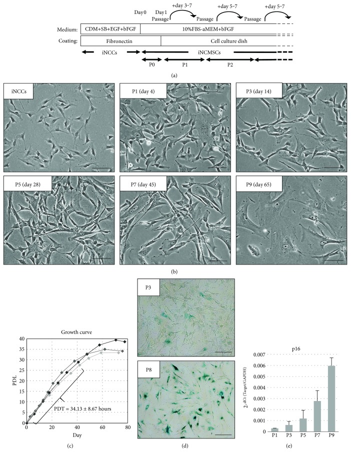
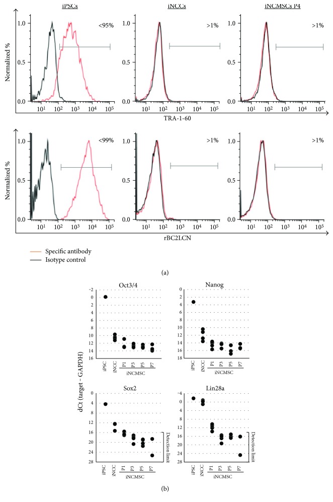
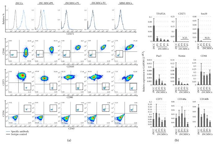
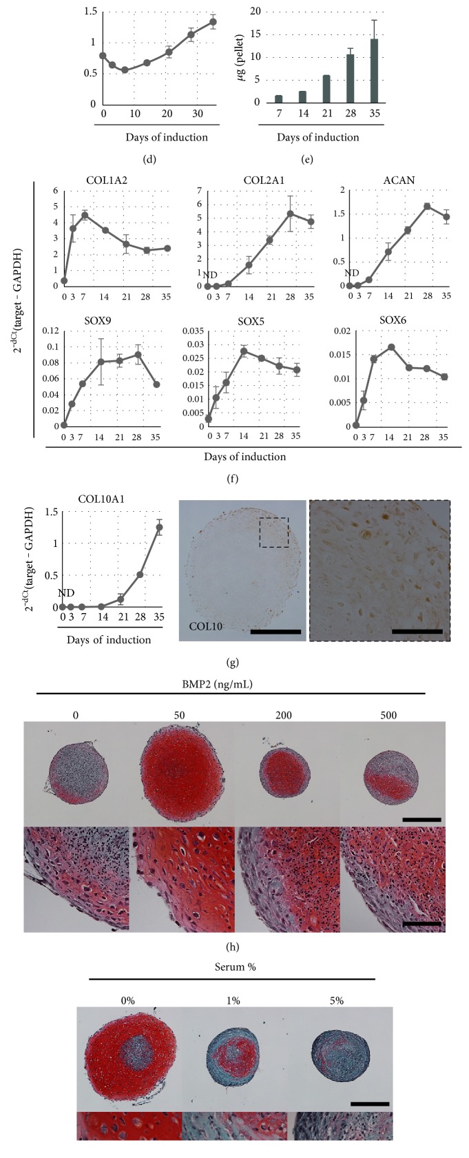
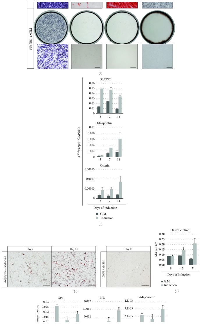
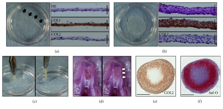
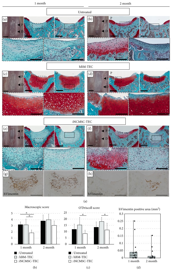
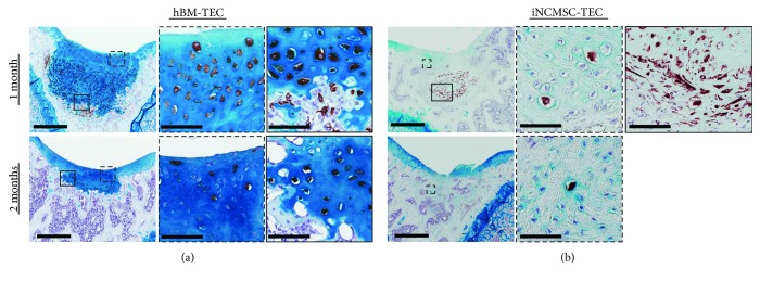
Mesenchymal stem cells (MSCs) derived from induced pluripotent stem cells (iPSCs) are a promising cell source for the repair of skeletal disorders. Recently, neural crest cells (NCCs) were reported to be effective for inducing mesenchymal progenitors, which have potential to differentiate into osteochondral lineages. Our aim was to investigate the feasibility of MSC-like cells originated from iPSCs via NCCs for osteochondral repair. Initially, MSC-like cells derived from iPSC-NCCs (iNCCs) were generated and characterized in vitro. These iNCC-derived MSC-like cells (iNCMSCs) exhibited a homogenous population and potential for osteochondral differentiation. No upregulation of pluripotent markers was detected during culture. Second, we implanted iNCMSC-derived tissue-engineered constructs into rat osteochondral defects without any preinduction for specific differentiation lineages. The implanted cells remained alive at the implanted site, whereas they failed to repair the defects, with only scarce development of osteochondral tissue in vivo. With regard to tumorigenesis, the implanted cells gradually disappeared and no malignant cells were detected throughout the 2-month follow-up. While this study did not show that iNCMSCs have efficacy for repair of osteochondral defects when implanted under undifferentiated conditions, iNCMSCs exhibited good chondrogenic potential in vitro under appropriate conditions. With further optimization, iNCMSCs may be a new source for tissue engineering of cartilage.


Figures

Figure 1
Figure 1
Transition to iNCMSCs from iNCCs and their expandability. (a) Schematic protocol for generation of iNCMSCs from iNCCs. (b) Cell morphology of iNCCs and iNCMSCs during expansion culture. Passage number and expanded days are presented in their inboxes. (c) Growth curve of iNCMSCs over two months (N = three lines). Each symbol represents each passage. PDT was calculated during first seven weeks (mean ± SD for three lines). (d) Senescence-associated β-galactosidase staining of iNCMSCs at passages 3 and 8. (e) Upregulation of p16 mRNA expression during expansion culture (means ± SD for three lines). Scale bars = 100 μm (b, d). Abbreviations: CDM: Chemical defined medium; SB: SB431452; PDL: Population doubling level; PDT: Population doubling time; iNCCs: Induced neural crest cells; iPSCs: Induced pluripotent stem cells; iNCMSCs: iNCC-derived mesenchymal stem cells.
Figure 2
Figure 2
Depletion of pluripotent markers in iNCMSCs. (a, b) FACS analysis of PSC-specific surface antigen and gene expression analysis of PSC-specific transcript factors in iPSCs, iNCCs, and iNCMSCs. 409B2-iPSCs were used for positive control of each assays. Abbreviations: PSCs: Pluripotent stem cells; iNCCs: Induced neural crest cells; iPSCs: Induced pluripotent stem cells; iNCMSCs: iNCC-derived mesenchymal stem cells.
Figure 3
Figure 3
Neural crest markers and mesenchymal stem cell markers. (a) FACS analysis of NC and MSC surface markers in iNCC and iNCMSC (P0, P1, and P2). The date are representative of four lines. Human bone marrow derived MSCs were used for assay control. (b) Gene expression of NC and MSC markers during expansion of iNCMSCs. (mean ± SD for four lines). Abbreviations: iNCCs: Induced neural crest cells; iPSCs: Induced pluripotent stem cells; iNCMSCs: Induced neural crest derived mesenchymal stem cells; hBM-MSCs: Human bone marrow-derived MSCs.
Figure 4
Figure 4
Chondrogenesis of iNCMSCs. The representative data from three lines is shown. (a) Safranin-O staining of chondrogenic pellet stimulated with TGFβ3 and BMP2 at day 28. High magnification image is shown in the right panel with the same frame. (b, c) Immunostaining for COL2 and SOX9 of chondrogenic pellet at day 28. (d) The pellet size of chondrogenic pellets during culture. (e) Deposition of sulfated glycosaminoglycan in chondrogenic pellets during culture. (f) The alteration of chondrogenesis-related gene expression. (g) mRNA expression levels for type 10 collagen in long-term culture and immunostaining for type 10 collagen at day 28 in chondrogenic pellets. (h) Saf-O images of chondrogenic pellet culture with TGFβ3 (10 ng/mL) and various concentrations of BMP2 (0–500 ng/mL) for 28 days. (i) Saf-O images of chondrogenic pellet cultures in TB media with addition of variable serum concentrations for 28 days. Scale bars = 500 μm (a, h, i) and 100 μm (b, c, and high-magnified images of a, h, i). Data are expressed as mean ± SD for three pellet replicates (d–g). Abbreviations: Saf-O: Safranin-O staining; COL2: Type 2 collagen; sGAG: Sulfated glycosaminoglycan; COL10: Type 10 collagen.
Figure 5
Figure 5
Osteo- and adipogenesis of iNCMSCs. The representative data for three lines is shown. (a) ALP staining at day 7 and calcium staining (alizarin red S and von Kossa staining) at day 14 of iNCMSCs cultured in osteogenic medium in 24-well plates. (b) Osteogenesis-related gene expression at days 3, 7, and 14. (c) Oil red staining of iNCMSCs cultured in adipogenesis medium at days 9 and 21. (d) Measurement of oil red positive droplets. (e) Adipogenesis-related gene expression at day 9, 15, and 21. Data are expressed as mean ± SD for three well replicate per group (b, d, e). Scale bars = 100 μm (a, c). Abbreviations: ALP: Alkaline phosphatase; G.M.: Growth medium.
Figure 6
Figure 6
In vitro development of tissue-engineered tissue from iNCMSCs. (a–c)ses gross appearance of iNCMSC-TEC developed in 35 mm dishes at day 7. Thin monolayer cell sheet was formed in the culture bottom surface after 7 days of high-density culture (a), and once artificially detached (black arrows), a thick sheet-like construct (iNCMSC-TEC) developed through active tissue contraction within a few minutes (b). HE staining and immunostaining for COL1 and COL2 of transverse section are shown in their corresponding right panels. Scale bars = 50 μm. The iNCMSC-TEC exhibited sufficient strength to handle with forceps (c). (d) Ex vivo implantation of iNCMSC-TEC into osteochondral defects of cadaver rats. iNCMSC-TEC readily filled the defect created in the femoral groove (white arrows). Scale bars = 1 mm. (e, f) Chondrogenic potential of iNCMSC-TEC. COL2 immunostaining (e) and Safranin-O staining (f) of iNCMSC-TEC cultured in chondrogenic medium for 1 month. Scale bars = 500 μm. Abbreviations: HE: Hematoxylin and eosin staining; COL1: Type 1 collagen; COL2: Type 2 collagen; Saf-O: Safranin-O.
Figure 7
Figure 7
In vivo transplantation of TEC to rat osteochondral defects. (a–f) Macroappearance and Safranin-O staining (Saf-O) for each group at 1 month and 2 months. Inset boxes in the upper panels are magnified in lower panels with the same frame. Representative data are shown (N = 5 knees for the empty group, N = 6 knees for the hBM-TEC group, and N = 7 knees for the iNCMSC-TEC group at each endpoint). Histological assessments were conducted at two different points as shown in the macroimages (“P” represents “proximal section” and “D” presents “distal section”). Scale bars = 1 mm (macroimage), 500 μm (upper panels of Saf-O), and 200 μm (lower panels of Saf-O). (g, h) Human vimentin immunostaining of the iNCMSC-TEC group at 1 month and 2 months. Scale bars = 500 μm. (i) Macroscopic score based on their gross appearance [57]. Averaged score with standard deviation is shown. P < 0.05 by the Steel-Dwass test. (j) Histological grading by the O'Driscoll scoring system based on Saf-O staining using two specimens (“P” and “D”) per each knee. Data are expressed as the mean ± SD for each group. P < 0.05 by the Steel-Dwass test. (k) The quantitation of transplanted iNCMSCs remaining at the implantation site was calculated from their hVimentin-DAB positive area. Data are shown as box-and-whisker plots and dot plots. Abbreviations: TEC: Tissue-engineered construct; hBM: Human bone marrow mesenchymal stem cell; iNCMSC: Induced neural crest-derived mesenchymal stem cells.
Figure 8
Figure 8
Vimentin and alcian blue staining of in vivo implanted TEC. (a, b) hVimentin/alcian blue staining for the hBM-TEC and iNCMSC-TEC groups at 1 month and 2 months. Right panels are magnification images of inset boxes of the left panels. Scale bars = 500 μm and 50 μm (high-magnified images). Abbreviations: TEC: Tissue-engineered construct; hBM: Human bone marrow-derived mesenchymal stem cells; iNCMSC: Induced neural crest-derived mesenchymal stem cells.

Similar articles

Cited by

References

    1. Huey D. J., Hu J. C., Athanasiou K. A. Unlike bone, cartilage regeneration remains elusive. Science. 2012;338(6109):917–921. doi: 10.1126/science.1222454. - DOI - PMC - PubMed
    1. Wagner W., Horn P., Castoldi M., et al. Replicative senescence of mesenchymal stem cells: a continuous and organized process. PloS One. 2008;3(5, article e2213) doi: 10.1371/journal.pone.0003578. - DOI - PMC - PubMed
    1. Kretlow J. D., Jin Y. Q., Liu W., et al. Donor age and cell passage affects differentiation potential of murine bone marrow-derived stem cells. BMC Cell Biology. 2008;9(1):p. 60. doi: 10.1186/1471-2121-9-60. - DOI - PMC - PubMed
    1. Wagner W., Bork S., Horn P., et al. Aging and replicative senescence have related effects on human stem and progenitor cells. PloS One. 2009;4(6, article e5846) doi: 10.1371/journal.pone.0005846. - DOI - PMC - PubMed
    1. Xin Y., Wang Y. M., Zhang H., et al. Aging adversely impacts biological properties of human bone marrow-derived mesenchymal stem cells: implications for tissue engineering heart valve construction. Artificial Organs. 2010;34(3):215–222. doi: 10.1111/j.1525-1594.2009.00824.x. - DOI - PubMed