

Aberrant Cx43 Expression and Mislocalization in Metastatic Human Melanomas
- PMID: 28607585
- PMCID: PMC5463425
- DOI: 10.7150/jca.18569
Aberrant Cx43 Expression and Mislocalization in Metastatic Human Melanomas
Abstract
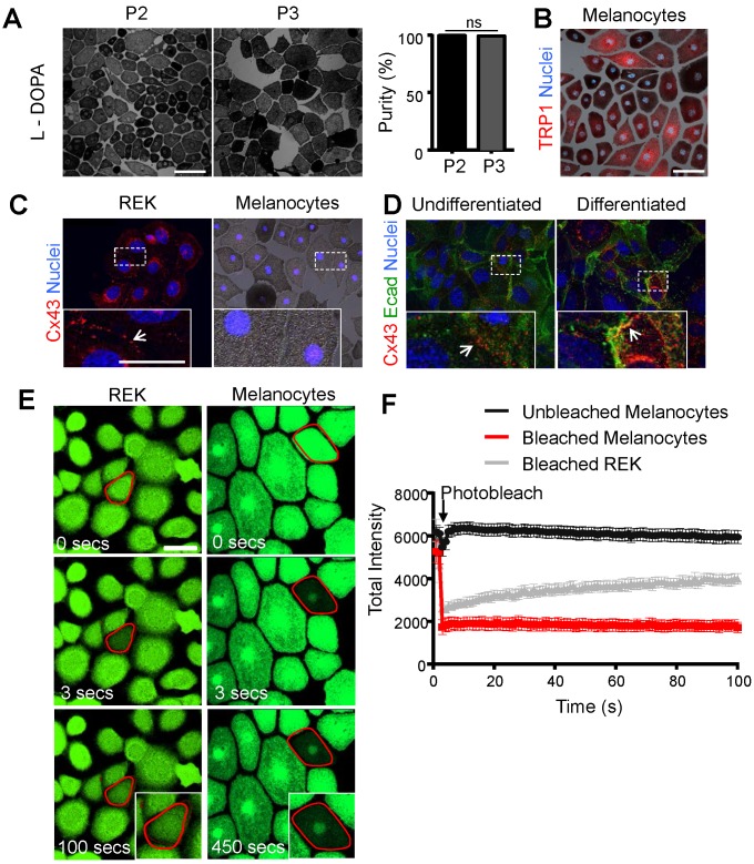
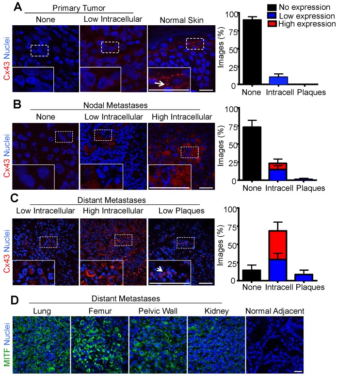
At present, it is unclear if melanocytes contain Cx43 gap junctions and whether Cx43 expression is regulated in melanoma onset and progression. To this end, we cultured pure populations of mouse melanocytes and found that they had no detectable Cx43 and exhibited an inability for dye transfer indicating they were devoid of functional gap junctions. Given the evidence that melanomas acquire the expression of other connexin isoforms during tumor progression, we assessed if Cx43 was also expressed and assembled into gap junctions at any stage of human melanoma onset and progression to distant metastases. Nearly all primary melanomas within the epidermis lacked Cx43. In contrast, nodal metastases expressed low levels of Cx43 which was markedly higher in distant metastases that had invaded vital organs. Importantly, in all stages of melanoma progression, Cx43 could be detected in intracellular compartments but was rarely assembled into gap junctions indicative of functional gap junction channels. Overall, these studies suggest that melanocytes do not form Cx43 homocellular gap junctions and even though Cx43 levels increase during melanoma progression, this connexin rarely assembles into gap junction structures.
Keywords: Cx43; connexin; gap junctional intercellular communication; gap junctions; melanoma..
Conflict of interest statement
Competing Interests: The authors have declared that no competing interest exists.
Figures
Similar articles
-
Exploring Differential Connexin Expression across Melanocytic Tumor Progression Involving the Tumor Microenvironment.Cancers (Basel). 2019 Feb 1;11(2):165. doi: 10.3390/cancers11020165. Cancers (Basel). 2019. PMID: 30717194 Free PMC article.
-
Functional formation of heterotypic gap junction channels by connexins-40 and -43.Channels (Austin). 2014;8(5):433-43. doi: 10.4161/19336950.2014.949188. Channels (Austin). 2014. PMID: 25483586 Free PMC article.
-
Expression of a connexin 43/beta-galactosidase fusion protein inhibits gap junctional communication in NIH3T3 cells.J Cell Biol. 1995 Jul;130(2):419-29. doi: 10.1083/jcb.130.2.419. J Cell Biol. 1995. PMID: 7542247 Free PMC article.
-
[Remodeling of cardiac gap junctions and arrhythmias].Sheng Li Xue Bao. 2011 Dec 25;63(6):586-92. Sheng Li Xue Bao. 2011. PMID: 22193455 Review. Chinese.
-
Connexin 43 (Cx43) in cancer: Implications for therapeutic approaches via gap junctions.Cancer Lett. 2019 Feb 1;442:439-444. doi: 10.1016/j.canlet.2018.10.043. Epub 2018 Nov 22. Cancer Lett. 2019. PMID: 30472182 Review.
Cited by
-
Hypoxic Melanoma Cells Deliver microRNAs to Dendritic Cells and Cytotoxic T Lymphocytes through Connexin-43 Channels.Int J Mol Sci. 2020 Oct 13;21(20):7567. doi: 10.3390/ijms21207567. Int J Mol Sci. 2020. PMID: 33066331 Free PMC article.
-
The role of connexins in breast cancer: from misregulated cell communication to aberrant intracellular signaling.Tissue Barriers. 2022 Jan 2;10(1):1962698. doi: 10.1080/21688370.2021.1962698. Epub 2021 Aug 6. Tissue Barriers. 2022. PMID: 34355641 Free PMC article. Review.
-
Cx43 mediates cross-talk of tumor cells and macrophage via cGAS-STING signaling.Med Oncol. 2025 May 28;42(7):223. doi: 10.1007/s12032-025-02773-7. Med Oncol. 2025. PMID: 40437141
-
Epithelial-Mesenchymal Transition: Role in Cancer Progression and the Perspectives of Antitumor Treatment.Acta Naturae. 2020 Jul-Sep;12(3):4-23. doi: 10.32607/actanaturae.11010. Acta Naturae. 2020. PMID: 33173593 Free PMC article.
-
Exploring Differential Connexin Expression across Melanocytic Tumor Progression Involving the Tumor Microenvironment.Cancers (Basel). 2019 Feb 1;11(2):165. doi: 10.3390/cancers11020165. Cancers (Basel). 2019. PMID: 30717194 Free PMC article.
References
-
- Sohl G, Willecke K. An update on connexin genes and their nomenclature in mouse and man. Cell Commun Adhes. 2003;10:173–80. - PubMed
-
- Goodenough DA, Goliger JA, Paul DL. Connexins, connexons, and intercellular communication. Annu Rev Biochem. 1996;65:475–502. - PubMed
-
- Churko JM, Laird DW. Gap junction remodeling in skin repair following wounding and disease. Physiology. 2013;28:190–8. - PubMed
-
- Hsu M, Andl T, Li G, Meinkoth JL, Herlyn M. Cadherin repertoire determines partner-specific gap junctional communication during melanoma progression. J Cell Sci. 2000;113:1535–42. - PubMed
LinkOut - more resources
Full Text Sources
Other Literature Sources
Miscellaneous
