

Molecular mechanisms underlying the pilsicainide-induced stabilization of hERG proteins in transfected mammalian cells
- PMID: 28607619
- PMCID: PMC5459418
- DOI: 10.1016/j.joa.2016.09.003
Molecular mechanisms underlying the pilsicainide-induced stabilization of hERG proteins in transfected mammalian cells
Abstract
Background: Pilsicainide, classified as a relatively selective Na+ channel blocker, also has an inhibitory action on the rapidly-activating delayed-rectifier K+ current (IKr ) through human ether-a-go-go-related gene (hERG) channels. We studied the effects of chronic exposure to pilsicainide on the expression of wild-type (WT) hERG proteins and WT-hERG channel currents, as well as on the expression of mutant hERG proteins, in a heterologous expression system.
Methods: HEK293 cells stably expressing WT or mutant hERG proteins were subjected to Western blotting, immunofluorescence microscopy and patch-clamp experiments.
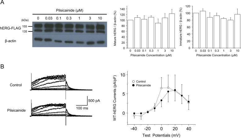
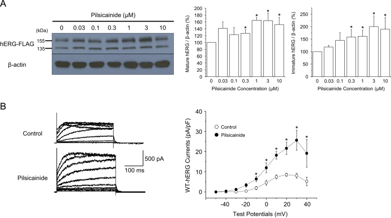
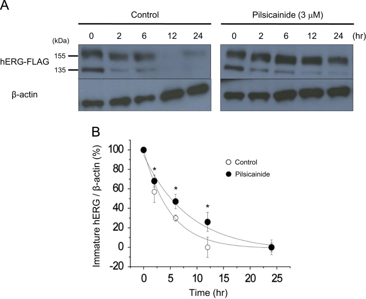
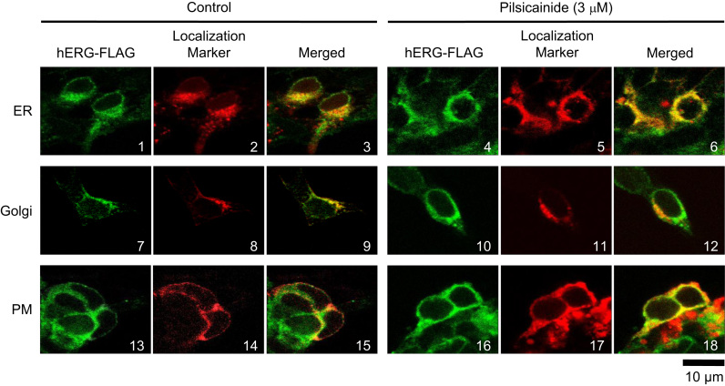
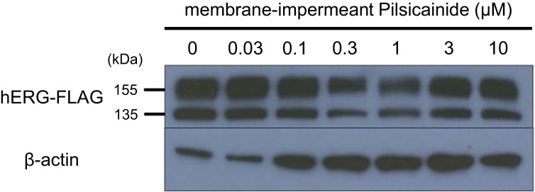
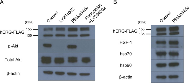
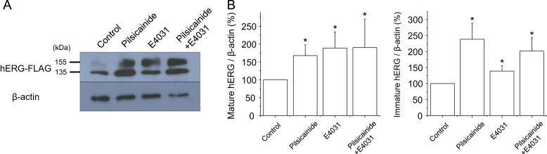
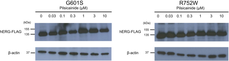
Results: Acute exposure to pilsicainide at 0.03-10 μM influenced neither the expression of WT-hERG proteins nor WT-hERG channel currents. Chronic treatment with 0.03-10 μM pilsicainide for 48 h, however, increased the expression of WT-hERG proteins and channel currents in a concentration-dependent manner. Chronic treatment with 3 μM pilsicainide for 48 h delayed degradation of WT-hERG proteins and increased the channels expressed on the plasma membrane. A cell membrane-impermeant pilsicainide derivative did not influence the expression of WT-hERG, indicating that pilsicainide stabilized the protein inside the cell. Pilsicainide did not influence phosphorylation of Akt (protein kinase B) or expression of heat shock protein families such as HSF-1, hsp70 and hsp90. E4031, a chemical chaperone for hERG, abolished the pilsicainide effect on hERG. Chronic treatment with pilsicainide could also increase the protein expression of trafficking-defective mutant hERG, G601S and R752W.
Conclusions: Pilsicainide penetrates the plasma membrane, stabilizes WT-hERG proteins by acting as a chemical chaperone, and enhances WT-hERG channel currents. This mechanism could also be applicable to modulations of certain mutant-hERG proteins.
Keywords: Chemical chaperone; Pilsicainide; hERG.
Figures
Similar articles
-
Effects of Na+ channel blocker, pilsicainide, on HERG current expressed in HEK-293 cells.J Cardiovasc Pharmacol. 2003 Sep;42(3):410-8. doi: 10.1097/00005344-200309000-00013. J Cardiovasc Pharmacol. 2003. PMID: 12960687
-
Role of the cytosolic chaperones Hsp70 and Hsp90 in maturation of the cardiac potassium channel HERG.Circ Res. 2003 Jun 27;92(12):e87-100. doi: 10.1161/01.RES.0000079028.31393.15. Epub 2003 May 29. Circ Res. 2003. PMID: 12775586
-
Characterization of the novel mutant A78T-HERG from a long QT syndrome type 2 patient: Instability of the mutant protein and stabilization by heat shock factor 1.J Arrhythm. 2016 Oct;32(5):433-440. doi: 10.1016/j.joa.2015.10.005. Epub 2015 Nov 25. J Arrhythm. 2016. PMID: 27761169 Free PMC article.
-
Inhibitory effects and mechanism of dihydroberberine on hERG channels expressed in HEK293 cells.PLoS One. 2017 Aug 1;12(8):e0181823. doi: 10.1371/journal.pone.0181823. eCollection 2017. PLoS One. 2017. PMID: 28763460 Free PMC article.
-
HERG channel trafficking.Novartis Found Symp. 2005;266:57-69; discussion 70-4, 95-9. Novartis Found Symp. 2005. PMID: 16050262 Review.
Cited by
-
Towards Bridging Translational Gap in Cardiotoxicity Prediction: an Application of Progressive Cardiac Risk Assessment Strategy in TdP Risk Assessment of Moxifloxacin.AAPS J. 2018 Mar 14;20(3):47. doi: 10.1208/s12248-018-0199-4. AAPS J. 2018. PMID: 29541956
-
A PAS-targeting hERG1 activator reduces arrhythmic events in Jervell and Lange-Nielsen syndrome patient-derived hiPSC-CMs.JCI Insight. 2025 Jan 9;10(4):e183444. doi: 10.1172/jci.insight.183444. JCI Insight. 2025. PMID: 39786967 Free PMC article.
References
-
- Sanguinetti M.C., Jiang C., Curran M.E., Keating M.T. A mechanistic link between an inherited and an acquired cardiac arrhythmia: HERG encodes the IKr potassium channel. Cell. 1995;81:299–307. - PubMed
-
- Surawicz B. Electrophysiologic substrate of torsade de pointes: dispersion of repolarization or early afterdepolarizations? J Am Coll Cardiol. 1989;14:172–184. - PubMed
-
- Li P., Ninomiya H., Kurata Y. Reciprocal control of hERG stability by hsp70 and hsc70 with implication for restoration of LQT2 mutant stability. Circ Res. 2011;108:458–468. - PubMed
LinkOut - more resources
Full Text Sources
Other Literature Sources
