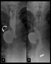
Mid-Esophageal Diverticulum Mimicking an Aortic Aneurysm on Chest Radiography
- PMID: 28607626
- PMCID: PMC5448610
- DOI: 10.12659/PJR.899248
Mid-Esophageal Diverticulum Mimicking an Aortic Aneurysm on Chest Radiography
Abstract
Background: Mid-esophageal region is an uncommon location of esophageal diverticula, a condition usually diagnosed in elderly individuals.
Case report: We report a case of an elderly male with incidental finding of mediastinal lesion, which was initially thought to be an aortic aneurysm. Further evaluation demonstrated a mid-esophageal diverticulum at the level of the carina. We present patient's medical history and imaging, followed by a discussion on symptoms and management.
Conclusions: Knowledge of benign conditions that might mimic a mediastinal vascular pathology is important for therapeutic and prognostic reasons, as they are often managed conservatively.
Keywords: Deglutition Disorders; Diverticulum, Esophageal; Esophageal Motility Disorders; Esophagectomy; Mediastinum.
Figures


Similar articles
-
Squamous cell carcinoma in an esophageal diverticulum below the aortic arch.Int J Surg Case Rep. 2012;3(11):574-6. doi: 10.1016/j.ijscr.2012.07.010. Epub 2012 Aug 7. Int J Surg Case Rep. 2012. PMID: 22940699 Free PMC article.
-
Combined thoracoscopic and endoscopic management of mid-esophageal benign lesions: use of the prone patient position : Thoracoscopic surgery for mid-esophageal benign tumors and diverticula.Surg Endosc. 2008 Jan;22(1):250-4. doi: 10.1007/s00464-007-9359-9. Epub 2007 May 19. Surg Endosc. 2008. PMID: 17514385
-
Esophageal diverticula: patient assessment.Semin Thorac Cardiovasc Surg. 1999 Oct;11(4):326-36. doi: 10.1016/s1043-0679(99)70077-8. Semin Thorac Cardiovasc Surg. 1999. PMID: 10535374 Review.
-
Giant esophageal diverticula after laparoscopic band placement.Ann Thorac Surg. 2012 Oct;94(4):1330-2. doi: 10.1016/j.athoracsur.2012.02.082. Ann Thorac Surg. 2012. PMID: 23006690
-
Severe dysphagia, dysmotility, and unusual saccular dilation (diverticulum) of the esophagus following excision of an asymptomatic congenital cyst.Am J Gastroenterol. 1996 Jun;91(6):1254-8. Am J Gastroenterol. 1996. PMID: 8651183 Review.
References
-
- Thacker PG, Mahani MG, Heider A, Lee EY. Imaging evaluation of mediastinal masses in children and adults: practical diagnostic approach based on a new classification system. J Thorac Imaging. 2015;30(4):247–67. - PubMed
-
- Gore RM, Levine MS. Textbook of gastrointestinal radiology. 3rd ed. Philadelphia: Saunders Elsevier; 2008. pp. 475–77.
-
- Constantini M, Zaninotto G. Oesophageal diverticula. Best Pract Res Clin Gastroenterol. 2004;18:3–17. - PubMed
-
- Thomas ML, Anthony AA, Fosh BG. Oesophageal diverticula. Br J Surg. 2001;88:629–42. - PubMed
-
- Khan N, Ismail F, Van de Werke IE. Oesophageal pouches and diverticula: A pictorial review. S Afr J Surg. 2012;50(3):71–75. - PubMed
Publication types
LinkOut - more resources
Full Text Sources
Other Literature Sources
