Review
. 2017:2017:4328015.
doi: 10.1155/2017/4328015. Epub 2017 Apr 30.

Could LC-NE-Dependent Adjustment of Neural Gain Drive Functional Brain Network Reorganization?

Affiliations
Review

Could LC-NE-Dependent Adjustment of Neural Gain Drive Functional Brain Network Reorganization?

Carole Guedj et al. Neural Plast. 2017.

Abstract

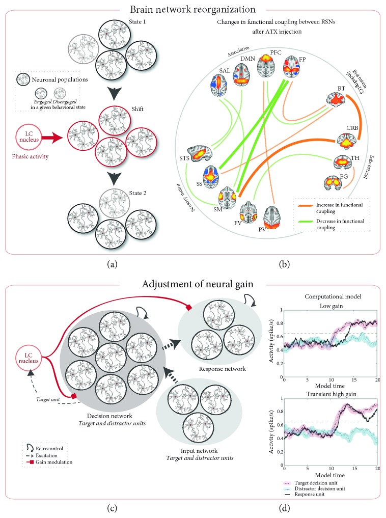
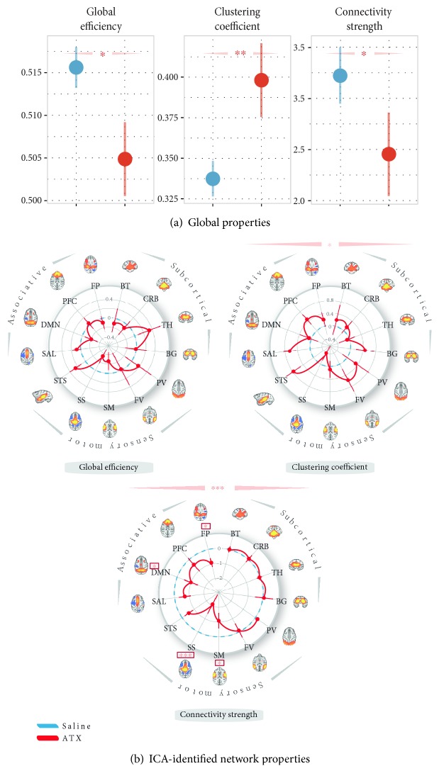
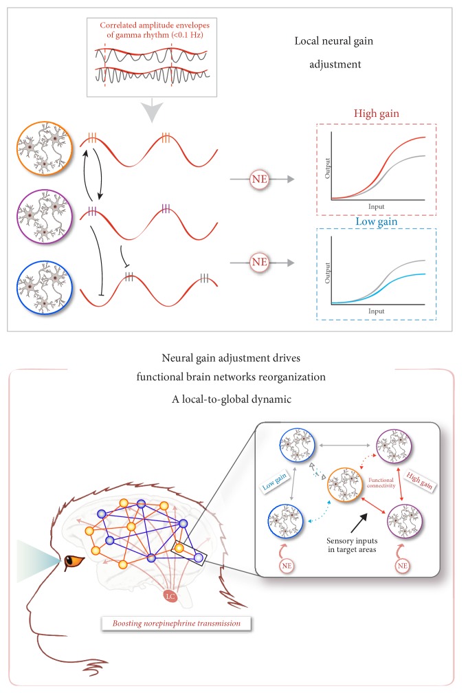
The locus coeruleus-norepinephrine (LC-NE) system is thought to act at synaptic, cellular, microcircuit, and network levels to facilitate cognitive functions through at least two different processes, not mutually exclusive. Accordingly, as a reset signal, the LC-NE system could trigger brain network reorganizations in response to salient information in the environment and/or adjust the neural gain within its target regions to optimize behavioral responses. Here, we provide evidence of the co-occurrence of these two mechanisms at the whole-brain level, in resting-state conditions following a pharmacological stimulation of the LC-NE system. We propose that these two mechanisms are interdependent such that the LC-NE-dependent adjustment of the neural gain inferred from the clustering coefficient could drive functional brain network reorganizations through coherence in the gamma rhythm. Via the temporal dynamic of gamma-range band-limited power, the release of NE could adjust the neural gain, promoting interactions only within the neuronal populations whose amplitude envelopes are correlated, thus making it possible to reorganize neuronal ensembles, functional networks, and ultimately, behavioral responses. Thus, our proposal offers a unified framework integrating the putative influence of the LC-NE system on both local- and long-range adjustments of brain dynamics underlying behavioral flexibility.


Figures

Figure 1
Figure 1
LC-NE system theoretical models ((a) and (b)) brain network reorganizations adapted from Bouret and Sara [33]. (a) A behavioral state is associated with a given functional network with a specific spatiotemporal pattern of neuronal activity. When a stimulus induces a behavioral shift, the LC activation immediately preceding this change modulates the underlying interactions between the neuronal populations via its simultaneous action on several of its target structures, promoting changes within and between functional networks (state 1 → state 2). (b) Overview of the functional coupling changes between 13 resting-state networks (RSNs) following ATX injection (from Guedj et al. [49]). Line thicknesses reflect the correlation strength of the ATX-induced changes. ATX injection modulated the functional coupling of the subcortical network including the LC and decreased the functional coupling between associative and sensory-motor networks. The frontoparietal network, negatively correlated with the brainstem network including the LC region in the saline condition, switched to a positive correlation under ATX ((c) and (d)) adjustment of neural gain. (c) Architecture of the computational model described by Usher et al. [38]. The LC inputs regulate the gain via a multiplier effect on the decision and the response networks. (d) Simulated time courses of activity for the response and decision model units under various neural gain levels (low and high gain). A transient increase of the neural gain induced by a LC phasic response improves the processing of the target stimulus, resulting in faster and sharper increase in response unit activity. Adapted from Usher et al. [38] and Gilzenrat et al. [60]. Circles represent a defined neuronal population. The red circle represents the population of LC neurons. ATX = atomoxetine, BG = basal ganglia, BT = brainstem, CRB = cerebellum, DMN = default-mode network, FP = frontoparietal, FV = foveal visual, PFC = prefrontal cortex, PV = peripheral visual, SAL = salience, SM = somatomotor, SS = somatosensory, STS = superior temporal sulcus, and TH = thalamus.
Figure 2
Figure 2
ATX effect on network architecture—(a) global graph properties. Global efficiency, clustering coefficient, and connectivity strength, under saline (blue) and ATX (red) pharmacological conditions. (b) ICA-identified network properties. The three spider plots represent the global efficiency, the clustering coefficient, and the connectivity strength computed for each ICA-identified resting-state networks (see Guedj et al. [49]). Importantly, these scores were expressed as a difference between the ATX condition and the saline control condition. Blue lines represent no difference between the two pharmacological conditions (difference equals to 0). Red stars indicate statistical differences between saline and ATX conditions: stars above the spider plots indicate a main effect of the pharmacological condition while stars above the networks indicated an interaction between the pharmacological condition and the ICA-identified network type (∗∗∗ = p value < 0.0001; ∗∗ = p value < 0.001; ∗ = p value < 0.05; • = p value < 0.1). Throughout this figure, the results are plotted as mean ± SEM. ATX = atomoxetine, BG = basal ganglia, BT = brainstem, CRB = cerebellum, DMN = default-mode network, FP = frontoparietal, FV = foveal visual, ICA = independent component analysis, PFC = prefrontal cortex, PV = peripheral visual, SAL = salience, SM = somatomotor, SS = somatosensory, STS = superior temporal sulcus, and TH = thalamus.
Figure 3
Figure 3
Mechanism proposed for the NE-dependent local-to-global neuronal dynamics—top panel—relationship between the amplitude envelope correlations in gamma rhythm and NE-dependent local neural gain adjustments. The orange and purple neuronal populations, whose amplitude envelopes cofluctuate, allow the increase in neural gain induced by the norepinephrine system activation to be effective and to spread (upper-right inset, red trace), whereas the purple and blue neuronal populations that exhibited a distinct temporal dynamic do not allow the increase in neural gain to be expressed locally, and this results in a decrease in neural gain (bottom-right inset, blue trace)—bottom panel—boosting norepinephrine transmission is thought to induce an increase in neural gain. The insert illustrates an example of the effect of neural gain adjustment on interactions between different neuronal populations. The five groups of neurons are anatomically interconnected. Sensory input signals influence the activity of target regions (purple and orange groups of neurons). Norepinephrine transmission also modulates the activity of groups of target neurons (violet and blue groups of neurons), increasing or decreasing the neural gain, respectively. The amplification of gain in the neuronal violet group then induces an increase in the functional connectivity between violet and orange groups (red arrows). Similarly, the reduction in neural gain in the blue neuronal group induces a decrease in functional connectivity between blue and orange groups (blue arrow and cross). Thus, under the influence of this local modulation of neural gain, neural networks reconfigure, creating a new organization of functional networks at the whole-brain scale.

References

    1. Aston-Jones G., Rajkowski J., Cohen J. Role of locus coeruleus in attention and behavioral flexibility. Biological Psychiatry. 1999;46(9):1309–1320. doi: 10.1016/S0006-3223(99)00140-7. - DOI - PubMed
    1. Kehagia A. A., Murray G. K., Robbins T. W. Learning and cognitive flexibility: frontostriatal function and monoaminergic modulation. Current Opinion in Neurobiology. 2010;20(2):199–204. doi: 10.1016/j.conb.2010.01.007. - DOI - PubMed
    1. Arnsten A. F. T., Wang M. J., Paspalas C. D. Neuromodulation of thought: flexibilities and vulnerabilities in prefrontal cortical network synapses. Neuron. 2012;76(1):223–239. doi: 10.1016/j.neuron.2012.08.038. - DOI - PMC - PubMed
    1. Sara S. J., Bouret S. Orienting and reorienting: the locus coeruleus mediates cognition through arousal. Neuron. 2012;76(1):130–141. doi: 10.1016/j.neuron.2012.09.011. - DOI - PubMed
    1. Markovic J., Anderson A. K., Todd R. M. Tuning to the significant: neural and genetic processes underlying affective enhancement of visual perception and memory. Behavioural Brain Research. 2014;259(1):229–241. doi: 10.1016/j.bbr.2013.11.018. - DOI - PubMed

LinkOut - more resources