

Immunohistochemical evaluation of vitamin D receptor (VDR) expression in cutaneous melanoma tissues and four VDR gene polymorphisms
- PMID: 28607807
- PMCID: PMC5444928
- DOI: 10.20892/j.issn.2095-3941.2017.0020
Immunohistochemical evaluation of vitamin D receptor (VDR) expression in cutaneous melanoma tissues and four VDR gene polymorphisms
Abstract
Objective: : Vitamin D receptor (VDR) mediates vitamin D activity. We examined whether VDR expression in excised melanoma tissues is associated with VDR gene (VDR) polymorphisms.
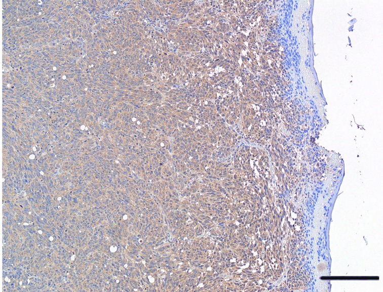
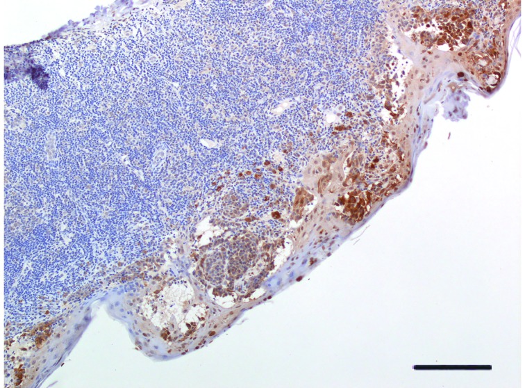
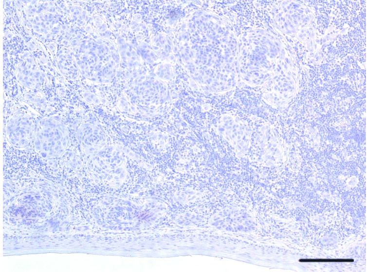
Methods: : We evaluated VDR protein expression (by monoclonal antibody immunostaining), melanoma characteristics, and carriage of VDR-FokI-rs2228570 (C>T),VDR-BsmI-rs1544410 (G>A),VDR-ApaI-rs7975232 (T>G), andVDR-TaqI-rs731236 (T>C) polymorphisms (by restriction fragment length polymorphism). Absence or presence of restriction site was denoted by a capital or lower letter, respectively: " F" and " f" for FokI, " B" and " b" for BsmI, " A" and " a" for ApaI, and " T" and " t" for TaqI endonuclease. Seventy-four Italian cutaneous primary melanomas (52.1±12.7 years old) were studied; 51.4% were stage I, 21.6% stage II, 13.5% stage III, and 13.5% stage IV melanomas. VDR expression was categorized as follows: 100% positivevs. <100%; over the median 20% (high VDR expression) vs. ≤20% (low VDR expression); absence vs. presence of VDR-expressing cells.
Results: : Stage I melanomas, Breslow thickness of <1.00 mm, level II Clark invasion, Aa heterozygous genotype, and AaTT combined genotype were more frequent in melanomas with high vs. low VDR expression. Combined genotypes BbAA, bbAa, AATt, BbAATt, and bbAaTT were more frequent in 100% vs. <100% VDR-expressing cells. Combined genotype AATT was more frequent in melanomas lacking VDR expression (odds ratio=14.5; P=0.025). VDR expression was not associated with metastasis, ulceration, mitosis >1, regression, tumor-infiltrating lymphocytes, tumoral infiltration of vascular tissues, additional skin and non-skin cancers, and melanoma familiarity.
Conclusions: : We highlighted that VDR polymorphisms can affect VDR expression in excised melanoma cells. Low VDR expression in AATT carriers is a new finding that merits further study. VDR expression possibly poses implications for vitamin D supplementation against melanoma. VDR expression and VDR genotype may become precise medicinal tools for melanoma in the future.
Keywords: FokI polymorphism; VDR polymorphism; VDR protein expression; Vitamin D receptor; cutaneous melanoma; metastatic melanoma; predictive biomarkers; skin cancer.
Figures
Similar articles
-
BsmI (rs1544410) and FokI (rs2228570) vitamin D receptor polymorphisms, smoking, and body mass index as risk factors of cutaneous malignant melanoma in northeast Italy.Cancer Biol Med. 2017 Aug;14(3):302-318. doi: 10.20892/j.issn.2095-3941.2017.0064. Cancer Biol Med. 2017. PMID: 28884047 Free PMC article.
-
VDR gene FokI polymorphism as a poor prognostic factor for papillary thyroid cancer.Tumour Biol. 2018 Nov;40(11):1010428318811766. doi: 10.1177/1010428318811766. Tumour Biol. 2018. PMID: 30486759
-
Association of Vitamin D Receptor Polymorphisms (FokI (Rs2228570), ApaI (Rs7975232), BsmI (Rs1544410), and TaqI (Rs731236)) with Gastric Cancer in a Kurdish Population from West of Iran.Rep Biochem Mol Biol. 2021 Jan;9(4):435-441. doi: 10.52547/rbmb.9.4.435. Rep Biochem Mol Biol. 2021. PMID: 33969137 Free PMC article.
-
Association of Vitamin D Receptor Gene Polymorphisms With Melanoma Risk: A Meta-analysis and Systematic Review.Anticancer Res. 2020 Feb;40(2):583-595. doi: 10.21873/anticanres.13988. Anticancer Res. 2020. PMID: 32014899
-
Relationship Between the ApaI (rs7975232), BsmI (rs1544410), FokI (rs2228570), and TaqI (rs731236) Variants in the Vitamin D Receptor Gene and Urolithiasis Susceptibility: An Updated Meta-Analysis and Trial Sequential Analysis.Front Genet. 2020 Apr 15;11:234. doi: 10.3389/fgene.2020.00234. eCollection 2020. Front Genet. 2020. PMID: 32346382 Free PMC article.
Cited by
-
High Levels of Circulating Type II Collagen Degradation Marker (CTx-II) Are Associated with Specific VDR Polymorphisms in Patients with Adult Vertebral Osteochondrosis.Int J Mol Sci. 2017 Sep 29;18(10):2073. doi: 10.3390/ijms18102073. Int J Mol Sci. 2017. PMID: 28961166 Free PMC article.
-
Clinical and genetic determinants of vitamin D receptor expression in cutaneous melanoma patients.Melanoma Res. 2024 Apr 1;34(2):125-133. doi: 10.1097/CMR.0000000000000929. Epub 2024 Feb 13. Melanoma Res. 2024. PMID: 38348498 Free PMC article.
-
Apa-I polymorphism in VDR gene is related to metabolic syndrome in polycystic ovary syndrome: a cross-sectional study.Reprod Biol Endocrinol. 2018 Apr 18;16(1):38. doi: 10.1186/s12958-018-0355-9. Reprod Biol Endocrinol. 2018. PMID: 29669566 Free PMC article.
-
BsmI (rs1544410) and FokI (rs2228570) vitamin D receptor polymorphisms, smoking, and body mass index as risk factors of cutaneous malignant melanoma in northeast Italy.Cancer Biol Med. 2017 Aug;14(3):302-318. doi: 10.20892/j.issn.2095-3941.2017.0064. Cancer Biol Med. 2017. PMID: 28884047 Free PMC article.
-
Anticancer Activity of Vitamin D, Lumisterol and Selected Derivatives against Human Malignant Melanoma Cell Lines.Int J Mol Sci. 2024 Oct 10;25(20):10914. doi: 10.3390/ijms252010914. Int J Mol Sci. 2024. PMID: 39456696 Free PMC article.
References
-
-
GLOBOCAN 2012: Estimated cancer incidence, mortality and prevalence worldwide in 2012. World Health Organization. International Agency for Research on Cancer (IARC). Available at: http://globocan.iarc.fr/Default.aspx; accessed March 27, 2017.
-
-
- Arnold M, Holterhues C, Hollestein LM, Coebergh JWW, Nijsten T, Pukkala E, et al. Trends in incidence and predictions of cutaneous melanoma across Europe up to 2015. J Eur Acad Dermatol Venereol. 2014;28:1170–8. - PubMed
-
-
EUCAN. Malignant melanoma of skin. Available at: http://eu-cancer.iarc.fr/EUCAN/Cancer.aspx?Cancer=20; accessed March 27, 2017.
-
-
- Vos T, Barber RM, Bell B, Bertozzi-Villa A, Biryukov S, Bolliger I, et al. Global, regional, and national incidence, prevalence, and years lived with disability for 301 acute and chronic diseases and injuries in 188 countries, 1990-2013: a systematic analysis for the Global Burden of Disease Study 2013. Lancet. 2015;386:743–800. - PMC - PubMed
-
-
ITACAN. AIRTUM. The Italian Association of Cancer Registries. I numeri del cancro in Italia - 2016. Available at: htttp://www.registri-tumori.it/itacan; accessed March 27, 2017.
-
LinkOut - more resources
Full Text Sources
Other Literature Sources