

Ultrasonographic Images of Nasal Bone Fractures with Water Used as the Coupling Medium
- PMID: 28607870
- PMCID: PMC5459653
- DOI: 10.1097/GOX.0000000000001350
Ultrasonographic Images of Nasal Bone Fractures with Water Used as the Coupling Medium
Abstract
Background: Ultrasonography can show local and superficial fractures of the nasal bone. However, it is difficult to see the whole nasal bone. We used water as the coupling medium for ultrasonography.
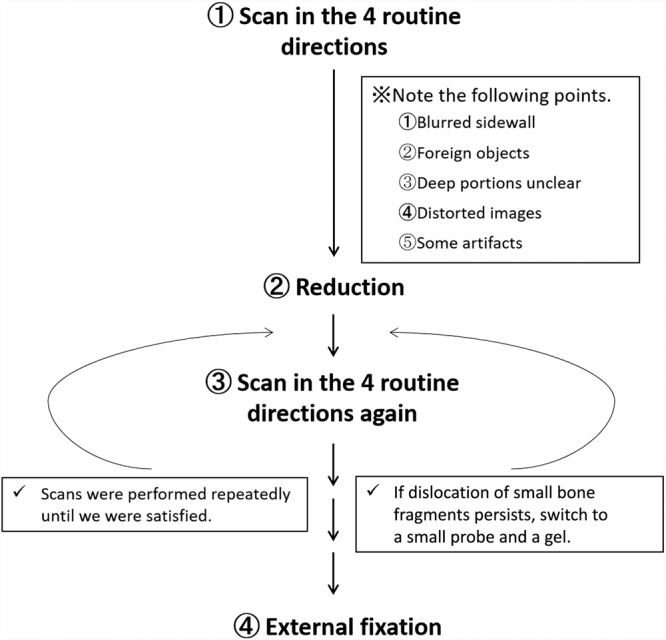
Methods: This method was used in 76 nasal bone fracture cases from July 2011 to March 2013, and we could obtain clear images of the entire nasal bone and surrounding bones. However, in some images, there were artifacts and blurred areas. The patterns of blurring were classified and their causes were analyzed.
Results: The 6 patterns of artifacts and blurred images were (1) Blurred side wall of the nasal bone in 68 cases, (2) air bubbles in the water in 68 cases, (3) unclear deep portions by attenuation in 23 cases, (4) distorted images caused by shaking of the probe in 44 cases, (5) parallel shadows due to multiple reflections in 18 cases, and (6) mysterious shadows caused by side lobes of the ultrasound beams in 55 cases. Almost all of them could be avoided by adding some small changes of techniques.
Conclusions: Our methods can provide whole clear images of the nasal bone and surrounding bones in 1 field. Almost all the artifacts and blurred images which occurred during the performance of our methods could be avoided by adding some small changes, for example, tilting the probe, pouring the water slowly, and moving the probe closer to the nose.
Figures


Similar articles
-
Ultrasonography in the diagnosis of nasal bone fractures: a comparison with conventional radiography and computed tomography.Eur Arch Otorhinolaryngol. 2016 Feb;273(2):413-8. doi: 10.1007/s00405-015-3595-8. Epub 2015 Mar 8. Eur Arch Otorhinolaryngol. 2016. PMID: 25749616
-
High-resolution sonography for nasal fracture in children.AJR Am J Roentgenol. 2007 Jan;188(1):W86-92. doi: 10.2214/AJR.05.1067. AJR Am J Roentgenol. 2007. PMID: 17179332
-
[Ultrasonography of the nose and paranasal sinuses].Pol Merkur Lekarski. 2007 Jan;22(127):32-5. Pol Merkur Lekarski. 2007. PMID: 17477087 Polish.
-
Management of nasal fractures.Arch Fam Med. 2000 Aug;9(8):738-42. doi: 10.1001/archfami.9.8.738. Arch Fam Med. 2000. PMID: 10927714 Review.
-
First-trimester three-dimensional ultrasonographic findings in a fetus with frontonasal malformation.J Matern Fetal Neonatal Med. 2004 Sep;16(3):187-97. doi: 10.1080/14767050400009139. J Matern Fetal Neonatal Med. 2004. PMID: 15590446 Review.
Cited by
-
Intraoperative Use of Ultrasonography in the Reduction of Zygomatico-Maxillary Complex Fractures.Craniomaxillofac Trauma Reconstr. 2022 Sep;15(3):229-236. doi: 10.1177/19433875211029145. Epub 2021 Jul 7. Craniomaxillofac Trauma Reconstr. 2022. PMID: 36081677 Free PMC article.
References
-
- Forrest CR, Lata AC, Marcuzzi DW, et al. The role of orbital ultrasound in the diagnosis of orbital fractures. Plast Reconstr Surg. 1993;92:28–34.. - PubMed
-
- Hirai T, Manders EK, Nagamoto K, et al. Ultrasonic observation of facial bone fractures: report of cases. J Oral Maxillofac Surg. 1996;54:776–779; discussion 779.. - PubMed
-
- Kiwanuka E, Smith SE, Frates MC, et al. Use of high-frequency ultrasound guidance for intraoperative zygomatic arch fracture reduction. J Craniofac Surg. 2013;24:2036–2038.. - PubMed
-
- Gateno J, Miloro M, Hendler BH, et al. The use of ultrasound to determine the position of the mandibular condyle. J Oral Maxillofac Surg. 1993;51:1081–1086; discussion 1086.. - PubMed
-
- Mohammadi A, Javadrashid R, Pedram A, et al. Comparison of ultrasonography and conventional radiography in the diagnosis of nasal bone fractures. Iran J Radiol Reconstr Surg. 2009;6:7–11..
LinkOut - more resources
Full Text Sources
Other Literature Sources
Medical