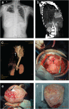
Analysis of giant thoracic neoplasms: Correlations between imaging, pathology and surgical management
- PMID: 28608450
- PMCID: PMC5582482
- DOI: 10.1111/1759-7714.12448
Analysis of giant thoracic neoplasms: Correlations between imaging, pathology and surgical management
Abstract
Background: A giant thoracic neoplasm is extremely rare and poorly understood. Our systemic study introduced computed tomography angiography (CTA) with three-dimensional (3D) reconstruction imaging and evaluated correlations between imaging, pathology, and surgical management.
Methods: Data from 45 patients undergoing surgery for giant thoracic neoplasm in our institution between May 2007 and November 2015 were collected. The clinical characteristics, imaging manifestations, preoperative biopsy, surgical management, postoperative pathology, and prognosis and their correlation were analyzed.
Results: The clinical characteristics, imaging manifestations, and pathological types were complicated. Four patients underwent CTA with 3D reconstruction imaging and feeding vessels were found in three cases. Twenty-four selected patients accepted preoperative biopsy, eight of which were inconsistent with postoperative pathology. Complete resection was performed in 39 cases, 20 of which underwent extended excision. The median survival duration of all patients was 58 months (range 3.0-118.0). The one, three, and five-year survival rates were 86.0%, 64.4%, and 47.0%, respectively. Univariate analyses showed tumor size and resection status were prognostic factors for survival (P = 0.003 and P < 0.001, respectively).
Conclusions: A giant thoracic neoplasm should preferably be treated in experienced centers for precise diagnosis and optimal therapy schemes with comprehensive consideration of clinical characters, imaging manifestations, pathology, surgical management, and prognosis. Innovative CTA with 3D reconstruction imaging together with preoperative biopsy are feasible and effective in therapeutic decision-making and surgical planning. Complete surgical resection remains the mainstay of curative therapy for all resectable tumors.
Keywords: Computed tomography angiography; giant thoracic neoplasm; surgical management; three-dimensional reconstruction.
© 2017 The Authors. Thoracic Cancer published by China Lung Oncology Group and John Wiley & Sons Australia, Ltd.
Figures


References
-
- Cardillo G, Carbone L, Carleo F et al. Solitary fibrous tumors of the pleura: An analysis of 110 patients treated in a single institution. Ann Thorac Surg 2009; 88: 1632–7. - PubMed
-
- Pusiol T, Piscioli I, Scialpi M, Hanspeter E. Giant benign solitary fibrous tumor of the pleura (> 15 cm): Role of radiological pathological correlations in management. Report of 3 cases and review of the literature. Pathologica 2013; 105: 77–82. - PubMed
-
- Akiba T. Utility of three‐dimensional computed tomography in general thoracic surgery. Gen Thorac Cardiovasc Surg 2013; 61: 676–84. - PubMed
-
- Kurenov SN, Ionita C, Sammons D, Demmy TL. Three‐dimensional printing to facilitate anatomic study, device development, simulation, and planning in thoracic surgery. J Thorac Cardiovasc Surg 2015; 149: 973–9.e1. - PubMed
-
- Hagiwara M, Shimada Y, Kato Y et al. High‐quality 3‐dimensional image simulation for pulmonary lobectomy and segmentectomy: Results of preoperative assessment of pulmonary vessels and short‐term surgical outcomes in consecutive patients undergoing video‐assisted thoracic surgery. Eur J Cardiothorac Surg 2014; 46: e120–6. - PubMed
Publication types
MeSH terms
LinkOut - more resources
Full Text Sources
Other Literature Sources

