. 2017 Sep;174(17):2842-2861.
doi: 10.1111/bph.13908. Epub 2017 Jul 18.

Heteromers of μ opioid and dopamine D1 receptors modulate opioid-induced locomotor sensitization in a dopamine-independent manner

Affiliations

Heteromers of μ opioid and dopamine D1 receptors modulate opioid-induced locomotor sensitization in a dopamine-independent manner

Yi-Min Tao et al. Br J Pharmacol. 2017 Sep.

Abstract

Background and purpose: Exposure to opiates induces locomotor sensitization in rodents, which has been proposed to correspond to the compulsive drug-seeking behaviour. Numerous studies have demonstrated that locomotor sensitization can occur in a dopamine transmission-independent manner; however, the underlying mechanisms are unclear.

Experimental approach: Co-immunoprecipitation, BRET and cross-antagonism assays were used to demonstrate the existence of receptor heterodimers. Function of heterodimers was evaluated by behavioural studies of locomotor sensitization.

Key results: The dopamine D1 receptor antagonist SCH23390 antagonized the signalling initiated by stimulation of μ opioid receptors with agonists in transfected cells expressing two receptors and in striatal tissues from wild-type but not D1 receptor knockout (KO) mice, suggesting that SCH23390 modified μ receptor function via receptor heteromers, as the ability of an antagonist of one of the receptors to inhibit signals originated by stimulation of the partner receptor was a characteristic of receptor heteromers. The existence of μ receptor-D1 receptor heterodimers was further supported by biochemical and biophysical assays. In vivo, when dopamine release was absent (by destruction of the dopaminergic projection from the ventral tegmental area to the striatum), SCH23390 still significantly inhibited μ receptor agonist-induced behavioural responses in rats. Additionally, we demonstrated that D1 or μ receptor KO mice and thus unable to form μ receptor-D1 receptor heterodimers, failed to show locomotor sensitization to morphine.

Conclusion and implications: Our results suggest that μ receptor-D1 receptor heterodimers may be involved in the dopamine-independent expression of locomotor sensitization to opiates.


Figures

Figure 1
Figure 1
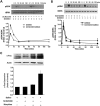
The D1 receptor antagonist SCH23390 attenuated μ receptor signalling mediated by selective μ receptor agonists DAMGO or morphine. (A, B) SCH23390 inhibited DAMGO or morphine‐stimulated [35S]GTPγS binding. Membranes prepared from CHO cells co‐expressing μ receptors (μOR) and D1 receptors (D1R) or expressing μ receptors alone were stimulated with various concentrations of DAMGO (A) or morphine (B) with or without 1 μM SCH23390, and the [35S]GTPγS binding was measured. Values are expressed as the mean ± SEM of three independent experiments in triplicate. (C) Effect of SCH23390 on basal [35S]GTPγS binding in the absence of DAMGO. (D, E) SCH23390 antagonized DAMGO or morphine‐inhibited adenylyl cyclase. CHO cells stably expressing μ receptors either alone or in combination with D1 receptors were treated with 1 μM DAMGO (D) or 10 μM morphine (E) for 1 h, and then 10 μM forskolin‐stimulated cAMP accumulation was measured. The results were normalized to 10 μM forskolin‐stimulated cAMP accumulation and expressed as the mean ± SEM of four independent experiments in triplicate.
Figure 2
Figure 2
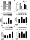
SCH23390 suppressed DAMGO‐mediated activation of ERK1/2 and morphine‐mediated expression of c‐Fos protein. (A, B) SCH23390 attenuated DAMGO‐induced increase in phosphorylated ERK1/2. HEK293 cells expressing μ receptors (μOR) and D1 receptors (D1R) (A) or expressing μ receptors alone (B) were treated with DAMGO (1 μM) for indicated time points in the absence or presence of SCH23390 (1 μM). The phosphorylation of ERK1/2 was determined by immunoblotting and quantified by densitometry. Upper panels show representative blots from Western blotting. Bottom panels show quantification of blots of phospho‐ERK1/2. Values are expressed as the mean ± SEM of three independent experiments. (C) SCH23390 inhibited morphine‐induced increase in c‐Fos protein levels. HEK293 cells expressing μ receptors and D1 receptors were treated with saline or morphine (10 μM) in the presence or absence of SCH23390 (1 μM). After 1 h, nuclear fractionation was isolated. The nuclear fraction (nuclei) and crude extract (total extract) were analysed by SDS‐PAGE and immunoblotted by using rabbit anti‐c‐Fos (1:500) and mouse anti‐actin (1:5000) respectively. The intensities of the immunoreactive bands were quantified by densitometry. Upper panels show representative blots of c‐Fos from Western blotting. Lower panels show quantification of blots of c‐Fos. All values are normalized against actin as a control protein. Values are expressed as the mean ± SEM of three independent experiments.
Figure 3
Figure 3
μ receptors and D1 receptors form functionally interacting heteromeric complexes in heterologous cells. (A) Complexes containing μ receptors (μOR) and D1 receptors (D1R) were detected in membranes from HEK293 cells expressing two receptors. Immunoprecipitation (IP) of cell lysates from HEK293 cells individually expressing either HA‐tagged μ receptors (HA‐μOR) or Flag‐tagged D1 receptors (Flag‐D1R), mixed cells individually expressing HA‐μ receptors or Flag‐D1 receptors or cells co‐expressing HA‐μ receptors and Flag‐D1 receptors was performed using anti‐HA antibodies or anti‐Flag antibodies. Western blotting (WB) of these immunocomplexes using anti‐HA antibodies shows a ~130 kDa protein representing the μ receptor‐D1 receptor heterodimer only in cells co‐expressing both HA‐μ receptors and Flag‐D1 receptors. Pretreatment of cells co‐expressing HA‐μ receptors and Flag‐D1 receptors with 50 mM DTT did not significantly alter the μ receptor‐D1 receptor heterodimer levels. (B) Significant BRET signals were detected in live HEK293 cells expressing μ receptors and D1 receptors by BRET. HEK293 cells were cotransfected with GFP‐ and Rluc‐fused μ receptor and D1 receptor constructs at the ratio of 1:1. BRET ratios were measured 24 h after transfection. The BRET signal was determined by the ratio of the light emitted by the receptor‐GFP (515 nm) over the light emitted by the receptor‐Rluc (460 nm). (C) Significant BRET signals were also detected in live HEK293 cells expressing μ receptors and D1 receptors at levels roughly comparable with those found in cells endogenously expressing these receptors (300–500 fmol·mg−1 protein) by BRET, and no significant BRET signals were detected in live cells co‐expressing μ receptors and muscarinic M2 receptors (M2R). (D) BRET saturation curves for μ receptor and D1 receptor homodimerization. HEK293 cells were co‐transfected with a fixing dose of Rluc‐fusion receptor construct and increasing doses of GFP‐fusion receptor construct. Twenty‐four hours after transfection, BRET signals were measured at room temperature. Co‐transfection of M2R‐Rluc did not induce any BRET signal. Results are the mean ± SEM of three or four independent experiments. (D) BRET saturation curves for μ receptor and D1 receptor homodimerization. HEK293 cells were co‐transfected with a fixing dose of Rluc‐fusion receptor construct and increasing doses of GFP‐fusion receptor construct. Twenty‐four hours after transfection, BRET signals were measured at room temperature. Co‐transfection of M2R‐Rluc did not induce any BRET signal. Results are the mean ± SEM of three or four independent experiments.
Figure 4
Figure 4
Immunocomplexes containing μ receptors (μOR) and D1 receptors (D1R) and functional interaction of two receptors were present in rodent striatum. (A) Heterodimeric complexes of μ receptors and D1 receptors were found in mouse striatal membranes. Solubilized striatal membranes from mice were subjected to immunoprecipitation (IP), using anti‐μ receptor or anti‐D1 receptor antibodies, as described. Immunocomplexes isolated by using anti‐μ receptor or anti‐D1 receptorantibody were immunoblotted (WB) by using anti‐D1 receptor or anti‐μ receptor antibodies. A band of 130 kDa is seen only upon administration of anti‐D1 receptor or anti‐μ receptor antibodies (lanes 2 and 4) and not in administration of non anti‐D1 receptor or anti‐μ receptor antibody vehicle control (lanes 1 and 3). (B) Effect of SCH23390 on DAMGO‐stimulated [35S]GTPγS binding to mouse striatal membranes prepared from wild‐type and D1 receptor KO mice. Striatal membranes prepared from wild‐type mice or D1 receptor KO mice were treated with 1 μM DAMGO with or without 1 μM SCH23390, and [35S]GTPγS binding was measured as described. Values are expressed as the mean ± SEM of three independent experiments performed in triplicate. (C) SCH23390 suppressed DAMGO‐induced activation of ERK. Cultured striatal neurons were treated with DAMGO (1 μM) for 15 min in the absence or presence of SCH23390 (1 μM). The phosphorylation of ERK1/2 was determined by immunoblotting and quantified by densitometry as described. Top panel shows representative blots from Western blotting (WB); lower panel shows quantification of blots of phospho‐ERK1/2. Values are expressed as the mean ± SEM of four independent experiments. (D) SCH23390 failed to attenuate morphine‐induced activation of ERK1/2 in D1 receptor KO mice. D1 receptor KO mice were treated with morphine (10 mg·kg−1, s.c.) with or without SCH23390 (0.03 mg·kg−1, i.p.) for 2 h. SCH23390 was given 10 min prior to morphine administration. (E, F) SCH23390 attenuated morphine‐induced c‐Fos expression in wild‐type but not D1 receptor KO mice. Wild‐type and D1 receptor KO mice were treated with morphine (10 mg·kg−1, s.c.) with or without SCH23390 (0.03 mg·kg−1, i.p.) for 2 h. SCH23390 was given 10 min prior to morphine administration. Extracts of mouse striatum (50 μg) were subjected to SDS‐PAGE and immunoblotted by using rabbit anti‐c‐Fos (1:500). The intensities of the immunoreactive bands were quantified by densitometry. Top panel shows representative blots from Western blotting. Lower panel shows quantification of blots of c‐Fos. All values are normalized by using actin as a control protein. Values are expressed as the mean ± SEM (n = 6). *P < 0.05, significantly different from saline‐treated control group; # P < 0.05, significantly different from morphine‐treated group.
Figure 5
Figure 5
Effects of SCH23390 on morphine‐induced locomotor activation and sensitization in mice. (A) SCH23390 inhibited morphine‐induced locomotor activation. Mice were treated with saline, SCH23390 (0.03 mg·kg−1, i.p.), morphine (10 mg·kg−1, s.c., twice daily) or morphine plus SCH23390 for 7 consecutive days. Locomotor activity (distance travelled over a period of 180 min in m) was measured on days 1, 3, 5 and 7. (B) SCH23390 prevented the induction of behavioural sensitization from chronic morphine treatment. Seven days after the last administration of morphine, an additional challenge injection of morphine (10 mg·kg−1, s.c.) was given to each group. (C) SCH23390 suppressed the expression of behavioural sensitization. The mice were chronically treated with morphine for 7 consecutive days. After another 7 days of abstinence, the animals were challenged with morphine with or without pretreatment with SCH23390. Values are expressed as the mean ± SEM (n = 10). *P < 0.05, significantly different from saline‐treated control group; # P < 0.05, significantly different from morphine alone‐treated group. One‐way ANOVA followed by Dunnett's post hoc tests.
Figure 6
Figure 6
Administration of SCH23390 or U0126 before challenge injection (Test inj) of morphine blocked the expression of behavioural sensitization to morphine. Rats were treated with morphine (10 mg·kg−1, i.p.). Two days later, these rats either were challenged with morphine (10 mg·kg−1, i.p.) or were injected with SCH23390 or U0126 before challenge with morphine. Locomotor responses (distance travelled over a period of 180 min in cm) were measured after challenge with morphine. (A) Systemic administration of SCH23390 blocked the expression of behavioural sensitization to morphine. SCH23390 (0.03 mg·kg−1, i.p.) was given 30 min prior to challenge with morphine. (B) Local administration of SCH23390 blocked the expression of behavioural sensitization to morphine. SCH23390 (0.1 μg/0.5 μL/side) or SCH23390 plus RS102221 (0.2 μg/0.5 μL/side) was microinjected into the NAc 30 min prior to challenge with morphine. (C) U0126 (0.1 μg/0.5 μL/side) was microinjected into the NAc 30 min prior to challenge with morphine. Values are expressed as the mean ± SEM (n = 8–10). * P < 0.05, significantly different from the first injection of morphine; # P < 0.05, significantly different from challenge injection of morphine without SCH23390 or U0126. One‐way ANOVA followed by Dunnett's post hoc tests. (D) Schematic representation of inj sites in the NAc for rats used in the experiments.
Figure 7
Figure 7
Intra‐NAc administration of SCH23390 or U0126 blocked systemic morphine or local DAMGO‐induced behavioural responses in rats that received lesions of the NAc with 6‐OHDA. (A, B) Intra‐NAc microinjection of SCH23390 or U0126 before challenge injection (Test inj) of morphine blocked the expression of behavioural sensitization to morphine. Rats that received lesions of the NAc with 6‐OHDA were treated with saline (Veh) or morphine (10 mg·kg−1, i.p.). Two days later, these rats either were challenged with morphine (10 mg·kg−1, i.p.) or were intra‐NAc microinjected with SCH23390 (0.1 μg/0.5 μL/side) or U0126 (0.1 μg/0.5 μL/side) 30 min before challenge injection of morphine. (C) Effect of administration of SCH23390 or U0126 before the challenge injection of morphine on behavioural sensitization expression in saline‐treated control mice. Rats were treated with saline; 2 days later, these rats were intra‐NAc microinjected with SCH23390 (0.1 μg/0.5 μL/side) or U0126 (0.1 μg/0.5 μL/side), 30 min later, followed by a challenge injection of morphine (10 mg·kg−1, i.p.). Locomotor responses (distance travelled over a period of 180 min in cm) were measured after the challenge injection of morphine. Values are expressed as the mean ± SEM (n = 7–9). *P < 0.05, significantly different from saline‐treated control group; # P < 0.05, significantly different from test injection of morphine without SCH23390 or U0126. One‐way ANOVA followed by Dunnett's post hoc tests. (D) Intra‐NAc microinjection of SCH23390 suppressed locomotor activity induced by direct administration of DAMGO into the NAc in rats that received lesions of the NAc with 6‐OHDA. DAMGO (0.2 μg/0.5 μL/side) was injected into NAc 10 min after intra‐NAc administration of SCH23390 (0.1 μg/0.5 μL/side) or saline. Locomotor responses (distance travelled over a period of 180 min in cm) were measured after 2 h after DAMGO administration. Values are expressed as the mean ± SEM (n = 8). *P < 0.05, significantly different from saline‐treated control group; # P < 0.05, significantly different from DAMGO‐treated group. One‐way ANOVA followed by Dunnett's post hoc tests. (E) Schematic representation of injection sites in the NAc for rats used in the experiments. (F) Photomicrographs of the tyrosine hydroxylase immunoreactive neurons in the striatum after bilateral intra‐NAc infusion of 6‐OHDA.
Figure 8
Figure 8
Mice lacking μ receptors (μOR) or D1 receptors (D1R) do not show locomotor sensitization to morphine or form complexes containing μ receptors and D1 receptors in the striatum. (A) D1 receptor KO mice (D1R−/−) were treated with morphine (10 mg·kg−1, s.c.), followed by a challenge injection (test injection) of morphine (10 mg·kg−1, s.c.) after 5 days of drug abstinence. Locomotor responses were measured during 180 min after the test injection of morphine. (B) complexes containing μ receptors and D1 receptors were detected only in wild‐type and heterozygous mice but not in D1 receptor‐deficient mice. Solubilized striatal membranes from wild‐type (D1+/+), heterozygous (D1+/‐) or homozygous (D1‐/‐) mice were subjected to immunoprecipitation(IP) by using anti‐D1 receptor or anti‐μ receptor antibodies, and immunocomplexes were immunoblotted (WB) by using anti‐μ receptor or anti‐D1 receptor antibodies as described. (C, D) KO mice (μOR−/−) did not show locomotor sensitization to morphine or form complexes containing μ receptors and D1 receptors in the striatum. Values are expressed as the mean ± SEM (n = 8–10). *P < 0.05, different from first injection of morphine; # P < 0.05, different from wild‐type and KO mice. Two‐way ANOVA followed by Dunnett's post hoc tests.

Similar articles

Cited by

References

    1. Alexander SPH, Davenport AP, Kelly E, Marrion N, Peters JA, Benson HE et al. (2015a). The Concise Guide to PHARMACOLOGY 2015/16: G protein‐coupled receptors. Br J Pharmacol 172: 5744–5869. - PMC - PubMed
    1. Alexander SPH, Fabbro D, Kelly E, Marrion N, Peters JA, Benson HE et al. (2015b). The Concise Guide to PHARMACOLOGY 2015/16: Enzymes. Br J Pharmacol 172: 6024–6109. - PMC - PubMed
    1. Amalric M, Koob GF (1985). Low doses of methylnaloxonium in the nucleus accumbens antagonize hyperactivity induced by heroin in the rat. Pharmacol Biochem Behav 23: 411–415. - PubMed
    1. Becker A, Grecksch G, Kraus J, Peters B, Schroeder H, Schulz S et al. (2001). Loss of locomotor sensitisation in response to morphine in D1 receptor deficient mice. Naunyn Schmiedebergs Arch Pharmacol 363: 562–568. - PubMed
    1. Berg KA, Rowan MP, Gupta A, Sanchez TA, Silva M, Gomes I et al. (2012). Allosteric interactions between δ and κ opioid receptors in peripheral sensory neurons. Mol Pharmacol 81: 264–272. - PMC - PubMed

MeSH terms