

In Vitro and In Vivo Studies of Alar-Nasal Cartilage Using Autologous Micro-Grafts: The Use of the Rigenera® Protocol in the Treatment of an Osteochondral Lesion of the Nose
- PMID: 28608799
- PMCID: PMC5490410
- DOI: 10.3390/ph10020053
In Vitro and In Vivo Studies of Alar-Nasal Cartilage Using Autologous Micro-Grafts: The Use of the Rigenera® Protocol in the Treatment of an Osteochondral Lesion of the Nose
Abstract
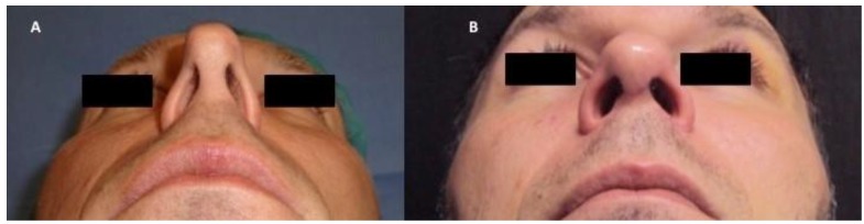
Cartilage defects represent a serious problem due to the poor regenerative properties of this tissue. Regarding the nose, nasal valve collapse is associated with nasal blockage and persistent airway obstruction associated with a significant drop in the quality of life for patients. In addition to surgical techniques, several cell-based tissue-engineering strategies are studied to improve cartilage support in the nasal wall, that is, to ameliorate wall insufficiency. Nevertheless, there are no congruent data available on the benefit for patients during the follow-up time. In this manuscript, we propose an innovative approach in the treatment of cartilage defects in the nose (nasal valve collapse) based on autologous micro-grafts obtained by mechanical disaggregation of a small portion of cartilage tissue (Rigenera® protocol). In particular, we first analyzed in vitro murine and human cartilage micro-grafts; secondly, we analyzed the clinical results of a patient with pinched nose deformity treated with autologous micro-grafts of chondrocytes obtained by Rigenera® protocol. The use of autologous micro-graft produced promising results in surgery treatment of cartilage injuries and could be safely and easily administrated to patients with cartilage tissue defects.
Keywords: Rigenera® protocol; autologous micro-grafts; chondrocytes; nasal valve collapse; tissue engineering approaches.
Conflict of interest statement
The authors declare no conflict of interest.
Figures


References
-
- Chan V.O., Moran D.E., Mwangi I., Eustace S.J. Prevalence and clinical significance of chondromalacia isolated to the anterior margin of the lateral femoral condyle as a component of patellofemoral disease: Observations at MR imaging. Skelet. Radiol. 2013;42:1127–1133. doi: 10.1007/s00256-013-1640-5. - DOI - PubMed
-
- Wang J., Han W., Wang X., Pan F., Liu Z., Halliday A., Jin X., Antony B., Cicuttini F., Jones G., et al. Mass effect and signal intensity alteration in the suprapatellar fat pad: Associations with knee symptoms and structure. Osteoarthr. Cartil. 2014;22:1619–1626. doi: 10.1016/j.joca.2014.05.018. - DOI - PubMed
-
- Lankhorst N.E., Damen J., Oei E.H., Verhaar J.A.N., Bierma-Zeinstra S.M.A., van Middelkoop M. Incidence, prevalence, natural course and prognosis of patellofemoral osteoarthritis; data of cohort hip and cohort knee study. Osteoarthr. Cartil. 2015;23:A42–A43. doi: 10.1016/j.joca.2015.02.095. - DOI - PubMed
LinkOut - more resources
Full Text Sources
Other Literature Sources
