. 2018 Jan/Feb;34(1-2):40-51.
doi: 10.1089/jop.2017.0023. Epub 2017 Jun 13.

Discovery and Preclinical Development of Netarsudil, a Novel Ocular Hypotensive Agent for the Treatment of Glaucoma

Affiliations

Discovery and Preclinical Development of Netarsudil, a Novel Ocular Hypotensive Agent for the Treatment of Glaucoma

Cheng-Wen Lin et al. J Ocul Pharmacol Ther. 2018 Jan/Feb.

Abstract

Purpose: Rho-associated protein kinase (ROCK) inhibitors lower intraocular pressure (IOP) by increasing aqueous outflow through the trabecular meshwork (TM). The preclinical characterization of netarsudil, a new ROCK/norepinephrine transporter (NET) inhibitor currently in clinical development, is presented herein.

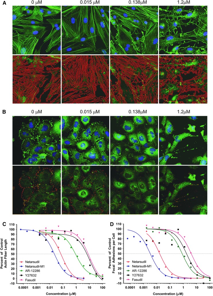
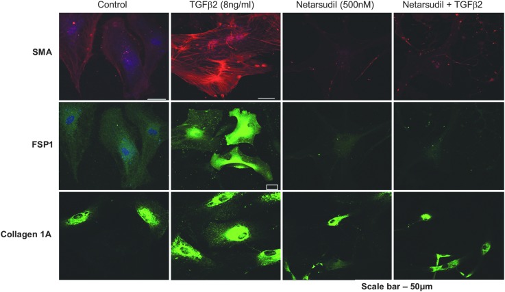
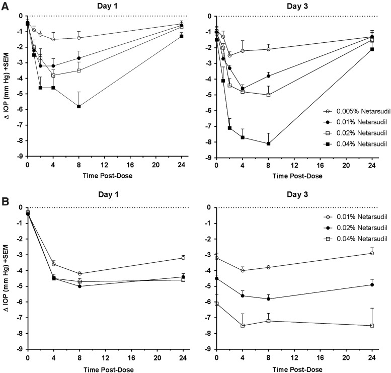
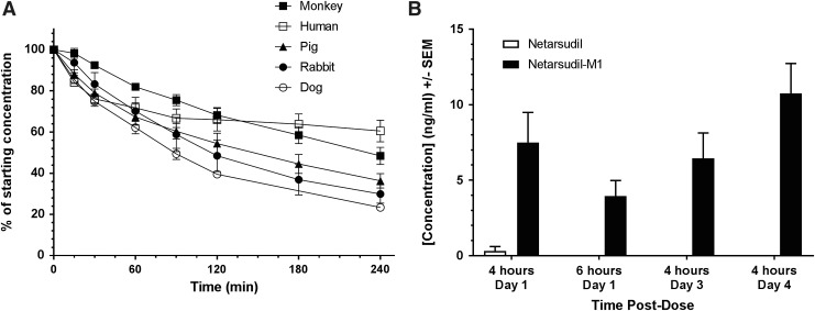
Methods: The kinase inhibitory activity of netarsudil was compared to its esterase metabolite, netarsudil-M1, and 3 other ROCK inhibitors using a commercially available kinase assay kit. Disruption of actin stress fibers was measured in primary porcine TM cells and disruption of focal adhesions in transformed human TM (HTM) cells. Induction of fibrosis markers after exposure to transforming growth factor-β2 (TGF-β2) was conducted in primary HTM cells. Ocular hypotensive activity and tolerability of topical formulations were evaluated in normotensive Dutch Belted rabbits and Formosan Rock monkeys. In vitro corneal metabolism assays were conducted using dog, pig, rabbit, monkey, and human corneas. In vivo ocular pharmacokinetics was studied in Dutch Belted rabbits.

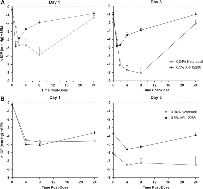
Results: Netarsudil inhibited kinases ROCK1 and ROCK2 with a Ki of 1 nM each, disrupted actin stress fibers and focal adhesions in TM cells with IC50s of 79 and 16 nM, respectively, and blocked the profibrotic effects of TGF-β2 in HTM cells. Netarsudil produced large reductions in IOP in rabbits and monkeys that were sustained for at least 24 h after once daily dosing, with transient, mild hyperemia observed as the only adverse effect.

Conclusion: Netarsudil is a novel ROCK/NET inhibitor with high potency in biochemical and cell-based assays, an ability to produce large and durable IOP reductions in animal models, and favorable pharmacokinetic and ocular tolerability profiles.

Keywords: Rho kinase; glaucoma; intraocular pressure; netarsudil; trabecular meshwork.


Conflict of interest statement

C.-W.L., B.S., L.A.M., C.L.L., M.A.d.L., and C.C.K. are employees of and stockholders in Aerie Pharmaceuticals, Inc. D.-W.L. and P.V.R. received research funding from Aerie Pharmaceuticals.

Figures

<b>FIG. 1.</b>
FIG. 1.
Structures of test compounds. NETi, norepinephrine transporter inhibitor; ROCKi, Rho-associated protein kinase inhibitor.
<b>FIG. 2.</b>
FIG. 2.
Disruption of actin stress fibers and focal adhesions by netarsudil versus other ROCK inhibitors. (A) Netarsudil dose-response in actin stress fiber assay. Primary PTM cells were incubated for 6 h in the presence of 0, 0.015, 0.138, or 1.2 μM netarsudil then fixed and stained with Alexa Fluor-488 phalloidin and Hoechst 33342 to reveal actin fibers and nuclei, respectively. Top panel: fluorescence images of stained cells. Bottom panel: False color images created by an automated, custom algorithm to identify stress fibers and calculate mean stress fiber length. (B) Netarsudil dose-response in focal adhesion assay. Immortalized HTM cells (TM-1) were incubated for 6 h in the presence of 0, 0.015, 0.138, or 1.2 μM netarsudil then fixed and stained with mouse anti-paxillin antibody/Alexa Fluor®488 goat-anti-mouse IgG and Hoechst 33342 to reveal focal adhesions and nuclei, respectively. Top panel: fluorescence images of stained cells. Bottom panel: False color images created by an automated, custom algorithm to identify focal adhesions and calculate the mean number of focal adhesions per cell. (C) Dose–response curves (n = 4) for netarsudil, netarsudil-M1, AR-12286, Y-27632, and fasudil in the PTM actin stress fiber length assay. Mean stress fiber length is presented as a percentage of the mean length of stress fibers measured in untreated control cells. (D) Dose–response curves (n = 4) for netarsudil, netarsudil-M1, AR-12286, Y-27632, and fasudil in the HTM focal adhesion assay. Mean number of focal adhesions per cell is presented as a percentage of the number of focal adhesions per cell measured in untreated control cells. HTM, human trabecular meshwork; PTM, porcine trabecular meshwork; ROCK, Rho-associated protein kinase.
<b>FIG. 3.</b>
FIG. 3.
Netarsudil blocks the profibrotic effects of TGF-β on HTM cells. Serum-starved primary HTM cells incubated for 24 h in the presence of either vehicle, 8 ng/mL human TGF-β2, 500 nM netarsudil, or 8 ng/mL TGF-β2 plus 500 nM netarsudil were fixed and stained for the fibrogenic markers α-SMA, fibroblast-specific protein 1 (FSP1), and Collagen 1A. α-SMA, α-smooth muscle actin; HTM, human trabecular meshwork; TGF-β2, transforming growth factor-β2.
<b>FIG. 4.</b>
FIG. 4.
Netarsudil dose-dependent lowering of IOP in rabbits and monkeys. Formulations containing 0.005% (rabbit only), 0.01%, 0.02%, or 0.04% netarsudil were administered once daily (AM) to 1 eye of each animal for 3 days, with the fellow untreated eye serving as the control. IOP was determined for both eyes before test article administration (time 0) and at times 1, 2, 4, 8, and 24 h (rabbits) or 4, 8, and 24 h (monkeys) after each morning dose on Day 1 and 3. (A) Changes in IOP for the treated eye relative to the untreated contralateral eye in Dutch Belted rabbits (n = 10/group). IOP reductions were statistically significant (P < 0.05) at all postdose time points. (B) Change in IOP in the netarsudil-treated eye relative to the untreated contralateral eye in Formosan Rock monkeys (n = 6/group). IOP reductions were statistically significant (P < 0.05) at all postdose time points. IOP, intraocular pressure.
<b>FIG. 5.</b>
FIG. 5.
IOP-lowering effect of netarsudil versus AR-12286 in rabbits and monkeys. Netarsudil 0.04% or AR-12286 0.5% was administered once daily (AM) to 1 eye of each animal for 3 days, with the fellow untreated eye serving as the control. IOP was determined for both eyes before test article administration (time 0) and at times 1, 2, 4, 8, and 24 h (rabbits) or 4, 8, and 24 h (monkeys) after each morning dose on Day 1 and 3. (A) Change in IOP in the treated eye relative to the untreated contralateral eye in Dutch Belted rabbits (n = 12/group). For netarsudil 0.04% and AR-12286 0.5%, IOP reductions were statistically significant (P < 0.01) at all postdose time points. (B) Change in IOP in the treated eye relative to the untreated contralateral eye in Formosan Rock monkeys (n = 6/group). For netarsudil 0.04% and AR-12286 0.5%, IOP reductions were statistically significant (P < 0.01) at all postdose time points. IOP, intraocular pressure.
<b>FIG. 6.</b>
FIG. 6.
Metabolism of netarsudil by ocular tissues. (A) Metabolism of netarsudil in the presence of corneal tissue isolated from Dutch Belted rabbit (n = 3), beagle dog (n = 3), pig (n = 3), cynomolgus monkey (n = 4), and human (n = 3) corneas. Corneal metabolism assays were initiated by adding netarsudil in assay buffer to wells containing individual corneal punches in assay buffer followed by incubation at 37°C. Samples were removed at specified time intervals and analyzed by HPLC to determine the percentage of netarsudil remaining at each time point. (B) Levels of netarsudil versus netarsudil-M1 in AH following topical ocular application of netarsudil 0.02% to Dutch Belted rabbits. Netarsudil was administered to both eyes of 3 male New Zealand rabbits in each of 4 groups. In groups 1 and 2, test article was dosed only once, and samples of AH were taken from each eye by paracentesis at 4 or 6 h postdose, respectively. In groups 3 and 4, test article was dosed once daily for 3 or 4 days, respectively, and samples of AH were taken 4 h after dosing. Levels of netarsudil and its metabolite netarsudil-M1 were measured in the samples by HPLC/mass spectrometry. AH, aqueous humor; HPLC, high performance liquid chromatography.

References

    1. Alward W.L. Medical management of glaucoma. N. Engl. J. Med. 339:1298–1307, 1998 - PubMed
    1. Casson R.J., Chidlow G., Wood J.P.M., et al. . Definition of glaucoma: clinical and experimental concepts. Clin. Exp. Ophthalmol. 40:341–349, 2012 - PubMed
    1. Vrabec J.P., and Levin L.A. The neurobiology of cell death in glaucoma. Eye. 21:S11–S14, 2007 - PubMed
    1. Schwartz K., and Budenz D. Current management of glaucoma. Curr. Opin. Ophthalmol. 15:119–126, 2004 - PubMed
    1. Stamer W.D., and Acott T.S. Current understanding of conventional outflow dysfunction in glaucoma. Curr. Opin. Ophthalmol. 23:135–143, 2012 - PMC - PubMed

MeSH terms