. 2017 Jun 13;8(1):144.
doi: 10.1186/s13287-017-0596-0.

Complete human serum maintains viability and chondrogenic potential of human synovial stem cells: suitable conditions for transplantation

Affiliations

Complete human serum maintains viability and chondrogenic potential of human synovial stem cells: suitable conditions for transplantation

Mitsuru Mizuno et al. Stem Cell Res Ther. .

Abstract

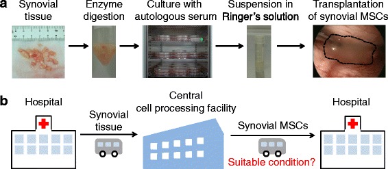
Background: In our clinical practice, we perform transplantations of autologous synovial mesenchymal stem cells (MSCs) for cartilage and meniscus regenerative medicine. One of the most important issues to ensuring clinical efficacy involves the transport of synovial MSCs from the processing facility to the clinic. Complete human serum (100% human serum) is an attractive candidate material in which to suspend synovial MSCs for their preservation during transport. The purpose of this study was to investigate whether complete human serum maintained MSC viability and chondrogenic potential and to examine the optimal temperature conditions for the preservation of human synovial MSCs.

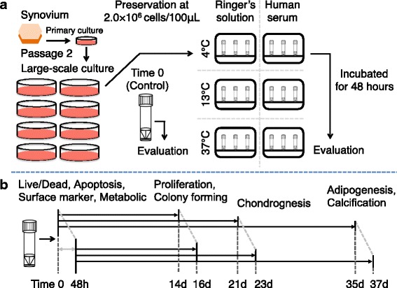
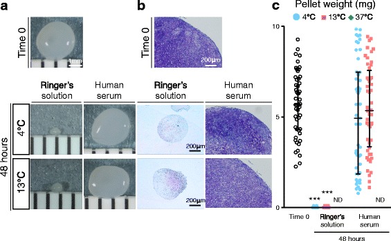
Methods: Human synovium was harvested from the knees of 14 donors with osteoarthritis during total knee arthroplasty. Passage 2 synovial MSCs were suspended at 2 million cells/100 μL in Ringer's solution or complete human serum at 4, 13, and 37 °C for 48 h. These cells were analyzed for live cell rates, cell surface marker expression, metabolic activity, proliferation, and adipogenic, calcification, and chondrogenic differentiation potentials before and after preservation.

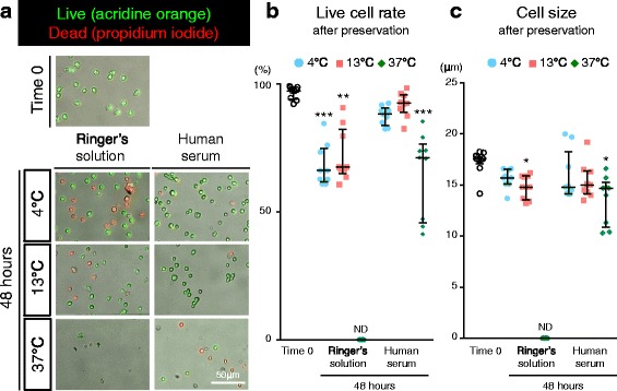
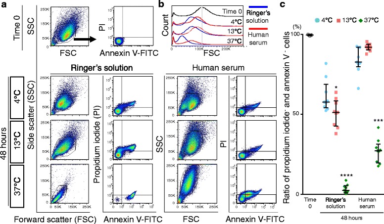
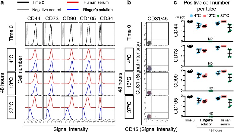
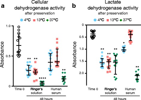
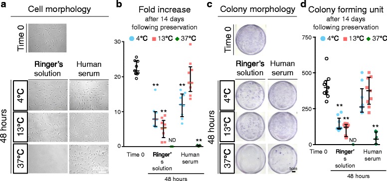
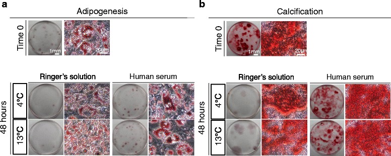
Results: After preservation, synovial MSCs maintained higher live cell rates in human serum than in Ringer's solution at 4 and 13 °C. Synovial MSCs preserved in human serum at 4 and 13 °C also maintained high ratios of propidium iodide- and annexin V- cells. MSC surface marker expression was not altered in cells preserved at 4 and 13 °C. The metabolic activities of cells preserved in human serum at 4 and 13 °C was maintained, while significantly reduced in other conditions. Replated MSCs retained their proliferation ability when preserved in human serum at 4 and 13 °C. Adipogenesis and calcification potential could be observed in cells preserved in each condition, whereas chondrogenic potential was retained only in cells preserved in human serum at 4 and 13 °C.

Conclusion: The viability and chondrogenic potential of synovial MSCs were maintained when the cells were suspended in human serum at 4 and 13 °C.

Keywords: Cell preservation; Human serum; Mesenchymal stem cells; Regenerative medicine; Synovium.


Figures

Fig. 1
Fig. 1
Scheme for regenerative medicine using synovial mesenchymal stem cells (MSCs). a Procedure for cartilage and meniscus regenerative medicine with autologous synovial MSCs. b Outline of transportation of synovial tissue and synovial MSCs between a hospital and a central cell-processing facility. Suitable conditions for transporting synovial MSCs from the central cell-processing facility to a hospital are essential
Fig. 2
Fig. 2
Scheme of the present study. a Two million passage 2 human synovial MSCs were suspended in glucose acetated Ringer’s solution or complete human serum. The cells were preserved at 4 °C, 13 °C, or 37 °C for 48 h, and then the cells were evaluated. b The cells were analyzed at time 0 and 48 h after preservation for live/dead, apoptosis, surface marker and metabolic assays. Then, the cells were cultured for 14 days for proliferation and colony forming abilities. The cells were also pelleted and cultured in chondrogenic medium for 21 days. Furthermore, the cells were cultured for 14 days to make cell colonies, then cultured in adipogenic medium and in calcification medium for 21 days
Fig. 3
Fig. 3
Live cell rate and size of synovial MSCs before and 48 h after preservation. a Representative images of live cells stained by AO (green) and dead cells stained by PI (red). b Live cell rate. c Cell size analyzed from fluorescence imaging. Median values and interquartile ranges are shown (n = 3). *p < .05, **p < .01, ***p < .001, compared with the value at Time 0 by Friedman test followed by Dunn’s multiple comparisons. ND not detected
Fig. 4
Fig. 4
Apoptosis of synovial MSCs 48 h after preservation. a Representative profiles of synovial MSCs by forward scatter (FSC) and side scatter (SSC), stained with fluorescein isothiocinate (FITC)-annexin V and propidium iodide (PI). Gates were placed around major cell populations. b Representative forward scatter histograms. c Apoptotic to normal cell ratios. Cells negative for annexin V and propidium iodide were considered nonapoptotic cells. Median values and interquartile ranges are shown (n = 3). *p < .05, ***p < .001, ****p < .0001, compared with the value at Time 0 by Kruskal-wallis test followed by Dunn’s multiple comparisons
Fig. 5
Fig. 5
Surface markers of synovial MSCs before and 48 h after preservation. a Representative histogram of synovial MSCs by CD44-PE-Cy7, CD73-V450, CD90-PE, CD105-APC, and CD34-PE-Cy5. b Representative dot plot of synovial MSCs by CD31-FITC and CD45-APC-H7 as negative markers. c Calculated live cell number per tube (2 million synovial MSCs). Median values and interquartile ranges are shown (n = 4). ND not detected
Fig. 6
Fig. 6
Metabolic activity of synovial MSCs before and 48 h after preservation. Passage 2 synovial MSCs before and 48 h after preservation were analyzed by colorimetric assay. a Cellular dehydrogenase activity as live cell metabolic activity. b Lactate dehydrogenase activity as dead cell metabolic activity. Median values and interquartile ranges are shown (n = 4). *p < .05, **p < .01, ****p < .001, compared with the value at Time 0 by Friedman test followed by Dunn’s multiple comparisons
Fig. 7
Fig. 7
Fold increase and colony formation unit of synovial MSCs before and 48 h after preservation. Passage 2 synovial MSCs before and 48 h after preservation were replated and cultured for 14 days. a Representative cell morphologies of synovial MSCs. b Fold increases after 14 days in culture before and 48 h after preservation. Median values and interquartile ranges are shown (n = 3). c Representative dishes stained with crystal violet. d Colony forming units after 14 days in culture before and 48 h after preservation. Median values and interquartile ranges are shown (n = 3). **p < .01, compared with the value at Time 0 by Friedman test followed by Steel’s multiple comparisons. ND not detected
Fig. 8
Fig. 8
Adipogenesis and calcification of synovial MSCs before and 48 h after preservation. Passage 2 synovial MSCs before and 48 h after preservation were cultured in differentiation medium for 21 days after colony formation. a Representative culture dishes and cell morphology stained with oil red-o after adipogenic differentiation. b Representative culture dishes and cell morphology stained with alizarin red after calcification
Fig. 9
Fig. 9
Chondrogenic ability of synovial MSCs before and 48 h after preservation. Passage 2 synovial MSCs before and 48 h after preservation were pelleted and cultured in chondrogenic medium for 21 days. a Representative macroscopic images of cartilage pellets. b Histological sections stained with toluidine blue. c Wet weight of cartilage pellets. Median values and interquartile ranges are shown (n = 10). ***p < .001, compared with the value at Time 0 by Kruskal-Wallis test followed by Dunn’s multiple comparisons. ND not detected

Similar articles

Cited by

References

    1. Sekiya I, Muneta T, Horie M, Koga H. Arthroscopic transplantation of synovial stem cells improves clinical outcomes in knees with cartilage defects. Clin Orthop Relat Res. 2015;473(7):2316–26. doi: 10.1007/s11999-015-4324-8. - DOI - PMC - PubMed
    1. Nakagawa Y, Muneta T, Kondo S, Mizuno M, Takakuda K, Ichinose S, et al. Synovial mesenchymal stem cells promote healing after meniscal repair in microminipigs. Osteoarthr Cartil. 2015;23(6):1007–17. doi: 10.1016/j.joca.2015.02.008. - DOI - PubMed
    1. Wiig H, Swartz MA. Interstitial fluid and lymph formation and transport: physiological regulation and roles in inflammation and cancer. Physiol Rev. 2012;92(3):1005–60. doi: 10.1152/physrev.00037.2011. - DOI - PubMed
    1. Weisberg HF. Osmotic pressure of the serum proteins. Ann Clin Lab Sci. 1978;8(2):155–64. - PubMed
    1. Mizuno N, Shiba H, Ozeki Y, Mouri Y, Niitani M, Inui T, et al. Human autologous serum obtained using a completely closed bag system as a substitute for foetal calf serum in human mesenchymal stem cell cultures. Cell Biol Int. 2006;30(6):521–4. doi: 10.1016/j.cellbi.2006.01.010. - DOI - PubMed

MeSH terms