. 2017 Sep 22;292(38):15758-15776.
doi: 10.1074/jbc.M117.794537. Epub 2017 Jun 13.

Global proteomic analysis of advanced glycation end products in the Arabidopsis proteome provides evidence for age-related glycation hot spots

Affiliations

Global proteomic analysis of advanced glycation end products in the Arabidopsis proteome provides evidence for age-related glycation hot spots

Tatiana Bilova et al. J Biol Chem. .

Abstract

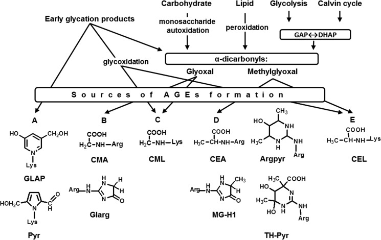
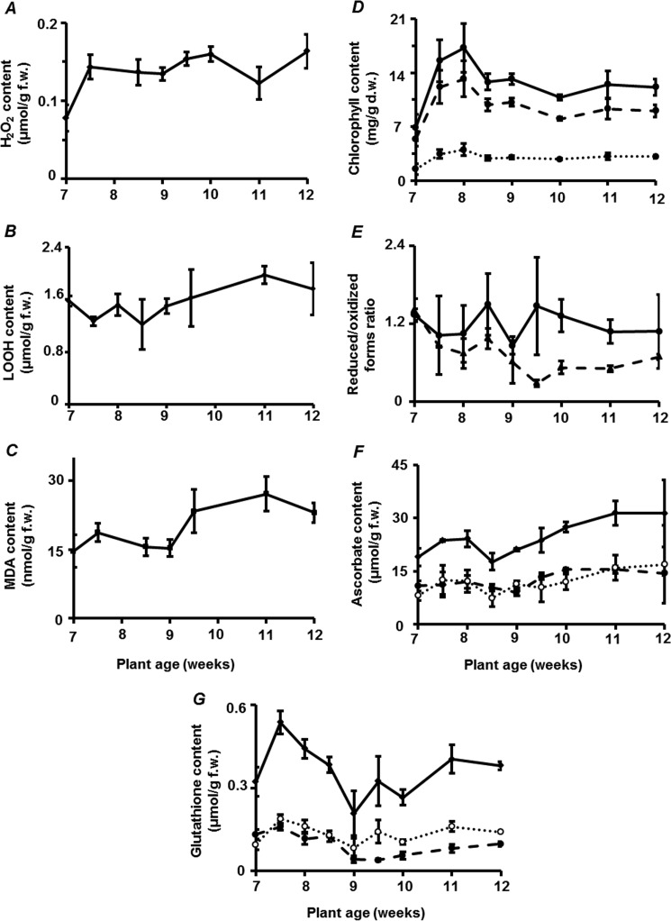
Glycation is a post-translational modification resulting from the interaction of protein amino and guanidino groups with carbonyl compounds. Initially, amino groups react with reducing carbohydrates, yielding Amadori and Heyns compounds. Their further degradation results in formation of advanced glycation end products (AGEs), also originating from α-dicarbonyl products of monosaccharide autoxidation and primary metabolism. In mammals, AGEs are continuously formed during the life of the organism, accumulate in tissues, are well-known markers of aging, and impact age-related tissue stiffening and atherosclerotic changes. However, the role of AGEs in age-related molecular alterations in plants is still unknown. To fill this gap, we present here a comprehensive study of the age-related changes in the Arabidopsis thaliana glycated proteome, including the proteins affected and specific glycation sites therein. We also consider the qualitative and quantitative changes in glycation patterns in terms of the general metabolic background, pathways of AGE formation, and the status of plant anti-oxidative/anti-glycative defense. Although the patterns of glycated proteins were only minimally influenced by plant age, the abundance of 96 AGE sites in 71 proteins was significantly affected in an age-dependent manner and clearly indicated the existence of age-related glycation hot spots in the plant proteome. Homology modeling revealed glutamyl and aspartyl residues in close proximity (less than 5 Å) to these sites in three aging-specific and eight differentially glycated proteins, four of which were modified in catalytic domains. Thus, the sites of glycation hot spots might be defined by protein structure that indicates, at least partly, site-specific character of glycation.

Keywords: Arabidopsis thaliana; advanced glycation end products (AGEs); aging; glycation; homology modeling; hot spots of glycation; metabolomics; oxidative stress; post-translational modification (PTM); proteomics.


Conflict of interest statement

The authors declare that they have no conflicts of interest with the contents of this article

Figures

Figure 1.
Figure 1.
Major pathways of advanced glycation end product (AGE) formation. The pathways are glycoxidation, i.e. oxidative degradation of early glycation products (Amadori and Heyns compounds and Schiff base intermediates), monosaccharide autoxidation, lipid peroxidation, conversion of glyceraldehyde phosphate (GAP) and dihydroxyacetone phosphate (DHAP), via degradation of the protein-bound and free sugar (A), formation of glyoxal (B), or both (C), as well as formation of methylglyoxal solely (D) or in parallel to the degradation of the protein-bound sugar (E). GLAP, glyceraldehyde-derived pyridinium compound; CEL, Nϵ-(carboxyethyl)lysine; CEA, Nδ-(carboxyethyl)arginine; CML, Nϵ-(carboxymethyl)lysine; CMA, Nδ-(carboxymethyl)arginine; MG-H1, methylglyoxal-derived hydroimidazolone 1, Nδ-(5-methyl-4-oxo-5-hydroimidazolinone-2-yl)-l-ornithine; Glarg, glyoxal-derived hydroimidazolone, 1-(4-amino-4-carboxybutyl)2-imino-5-oxo-imidazolidine; Pyr, 3-deoxyglucosone-derived pyrraline, 2′-formyl-5′-hydroxymethyl-pyrrolyl)norleucine; Argpyr, argpyrimidine, Nδ-(5-hydroxy-4,6-dimethylpyrimidin-2-yl)-l-ornithine; TH-Pyr, tetrahydropyrimidine, 4-carboxy-4,6-dimethyl-5,6-dihydroxy-1,4,5,6-tetrahydropyrimidin-2-yl)ornithine.
Figure 2.
Figure 2.
Time-course curves of the age-related changes in the leaf contents of stress markers, chlorophylls, and antioxidants. Shown are hydrogen peroxide (H2O2, A), LOOH (B), TBA-reactive substances (expressed as malondialdehyde equivalents, C), chlorophylls (D, solid, dashed and dotted lines represent total chlorophyll, its a and b isoforms, respectively), the Asc/DHA and GSH/GSSG ratios (E, solid and dashed lines, respectively), the contents of total ascorbate, Asc and DHA (F, solid, dashed, and dotted lines, respectively), as well as total glutathione, GSH, and GSSG (G, solid, dashed, and dotted lines, respectively). The A. thaliana plants were grown for 7–12 weeks under 8-h light/16-h dark cycle. All analyses were performed in three biological replicates (three plants per replicate) and one technical replicate.
Figure 3.
Figure 3.
Time-course curves of the age-related changes in the relative expression levels of APX1, GRcyt (A, solid and dashed lines, respectively), GLX1 and GLX2 (B, solid and dashed lines, respectively), and glyoxalase 2 activity (C). The A. thaliana plants were grown for 7–12 weeks under 8-h light/16-h dark cycle. All analyses were performed in three biological replicates (three plants per replicate) and one technical replicate.
Figure 4.
Figure 4.
Age-related changes in A. thaliana metabolic profiles. A, increase of primary metabolite leaf contents observed in 9- (white) and 12 (gray)-week-old A. thaliana plants in comparison with the 7-week-old ones. B, α-Dicarbonyl content (glyoxal (white) and methylglyoxal (gray)) in A. thaliana leaves of 7-, 9-, and 12-week-old plants. * and **, differences were significant at the confidence level of p ≤ 0.05 or 0.01, respectively. The * and ** for erythronic acid, galactose, xylose, fructose, and mannose were additionally confirmed with Benjamini-Hochberg-corrected p ≤ 0.05. All analyses were performed in three biological replicates (three plants per replicate) and one technical replicate.
Figure 5.
Figure 5.
MS/MS data confirming protein glycated sites. A, tandem mass spectra of m/z 655.81 corresponding to the [M + 2H]2+ of the tryptic AGE-modified peptide QYOxVAEIASMOxG[MG-H]405, representing lectin protein kinase; B, m/z 1003.46 corresponding to the [M + 2H]2+ of the tryptic hexose-modified peptide EYOxKHexosylLTYYTPEYETK32, representing Rubisco large chain subunit; C and D, annotation of these by the corresponding XICs (insets) at the characteristic tR, obtained from corresponding total ion chromatogram (TICs): TIC and XIC 1 (for m/z 655.81 ± 0.02, tR 32.7) (C); TIC and XIC 2 (for m/z 1003.46 ± 0.02, tR 25.4) (D).
Figure 6.
Figure 6.
Age-related quantitative changes in A. thaliana glycated proteome. Numbers of individual AGE-modified sites (sorted by individual AGE classes) demonstrate significant abundance changes of at least 1.5-fold (t test, p < 0.05) identified in proteins from 9- (white) and 12 (gray)-week-old A. thaliana plants (A), and the degree of these quantitative alterations expressed in scale of fold changes in 9- (B), and 12-week-old plants (C). In detail, the AGE-carrying sites (TH-Pyr, dark brown; Argpyr, light brown; CEA, pink; MG-H, red; Glarg, light blue; CMA, green; CML, yellow; CEL, white; pyrraline, dark blue; GLAP, purple) are distributed in n-fold change groups according their accumulation in 9- (B) and 12 (C)-week-old plants. The full names of the AGE modification are indicated in Fig. 1. The upper panels in A–C represent the count of modified sites, which increased in their abundance, and the lower panel shows glycated sites with decreased abundance.
Figure 7.
Figure 7.
Consensus sequences of A. thaliana proteins (25 residues up- and downstream) built for 51 arginyl (A) and 24 lysyl (B) AGE-modified residues (R* and K*, respectively) demonstrating significant changes in glycation rates. The consensus analysis was performed with the WebLogo Beta software tool. The position-specific occurrence frequencies of individual residues are expressed as informational unit bits. The bit axis was zoomed to provide better access to the residues in close proximity to the glycation site. The unprocessed graphs are available as supplemental Fig. S1-5.
Figure 8.
Figure 8.
Characterization of AGE-modified sites in 14 proteins, demonstrating significantly increased advanced glycation rates (at least 1.5-fold, t test, p < 0.05) in 9- (white) and 12 (gray)-week-old A. thaliana plants. Counts of these individual AGE-modified sites (sorted by individual AGE classes (A); the degree of corresponding quantitative alterations at AGE-sites (Argpyr, light brown; MG-H, red; CMA, green; CML, yellow; pyrraline, dark blue; GLAP, purple) expressed in scale of n-fold changes for 9- (B) and 12 (C)-week-old plants, and distributions of these AGE-sites in the proteins isolated from 9- (D) and 12 (E)-week old plants according to the most probable metabolic origin. The full names of the AGE modifications are given in Fig. 1.
Figure 9.
Figure 9.
Consensus sequences of A. thaliana proteins (25 residues up- and downstream) built for 13 arginyl (A) and 13 lysyl (B) residues from six age-specifically glycated proteins and 14 proteins with a significant age-dependent increase in glycation levels. The consensus analysis was performed with the WebLogo Beta software tool. The frequencies of the position-specific occurrences of individual residues are expressed as informational unit bits. The bit axis was zoomed to provide better access to the residues in close proximity to the glycation site. The unprocessed graphs are available as supplemental Fig. S1-6.
Figure 10.
Figure 10.
Structure homology modeling of aging-specific glycated proteins. A, isoform 2 of β-carbonic anhydrase 2, carbamidomethylated by the arginine 202 which is located in an α-helix at the surface of the protein. The conformation is stabilized by hydrogen bonds of the guanidine moiety with Glu-190 and between the carboxymethyl group (magenta carbon atoms) and the side chain of Asp-198. The ubiquitin C-terminal hydrolase 12 contains MG-H residue in position 711, located in a β-sheet at the surface of the protein (B). The conformation is stabilized by hydrogen bonds of the guanidine moiety with Glu-673, as well as between the hydroimidazolone group (magenta carbon atoms) and the side chain of Tyr-708.

References

    1. Vistoli G., De Maddis D., Cipak A., Zarkovic N., Carini M., and Aldini G. (2013) Advanced glycoxidation and lipoxidation end products (AGEs and ALEs): an overview of their mechanisms of formation. Free Radic. Res. 47, 3–27 - PubMed
    1. Heyns K., and Beilfuss W. (1970) Ketosylamine rearrangement of d-threo-pentulose (d-xylulose) with α-amino acids. Chem. Ber. 103, 2873–2876 - PubMed
    1. Hodge J. E. (1955) The Amadori rearrangement. Adv. Carbohydr. Chem. 10, 169–205 - PubMed
    1. Greifenhagen U., Frolov A., and Hoffmann R. (2015) Oxidative degradation of N(ϵ)-fructosylamine-substituted peptides in heated aqueous systems. Amino Acids 47, 1065–1076 - PubMed
    1. Hunt J. V., Dean R. T., and Wolff S. P. (1988) Hydroxyl radical production and autoxidative glycosylation. Glucose autoxidation as the cause of protein damage in the experimental glycation model of diabetes mellitus and ageing. Biochem. J. 256, 205–212 - PMC - PubMed

Publication types