. 2017 Jun 27;114(26):E5077-E5084.
doi: 10.1073/pnas.1705757114. Epub 2017 Jun 13.

Immunoengineering nerve repair

Affiliations

Immunoengineering nerve repair

Nassir Mokarram et al. Proc Natl Acad Sci U S A. .

Abstract

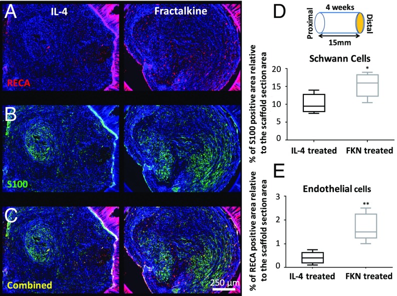
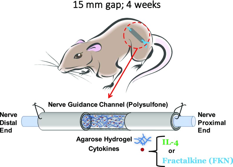
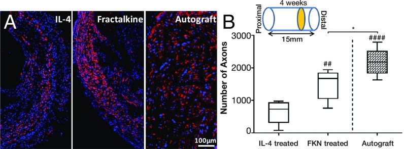
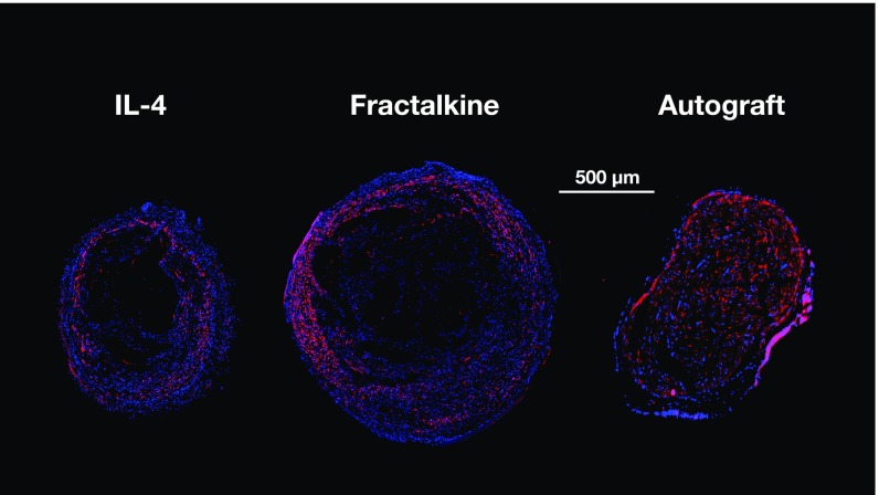
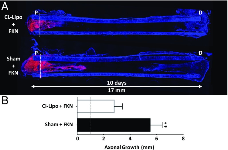
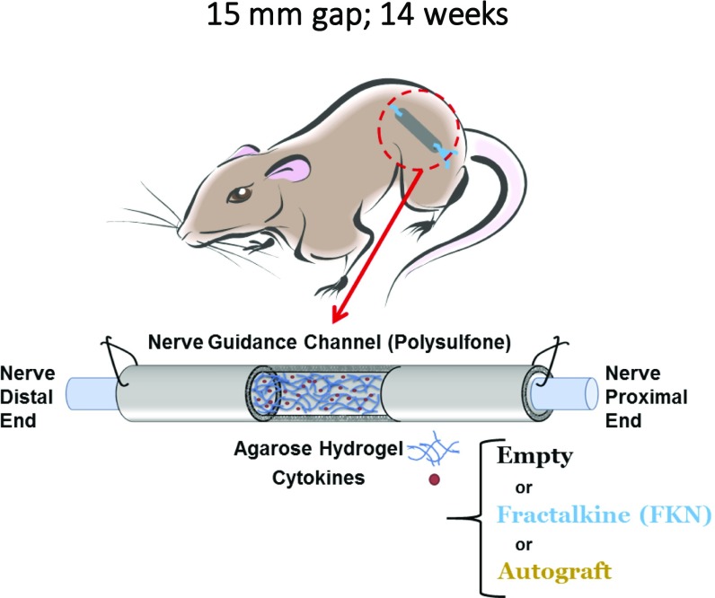
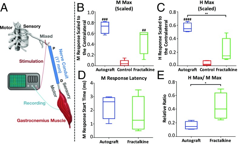
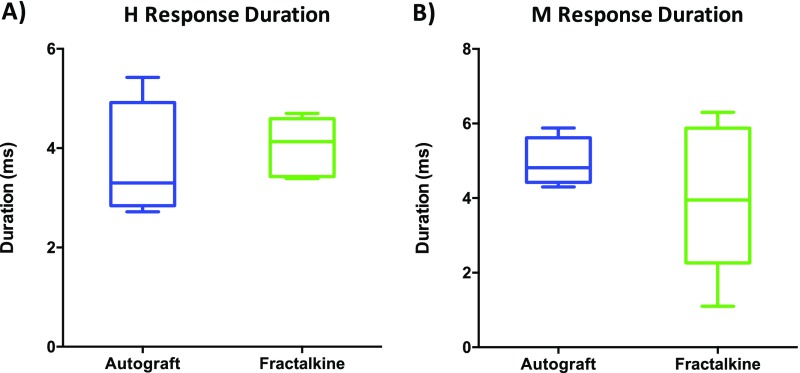
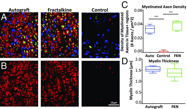
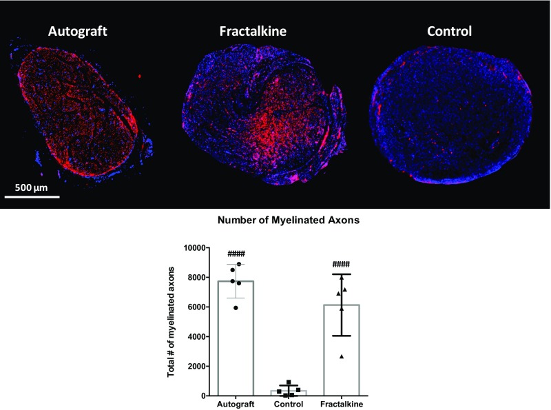
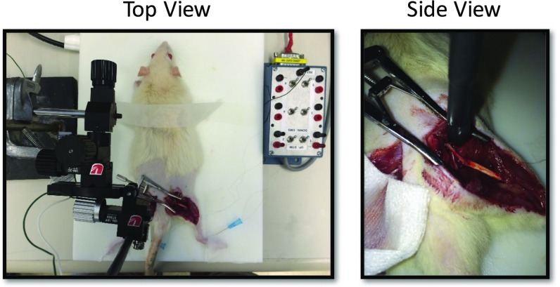
Injuries to the peripheral nervous system are major sources of disability and often result in painful neuropathies or the impairment of muscle movement and/or normal sensations. For gaps smaller than 10 mm in rodents, nearly normal functional recovery can be achieved; for longer gaps, however, there are challenges that have remained insurmountable. The current clinical gold standard used to bridge long, nonhealing nerve gaps, the autologous nerve graft (autograft), has several drawbacks. Despite best efforts, engineering an alternative "nerve bridge" for peripheral nerve repair remains elusive; hence, there is a compelling need to design new approaches that match or exceed the performance of autografts across critically sized nerve gaps. Here an immunomodulatory approach to stimulating nerve repair in a nerve-guidance scaffold was used to explore the regenerative effect of reparative monocyte recruitment. Early modulation of the immune environment at the injury site via fractalkine delivery resulted in a dramatic increase in regeneration as evident from histological and electrophysiological analyses. This study suggests that biasing the infiltrating inflammatory/immune cellular milieu after injury toward a proregenerative population creates a permissive environment for repair. This approach is a shift from the current modes of clinical and laboratory methods for nerve repair, which potentially opens an alternative paradigm to stimulate endogenous peripheral nerve repair.

Keywords: fractalkine; immunomodulation; macrophage; monocyte; nerve repair.


Conflict of interest statement

The authors declare no conflict of interest.

Figures

Fig. 1.
Fig. 1.
Monocyte subtypes, receptors, markers, population percentage, and function.
Fig. 2.
Fig. 2.
Schematic for immunoengineering nerve repair: recruitment and differentiation. This figure illustrates the response of circulating monocytes to the delivery of fractalkine at the site of nerve injury. A scaffold tube is sutured to the injured ends of the axotomized nerve and begins elution of fractalkine. Fractalkine recruits circulating monocytes, specifically reparative ones, to the site of injury, where further biochemical cues will potentially instruct the monocytes to differentiate into one of three potential prohealing macrophage phenotypes that enhance nerve repair: CD68+CD206+CD86+ (M2b) or CD68+Arg1+/CD206+(M2a or M2c).
Fig. 3.
Fig. 3.
The effect of early fractalkine release on SCs and endothelial cells. (A) Combined RECA (red), and DAPI (blue) staining. (B) Combined S100 (green) and DAPI (blue) staining. (C) Combined S100 (green), RECA (red), and DAPI (blue) staining. (D) Quantitative analysis of S100+ staining of SCs at the distal end of the conduit (P = 0.0243, two-tailed t test). (E) Quantitative analysis of RECA+ staining of endothelial cells at the distal end of the conduit (P = 0.0016, two-tailed t test) (n = 5). The fractalkine-treated scaffold significantly enhanced both SC infiltration and endothelial cell presence inside the nerve conduit in comparison with the IL-4–treated scaffold 4 wk after implantation. FKN, fractalkine.
Fig. S1.
Fig. S1.
Schematic of the rat 4-wk study.
Fig. S2.
Fig. S2.
Cytokine release profile from 1% SeaPrep.
Fig. S3.
Fig. S3.
Magnified fluorescent images of (A) RECA-S100, (B) CD68-arginase, and (C) CD86-CD206.
Fig. 4.
Fig. 4.
The effect of early fractalkine release on the number and phenotype of macrophages. (A) Combined CD68 (red), and DAPI (blue) staining. (B) Combined arginase (green) and DAPI (blue) staining. (C) Combined arginase (green), CD68 (red), and DAPI (blue) staining. (D) Quantitative analysis of total macrophage numbers at the distal end by using the CD68 marker (P = 0.015, two-tailed t test). (E) Quantitative analysis of prohealing macrophages at the distal end by double staining with CD68 and arginase (P = 0.288, two-tailed t test). (F) The ratio of prohealing macrophages to the total number of macrophages (P = 0.0019) (n = 5). Four weeks after implantation the fractalkine-treated scaffold has significantly fewer macrophages but a higher ratio of prohealing macrophage than the IL-4–treated scaffold. This figure is reproduced in part from ref. . FKN, fractalkine.
Fig. 5.
Fig. 5.
The effect of early fractalkine release on mannose receptor expression and regulatory macrophages. (A) Combined CD86 (red) and DAPI (blue) staining. (B) Combined CD206 (green) and DAPI (blue) staining. (C) Combined CD86 (red), CD206 (green), and DAPI (blue) staining. (D) Quantitative analysis of mannose receptor-expressing cells at the distal end assessed by the marker CD206 (P = 0.022, two-tailed t test). (E) Quantitative analysis of regulatory macrophages (M2b) at the distal end by double staining with CD86 and mannose receptor (P = 0.003, two-tailed t test) (n = 5). Four weeks after implantation, the fractalkine-treated scaffold contains a higher number of mannose receptor-expressing cells and regulatory macrophages than the IL-4–treated scaffold. FKN, fractalkine.
Fig. 6.
Fig. 6.
The effect of fractalkine release on axonal growth. (A) Immunohistochemical staining of axons (red) and DAPI (blue) at the distal end of nerve stump. (B) The number of regenerated axons at the distal end of the fractalkine- vs. the IL-4–treated scaffold in comparison with the autograft 4 wk after injury. *P < 0.05; ##P < 0.01; ####P < 0.0001 (one-way ANOVA) (n = 5). Fractalkine-treated scaffold dramatically increases the number of regenerated axons relative to IL-4, reaching very close to the number of regenerated axons in autograft.
Fig. S4.
Fig. S4.
Low-magnification fluorescent images of NF160-DAPI staining, 4-wk study.
Fig. 7.
Fig. 7.
CL-Lipo study. (A) Axonal regeneration (stained red with NF160 antibody) is demonstrated using longitudinally sectioned scaffolds in the CL-Lipo–treated animal vs. the nontreated animal (n = 4). Both scaffolds contain fractalkine. (B) Quantification of the length of axonal growth 10 d after scaffold implantation (P = 0.003, two-tailed t test). Dotted line in both A and B indicates the position of nerve on day 0. Partial depletion of monocytes using a single injection of CL-Lipo 48 h before nerve conduit implantation significantly reduces the amount of axonal growth, indicating the central role that monocyte presence plays in the fractalkine-treated scaffold (blue = DAPI). This image is reproduced in part from ref. . FKN, fractalkine.
Fig. S5.
Fig. S5.
Schematic of the rat 14-wk study.
Fig. 8.
Fig. 8.
Electrophysiological analysis. (A) Schematic demonstrating proximal stimulation of sciatic nerve motor axons and recording of the EMG signal from reinnervated gastrocnemius muscle 14 wk after scaffold implantation. (B and C) M Max (B) and H Max (C) amplitudes in the autograft group vs. the control scaffold and the fractalkine-loaded scaffold groups. (D and E) M-response latency (D) and H Max/M Max ratio (E) in the autograft vs. the fractalkine-loaded scaffold groups. *P < 0.05; ##P < 0.01; ###P < 0.001; ####P < 0.0001 (one-way ANOVA or two-tailed t test) (n = 6). M Max in rats with the fractalkine-loaded scaffold is significantly larger than in control rats and very closely approaches the autograft performance. H Max in rats with the fractalkine-loaded scaffold is still significantly lower than autograft, whereas the H Max/M Max ratio in rats with the fractalkine-loaded scaffold is significantly larger than autograft.
Fig. S6.
Fig. S6.
Graphs of H latency (A) and M latency (B).
Fig. 9.
Fig. 9.
Histological analysis of myelin and axon density at the late time point. (A) Representative pseudocolored fluorescent images showing NF160 (green), FluoroMyelin (red), and DAPI (blue) staining of scaffold cross-sections for autograft, fractalkine, and control groups. (B) Representative pseudocolored fluorescent images showing FluoroMyelin (red) staining of scaffold cross-sections at the 14-wk time point for autograft, fractalkine, and control groups, respectively. (C) Density of myelinated axons (normalized to the combined area of tissues positively stained for any of NF160, FluoroMyelin, or DAPI, not to the total image area) comparing autograft, fractalkine, and control (n = 8). The densities in the autograft and fractalkine groups are not significantly different, but both are significantly higher than in the control group (****P < 0.0001). (D) Comparison of myelin thickness surrounding regenerated axons in autograft and fractalkine (n = 8). The average thickness of myelin is greater in the autograft group than in the fractalkine group, but these differences are not statistically significant. FKN, fractalkine.
Fig. S7.
Fig. S7.
(Upper) Low-magnification fluorescent images of NF-160-DAPI staining. (Lower) Number of regenerated axons in the autograft, control, and fractalkine groups: 14-wk study. Statistical significance was observed for both autograft and fractalkine relative to control, but differences were not statistically significant between autograft and fractalkine. ####P < 0.0001.
Fig. S8.
Fig. S8.
Electrophysiology experiment setup.

References

    1. Belkas JS, Shoichet MS, Midha R. Peripheral nerve regeneration through guidance tubes. Neurol Res. 2004;26:151–160. - PubMed
    1. Millesi H. Factors affecting the outcome of peripheral nerve surgery. Microsurgery. 2006;26:295–302. - PubMed
    1. Bellamkonda RV. Peripheral nerve regeneration: An opinion on channels, scaffolds and anisotropy. Biomaterials. 2006;27:3515–3518. - PubMed
    1. Giusti G, et al. Return of motor function after segmental nerve loss in a rat model: Comparison of autogenous nerve graft, collagen conduit, and processed allograft (AxoGen) J Bone Joint Surg Am. 2012;94:410–417. - PubMed
    1. Wood MD, et al. Rat-derived processed nerve allografts support more axon regeneration in rat than human-derived processed nerve xenografts. J Biomed Mater Res A. 2014;102:1085–1091. - PubMed

Publication types