

M2-like Kupffer cells in fibrotic liver may protect against acute insult
- PMID: 28611518
- PMCID: PMC5449422
- DOI: 10.3748/wjg.v23.i20.3655
M2-like Kupffer cells in fibrotic liver may protect against acute insult
Abstract
Aim: To investigate the mechanism of hepatoprotection conferred by liver fibrosis through evaluating the activation phenotype of kupffer cells.
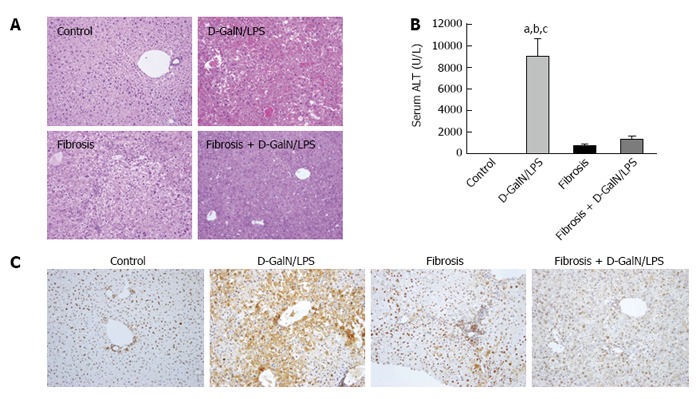
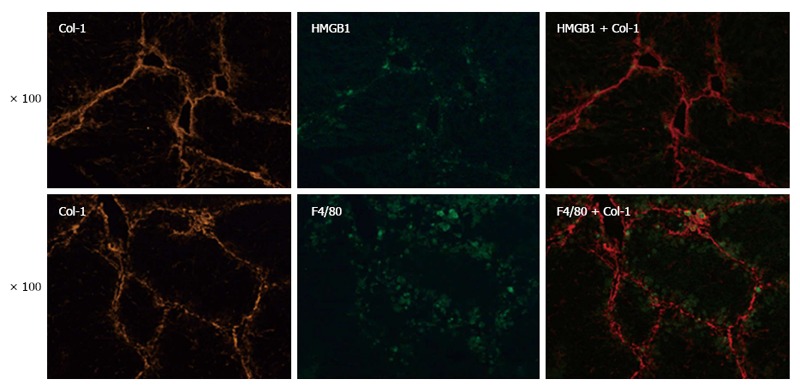
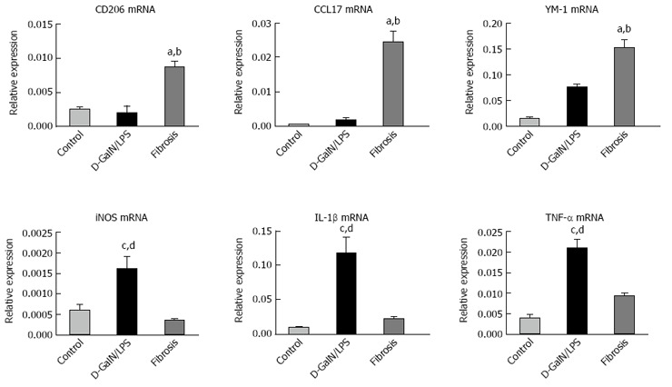
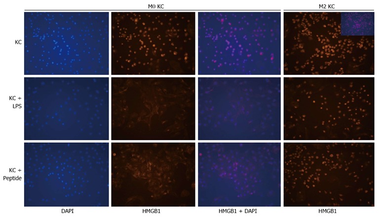
Methods: Control and fibrotic mice were challenged with a lethal dose of D-GalN/lipopolysaccharide (LPS), and hepatic damage was assessed by histology, serum alanine transferase (ALT) levels, and hepatic expression of HMGB1, a potent pro-inflammatory mediator. The localization of F4/80 (a surrogate marker of KCs), HMGB1, and type I collagen (Col-1) was determined by immunofluorescence staining. The phenotype of KCs was characterized by real-time PCR. KCs isolated from control or fibrotic mice were challenged with LPS or HMGB1 peptide, and HMGB1 translocation was analyzed.
Results: Liver fibrosis protected mice against D-GalN/LPS challenge, as shown by improved hepatic histology and reduced elevation of ALT compared with the normal mice treated in the same way. This hepatoprotection was also accompanied by inhibition of HMGB1 expression in the liver. Co-localization of F4/80, HMGB1, and Col-1 was found in fibrotic livers, indicating the close relationship between KCs, HMGB1 and liver fibrosis. KCs isolated from fibrotic mice predominantly exhibited an M2-like phenotype. In vitro experiments showed that HMGB1 was localized in the nucleus of the majority of M2-like KCs and that the translocation of HMGB1 was inhibited following stimulation with LPS or HMGB1 peptide, while both LPS and HMGB1 peptide elicited translocation of intranuclear HMGB1 in KCs isolated from the control mice.
Conclusion: M2-like Kupffer cells in fibrotic liver may exert a protective effect against acute insult by inhibiting the translocation of HMGB1.
Keywords: Injury resistance; Kupffer cell activation; Liver fibrosis; Translocation; high-mobility group box 1.
Conflict of interest statement
Conflict-of-interest statement: The authors declare that they have no conflict of interest related to this study.
Figures
References
-
- Friedman SL. Liver fibrosis -- from bench to bedside. J Hepatol. 2003;38 Suppl 1:S38–S53. - PubMed
-
- Ramachandran P, Iredale JP. Macrophages: central regulators of hepatic fibrogenesis and fibrosis resolution. J Hepatol. 2012;56:1417–1419. - PubMed
-
- Friedman SL. Evolving challenges in hepatic fibrosis. Nat Rev Gastroenterol Hepatol. 2010;7:425–436. - PubMed
MeSH terms
Substances
LinkOut - more resources
Full Text Sources
Other Literature Sources
Medical
