

Case Reports
doi: 10.1159/000458406.
eCollection 2017 Jan-Apr.
Necrolytic Acral Erythema in Seronegative Hepatitis C
Affiliations
- PMID: 28611625
- PMCID: PMC5465673
- DOI: 10.1159/000458406
Item in Clipboard
Case Reports
Necrolytic Acral Erythema in Seronegative Hepatitis C
Case Rep Dermatol.
.
Abstract
Necrolytic acral erythema (NAE) is a distinctive skin disorder. The exact cause and pathogenesis is still unclear. Most studies report an association of NAE with hepatitis C virus (HCV) infection. We report a 64-year-old woman who presented with chronic mildly pruritic brownish to erythematous rashes on both lateral malleoli for 7 months. The clinical and histopathological findings were compatible with NAE. However, the serologic marker for HCV was negative.
Keywords: Acral involvement; Acrodermatitis enteropathica; Glucagonoma; Hepatitis C; Necrolytic acral erythema; Pellagra.
Figures
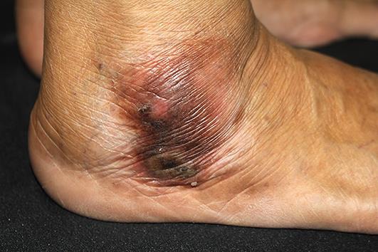
Well-defined brownish hyperkeratotic plaque with an erythematous rim located on the right lateral malleolus.

Histopathological findings revealed papillated psoriasiform epidermal hyperplasia, compact hyperkeratosis, and mounds of parakeratosis with neutrophils. There was absence of a granular layer, with pale and vacuolated keratinocytes in the superficial epidermal layer and scattered necrotic keratinocytes. Hematoxylin-eosin. Original magnification ×100.

Dense inflammatory cell infiltrate of mainly lymphocytes and extravasated erythrocytes, with dilated capillaries in the papillary dermis. Hematoxylin-eosin. Original magnification ×400.
References
-
- el Darouti M, Abu el Ela M. Necrolytic acral erythema: a cutaneous marker of viral hepatitis C. Int J Dermatol. 1996;35:252–256. - PubMed
-
- Liu A, Erickson CP, Cockerell CJ, Hsu S. Necrolytic acral erythema: a case not associated with hepatitis C infection. Dermatol Online J. 2008;14:10. - PubMed
-
- Wu YH, Tu ME, Lee CS, Lin YC. Necrolytic acral erythema without hepatitis C infection. J Cutan Pathol. 2009;36:355–358. - PubMed
-
- Nikam BP. Necrolytic acral erythema seronegative for hepatitis C virus – two cases from India treated with oral zinc. Int J Dermatol. 2009;48:1096–1099. - PubMed
Publication types
LinkOut - more resources
Full Text Sources
Other Literature Sources
