. 2017 May 30:8:321.
doi: 10.3389/fphar.2017.00321. eCollection 2017.

CD24 Expression Is Increased in 5-Fluorouracil-Treated Esophageal Adenocarcinoma Cells

Affiliations

CD24 Expression Is Increased in 5-Fluorouracil-Treated Esophageal Adenocarcinoma Cells

Pilar Jiménez et al. Front Pharmacol. .

Abstract

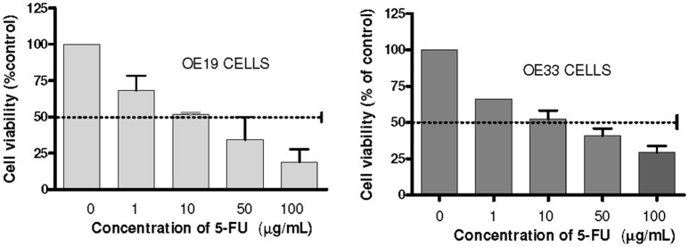
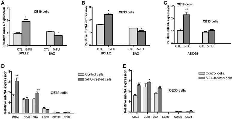
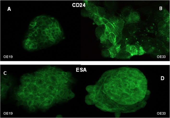
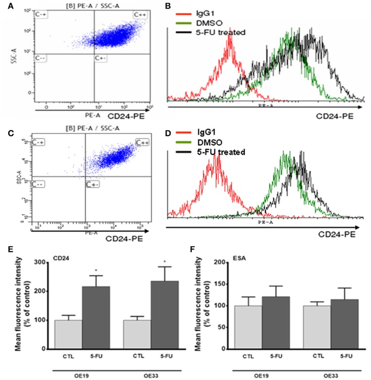
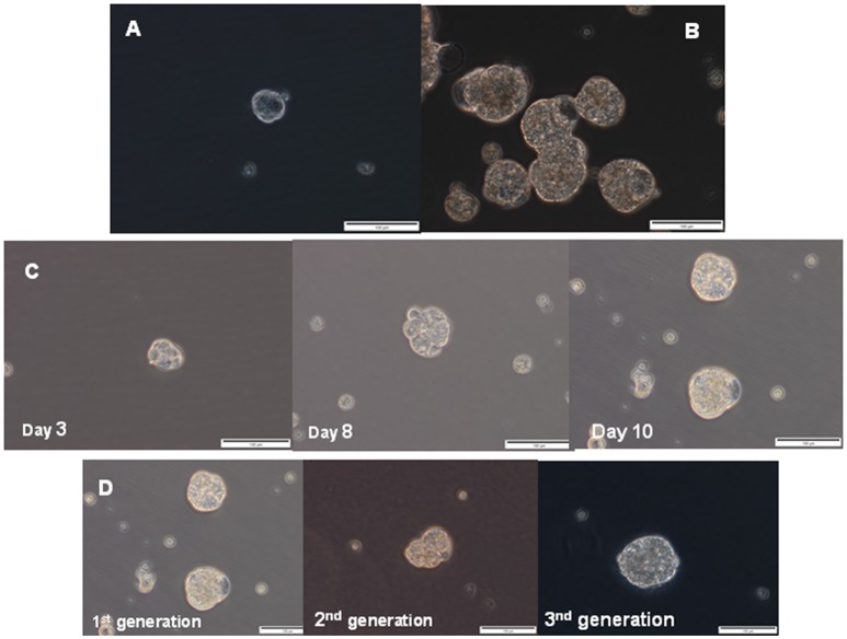
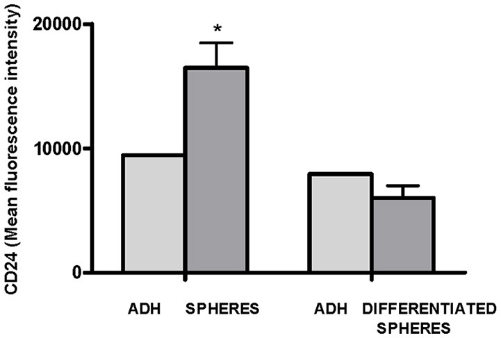
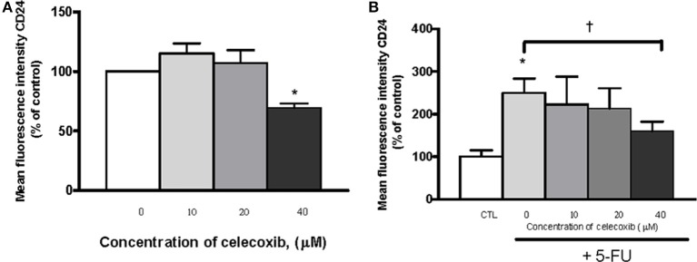
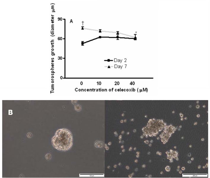
The cancer stem cell (CSC) model suggests that there are subsets of cells within a tumor with increased proliferation and self-renewal capacity, which play a key role in therapeutic resistance. The importance of cyclooxygenase-2 (COX-2) in carcinogenesis has been previously established and the use of COX-2 inhibitors as celecoxib has been shown to exert antitumor effects. The present study investigated whether treatment of esophageal adenocarcinoma (EAC) cells with 5-fluorouracil (5-FU) or the growth of tumor spheres increased the proportion of CSCs and also if treatment with celecoxib was able to reduce the putative CSC markers in this tumor. OE19 and OE33 EAC cells surviving 5-FU exposure exhibited an increase in CSC markers CD24 and ABCG2 and also an increased resistance to apoptosis. EAC cell lines had the capacity to form multiple spheres displaying typical CSC functionalities such as self-renewal and increased CD24 levels. In addition, after the induction of differentiation, cancer cells reached levels of CD24 similar to those observed in the parental cells. Treatment with celecoxib alone or in combination with 5-FU also resulted in a reduction of CD24 expression. Moreover, celecoxib inhibited the growth of tumor spheres. These findings showing a reduction in CSC markers induced by celecoxib suggest that the COX-2 inhibitor might be a candidate for combined chemotherapy in the treatment of EAC. However, additional clinical and experimental studies are needed.

Keywords: 5-fluorouracil; CD24; cancer stem cells; celecoxib; esophageal adenocarcinoma.


Figures

Figure 1
Figure 1
Analysis of cells chemosensitivity to 5-FU. Cell viability was measured using an MTT assay kit. The results are expressed as the mean ± SEM of cell viability percentage in 5-FU treated cells with respect to control cells of at least three independent experiments.
Figure 2
Figure 2
Analysis of mRNA levels of different markers related to stem phenotype. OE33 and OE19 EAC cell lines were exposed to 5-FU for 72 h. Relative gene expression analysis was developed in surviving cells vs. untreated cells. mRNA levels of the antiapoptotic gene BCLL2 and the proapoptotic gene BAX in OE19 (A) and OE33 (B) cells. mRNA levels of the drug transporter ABCG2 (C). mRNA levels of CSC markers CD24, CD34, CD44, CD133, ESA, LGR5 in OE19 (D) and OE33 (E) cells. Each bar represents the relative gene expression obtained after applying the following ratio: Normalized gene expression value/Kan-r gene value. Significant differences from the respective control values: *p < 0.05; **p < 0.01.
Figure 3
Figure 3
Cellular distribution of CD24 and ESA markers. Immunofluorescent staining of CD24 in OE19 (A) and OE33 (B) cells and ESA in OE19 (C) and OE33 (D) cell lines.
Figure 4
Figure 4
Analysis of CD24 and ESA expression after 5-FU treatment. Representative image of flow cytometry dot plot of CD24 expression in OE33 (A) and OE19 (C) tumor cells. Representative flow cytometry histograms displaying CD24 expression in OE33 (B) and OE19 (D) cells treated with 5-FU (10 μg/mL) for 72 h (black line) or in cells treated with the vehicle alone (green line). Mean fluorescence intensity of CD24 (E) and ESA (F) in 5-FU treated or in control cells. All data are expressed as the mean ± SEM of mean fluorescence units in treated cells relative to untreated cells of at least three independent experiments. Significant differences with respect to control cells: *p < 0.05.
Figure 5
Figure 5
Sphere formation assay. Representative phase contrast photomicrographs of tumor spheres generated by suspension culture of OE19 (A) and OE33 (B) EAC cells after 14 days of culture. Representative images of first-generation spheres' growth, established from adherent cultures (C). Representative images of the first-generation of spheres (corresponding to spheres 10 days after seeding, as previously shown in Figure 4C) and two successive generations of spheres (D).
Figure 6
Figure 6
Flow cytometric analysis of CD24 expression in adherent OE33 cells, OE33 spheres and differentiated spheres. All the results were expressed as mean ± SEM of mean fluorescence units of three independent experiments. *p < 0.05 with respect to adherent cells.
Figure 7
Figure 7
Effects of celecoxib and/or 5-FU on CD24 expression. The effects of the treatment were expressed as mean fluorescence units of OE33 cells treated with different concentrations of celecoxib (0–40 μM) alone (A) or in combination with 5-FU (10 μg/mL) (B) with respect to control (untreated) cells. All the results were expressed as mean ± SEM of mean fluorescence units in treated cells relative to untreated cells of three independent experiments. *p < 0.05 vs. control; p < 0.05 vs. 5-FU alone.
Figure 8
Figure 8
Effects of celecoxib on of esophageal tumor spheres' growth. Diameter of OE33 spheres after exposure to celecoxib (0–40 μM) for 2 or 7 days (A). All the results were expressed as mean ± SEM of the diameter of spheres of three independent experiments. p < 0.05, diameter of untreated spheres at day 2 vs. diameter of untreated spheres at day 7; *p < 0.05, diameter of spheres treated with celecoxib (40 μM) vs. untreated control at day 7. A representative photograph showing the disintegration of OE33 spheres after incubation with 40 μM celecoxib for 7 days (B, right) (200 × magnification).

Similar articles

Cited by

References

    1. Abnet C. C., Freedman N. D., Kamangar F., Leitzmann M. F., Hollenbeck A. R., Schatzkin A. (2009). Non-steroidal anti-inflammatory drugs and risk of gastric and oesophageal adenocarcinomas: results from a cohort study and a meta-analysis. Br. J. Cancer 100, 551–557. 10.1038/sj.bjc.6604880 - DOI - PMC - PubMed
    1. Abubaker K., Latifi A., Luwor R., Nazaretian S., Zhu H., Quinn M. A., et al. . (2013). Short-term single treatment of chemotherapy results in the enrichment of ovarian cancer stem cell-like cells leading to an increased tumor burden. Mol. Cancer 12:24. 10.1186/1476-4598-12-24 - DOI - PMC - PubMed
    1. Aigner S., Ramos C. L., Hafezi-Moghadam A., Lawrence M. B., Friederichs J., Altevogt P., et al. . (1998). CD24 mediates rolling of breast carcinoma cells on P-selectin. FASEB J. 12, 1241–1251. - PubMed
    1. Al-Hajj M., Wicha M. S., Benito-Hernandez A., Morrison S. J., Clarke M. F. (2003). Prospective identification of tumorigenic breast cancer cells. Proc. Natl. Acad. Sci. U.S.A. 100, 3983–3988. 10.1073/pnas.0530291100 - DOI - PMC - PubMed
    1. Altorki N. K., Christos P., Port J. L., Lee P. C., Mirza F., Spinelli C., et al. . (2011). Preoperative taxane-based chemotherapy and celecoxib for carcinoma of the esophagus and gastroesophageal junction: results of a phase 2 trial. J. Thorac. Oncol. 6, 1121–1127. 10.1097/JTO.0b013e31821529a9 - DOI - PMC - PubMed