

Monitoring Notch Signaling-Associated Activation of Stem Cell Niches within Injured Dental Pulp
- PMID: 28611689
- PMCID: PMC5447770
- DOI: 10.3389/fphys.2017.00372
Monitoring Notch Signaling-Associated Activation of Stem Cell Niches within Injured Dental Pulp
Abstract
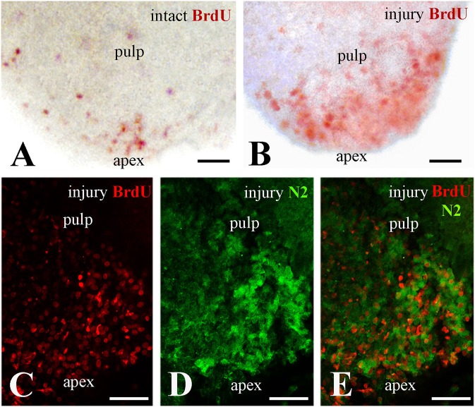
Dental pulp stem/progenitor cells guarantee tooth homeostasis, repair and regeneration throughout life. The decision between renewal and differentiation of these cells is influenced by physical and molecular interactions with stromal cells and extracellular matrix molecules forming the specialized microenvironment of dental pulp stem cell niches. Here we study the activation of putative pulp niches after tooth injury through the upregulation of Notch signaling pathway. Notch1, Notch2, and Notch3 molecules were used as markers of dental pulp stem/progenitor cells. Upon dental injury, Notch1 and Notch3 are detected in cells related to vascular structures suggesting a role of these proteins in the activation of specific pulpal perivascular niches. In contrast, a population of Notch2-positive cells that are actively proliferative is observed in the apical part of the pulp. Kinetics of these cells is followed up with a lipophilic DiI labeling, showing that apical pulp cells migrate toward the injury site where dynamic regenerative/repair events occur. The knowledge of the activation and regulation of dental pulp stem/progenitor cells within their niches in pathologic conditions may be helpful for the realization of innovative dental treatments in the near future.
Keywords: Notch signaling; Notch2; cell proliferation; dental pulp; niches; regeneration; stem cells; tooth injury.
Figures


Similar articles
-
Dental pulp stem cells, niches, and notch signaling in tooth injury.Adv Dent Res. 2011 Jul;23(3):275-9. doi: 10.1177/0022034511405386. Adv Dent Res. 2011. PMID: 21677078 Review.
-
Notch signaling in the dynamics of perivascular stem cells and their niches.Stem Cells Transl Med. 2021 Oct;10(10):1433-1445. doi: 10.1002/sctm.21-0086. Epub 2021 Jul 6. Stem Cells Transl Med. 2021. PMID: 34227747 Free PMC article.
-
Distinct Expression Patterns of Cxcl12 in Mesenchymal Stem Cell Niches of Intact and Injured Rodent Teeth.Int J Mol Sci. 2021 Mar 16;22(6):3024. doi: 10.3390/ijms22063024. Int J Mol Sci. 2021. PMID: 33809663 Free PMC article.
-
Activation of the Notch signaling pathway in response to pulp capping of rat molars.Eur J Oral Sci. 2005 Aug;113(4):312-7. doi: 10.1111/j.1600-0722.2005.00221.x. Eur J Oral Sci. 2005. PMID: 16048523
-
Differentiation potential of dental papilla, dental pulp, and apical papilla progenitor cells.J Endod. 2010 May;36(5):781-9. doi: 10.1016/j.joen.2010.02.006. Epub 2010 Mar 24. J Endod. 2010. PMID: 20416419 Review.
Cited by
-
Ameloblastomas Exhibit Stem Cell Potential, Possess Neurotrophic Properties, and Establish Connections with Trigeminal Neurons.Cells. 2020 Mar 6;9(3):644. doi: 10.3390/cells9030644. Cells. 2020. PMID: 32155948 Free PMC article.
-
The Genes Involved in Dentinogenesis.Organogenesis. 2022 Dec 31;18(1):1-19. doi: 10.1080/15476278.2021.2022373. Epub 2022 Jan 13. Organogenesis. 2022. PMID: 35023442 Free PMC article.
-
Mutanobactin-D, a Streptococcus mutans Non-Ribosomal Cyclic Lipopeptide, Induces Osteogenic/Odontogenic Differentiation of Human Dental Pulp Stem Cells and Human Bone Marrow Stem Cells.Int J Mol Sci. 2025 Jan 28;26(3):1144. doi: 10.3390/ijms26031144. Int J Mol Sci. 2025. PMID: 39940912 Free PMC article.
-
Three-Dimensional Culture Systems for Dissecting Notch Signalling in Health and Disease.Int J Mol Sci. 2021 Nov 19;22(22):12473. doi: 10.3390/ijms222212473. Int J Mol Sci. 2021. PMID: 34830355 Free PMC article. Review.
-
Histology-based profile of inflammatory mediators in experimentally induced pulpitis in a rat model: screening for possible biomarkers.Int Endod J. 2021 Aug;54(8):1328-1341. doi: 10.1111/iej.13514. Epub 2021 Mar 31. Int Endod J. 2021. PMID: 33715185 Free PMC article.
References
-
- Artavanis-Tsakonas S., Muskavitch M. A. (2010). Notch: the past, the present, and the future, in Current Topics in Developmental Biology, ed Kopan R. (Amsterdam: Elsevier Inc; ), 1–29. - PubMed
-
- Aurrekoetxea M., Garcia-Gallastegui P., Irastorza I., Luzuriaga J., Uribe-Etxebarria V., Unda F., et al. . (2015). Dental pulp stem cells as a multifaceted tool for bioengineering and the regeneration of craniomaxillofacial tissues. Front. Physiol. 6:289. 10.3389/fphys.2015.00289 - DOI - PMC - PubMed
LinkOut - more resources
Full Text Sources
Other Literature Sources
Miscellaneous