. 2017 May 29:8:894.
doi: 10.3389/fpls.2017.00894. eCollection 2017.

Proteomic Analysis of Lipid Droplets from Arabidopsis Aging Leaves Brings New Insight into Their Biogenesis and Functions

Affiliations

Proteomic Analysis of Lipid Droplets from Arabidopsis Aging Leaves Brings New Insight into Their Biogenesis and Functions

Lysiane Brocard et al. Front Plant Sci. .

Abstract

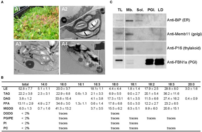
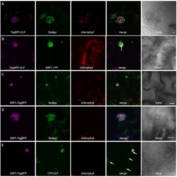
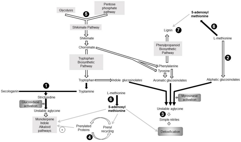
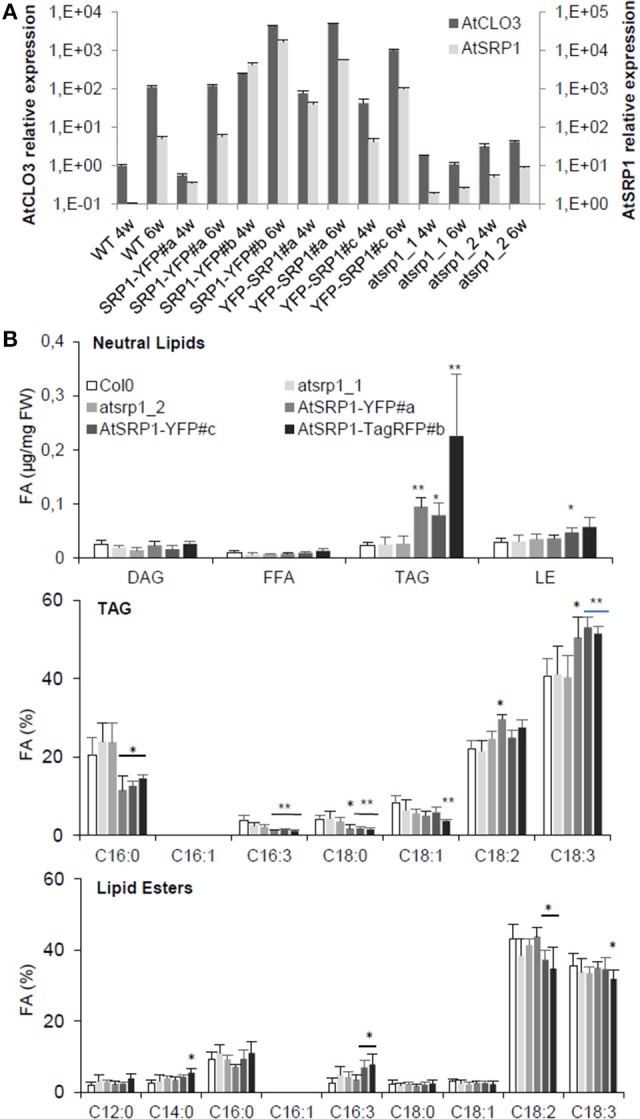
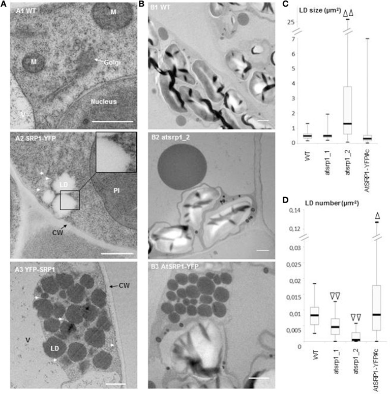
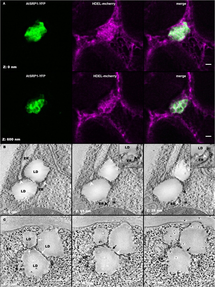
Lipid droplets (LDs) are cell compartments specialized for oil storage. Although their role and biogenesis are relatively well documented in seeds, little is known about their composition, structure and function in senescing leaves where they also accumulate. Here, we used a label free quantitative mass spectrometry approach to define the LD proteome of aging Arabidopsis leaves. We found that its composition is highly different from that of seed/cotyledon and identified 28 proteins including 9 enzymes of the secondary metabolism pathways involved in plant defense response. With the exception of the TRIGALACTOSYLDIACYLGLYCEROL2 protein, we did not identify enzymes implicated in lipid metabolism, suggesting that growth of leaf LDs does not occur by local lipid synthesis but rather through contact sites with the endoplasmic reticulum (ER) or other membranes. The two most abundant proteins of the leaf LDs are the CALEOSIN3 and the SMALL RUBBER PARTICLE1 (AtSRP1); both proteins have structural functions and participate in plant response to stress. CALEOSIN3 and AtSRP1 are part of larger protein families, yet no other members were enriched in the LD proteome suggesting a specific role of both proteins in aging leaves. We thus examined the function of AtSRP1 at this developmental stage and found that AtSRP1 modulates the expression of CALEOSIN3 in aging leaves. Furthermore, AtSRP1 overexpression induces the accumulation of triacylglycerol with an unusual composition compared to wild-type. We demonstrate that, although AtSRP1 expression is naturally increased in wild type senescing leaves, its overexpression in senescent transgenic lines induces an over-accumulation of LDs organized in clusters at restricted sites of the ER. Conversely, atsrp1 knock-down mutants displayed fewer but larger LDs. Together our results reveal that the abundancy of AtSRP1 regulates the neo-formation of LDs during senescence. Using electron tomography, we further provide evidence that LDs in leaves share tenuous physical continuity as well as numerous contact sites with the ER membrane. Thus, our data suggest that leaf LDs are functionally distinct from seed LDs and that their biogenesis is strictly controlled by AtSRP1 at restricted sites of the ER.

Keywords: ER contact site; Small rubber particle protein1; electron tomography; label free proteomics; leaf senescence; lipid droplet; secondary metabolism; ultrastructure.


Figures

Figure 1
Figure 1
Preparation of aging leaf lipid droplet fraction. (A) Six week old Arabidopsis leaves accumulate lipid droplets. (A1) 6 week old leaves grown under long day conditions; (A2–A4): details of the ultrastructure observed by TEM of a parenchyma cell from green (A2), purple (A3), and red (A4) leaves showing lipid droplet in close proximity to chloroplast. Insert in (A4) shows the plastid—LD contact site. Scale bar: 1 μm, except in insert: 200 nm. LD, lipid droplet. (B) Lipid composition of Arabidopsis leaf lipid droplets (in % of total fatty acids) n = 5. Lipids were extracted from LD fraction, separated by thin layer chromatography, and quantified by GC-FID after transesterification. LE, lipid ester; TAG, triacylglycerol; DAG, Diacylglycerol; FFA, free fatty acid; MGDG, monogalactosyldiacylglycerol; DGDG, digalactosydiacylglycerol; PG, phosphatidylglycerol; PE, phosphatidylethanolamine; PI, phosphatidylinositol; PC, phosphatidylcholine. (C) Immunoblot analysis of cell fractions using marker antibodies. Proteins for total leaf (TL), membranous (Mb.), soluble (Sol.), plastoglobules (PGl.), and lipid droplet (LD) fractions were precipitated and 10 μg from each fraction were loaded on SDS-PAGE. After transfer to nitrocellulose, proteins were probed with sera raised against Binding immunoglobulin protein (BiP) as ER marker, Membrin 11 (Memb11) as Golgi marker, P16 as thylakoid marker, and Fibrillin 1a (FBN1a) as plastoglobule marker. Two lines of the blots were cropped (dashed line) for clarity reason, but brightness and contrast balance were applied to every pixels of the whole blots before cropping.
Figure 2
Figure 2
Localization of AtULP and AtSRP1-fluorescent fusions in lipid droplets. Arabidopsis cotyledons (A,C–E), or N. benthamiana leaves (B) expressing TagRFP-AtULP (A,B) AtSRP1-YFP (B), AtSRP1-TagRFP (C–E), or YFP-AtULP (E) were visualized by confocal laser scanning microscopy. Arabidopsis plantlets were co-labeled with neutral lipid specific dye BODIPY493/503(A,C,D). Co-labeling of AtULP or AtSRP1 targeted structures by BODIPY493/503 confirmed that these structures are lipid droplets. Bodipy, chlorophyll, and trans indicate Bodipy fluorescence, chlorophyll autofluorescence and transmission microscopy image respectively. Merge indicates merge of TagRFP, Bodipy and chlorophyll fluorescences (A,C,D) or YFP and TagRFP fluorescence (B,E). Bar: 5 μm. White arrows indicate specific localization of AtULP in cells expressing both AtSRP1 and AtULP transgenes.
Figure 3
Figure 3
Seven proteins identified in the aging leaf lipid droplet (LD) core proteome participate to secondary metabolism pathways. Enzymes identified in leaf LDs are represented in black circles. Pathways are represented by gray rectangles. Black arrows indicate direct reactions, white arrows represent indirect reactions, and gray arrows depict involvement of one component in a pathway. (1): 3-deoxy-d-arabino-heptulosonate 7-phosphate synthase (DAHP; AT1G22410), (2): Strictosidine synthase (AT1G74010; AT1G74020), (3): Farnesylcysteine lyase (AT5G63910), (4): Methionine adenosyltransferase3 (AT2G36880), (5): 2-oxoglutarate (2OG) and Fe(II)-dependent oxygenase superfamily protein (AT2G25450), (6): Nitrile specifier protein5 (AT5G48180), (7): Peroxidase superfamily protein (Peroxidase70; AT5G64110). DMAPP, Dimethylallyl diphosphate; IPP, Isopentenyl diphosphate; MEP, Methyl-D-erythritol 4-phosphate; MIA, Monoterpenoid indole alkaloid biosynthetic pathway; PP, Diphosphate.
Figure 4
Figure 4
Impact of AtSRP1 misexpression on AtCLO3 expression and neutral lipid metabolism. (A) Relative quantification of AtCLO3 and AtSRP1 expression in wild-type (WT), AtSRP1 overexpressors (SRP1-YFP and YFP-SRP1), and atsrp1 knock-down (atsrp1_1 and atsrp1_2) lines. Relative expression levels of AtSRP1 were determined by quantitative RT-PCR in 4 (4w) and 6 (6w) week old plants, and normalized to three reference genes: actin, GAPDH, and eIF4A-1. Relative expression quantities are represented related to wild type level at 4 weeks, which was set to one. Error bars represent standard deviation of technical replicates. Similar results were obtained when repeated with different plants (see Figure S8). (B) Neutral lipid analysis of AtSRP1 knock-down or overexpressing leaves. Lipids from 6 week-old leaves were quantified by GC-FID after transesterification. Neutral lipid quantification (in μg of fatty acid (FA)/mg fresh weight) is represented in top panel, triacylglycerol (TAG) fatty acid composition (in % of total FA) in middle, and lipid ester fatty acid composition (in % of total FA) in down panel. Values represent average and SD from six biological replicates. Statistically significant differences from the wild type are indicated by *p < 0.05 or **p < 0.01 as determined by Wilcoxon's-test.
Figure 5
Figure 5
Ultrastructure study of AtSRP1 knock-down and overexpressing leaves. (A) Ultrastructure of lipid droplet (LD) clusters in AtSRP1 overexpressing leaves. Cryo-fixed and cryo-substituted first leaves of 6 day old wild type (A1) or pUB10::AtSRP1-YFP (A2) transgenic plantlets, and chemically fixed leaves of 20 day old pUB10::YFP-AtSRP1 plantlets (A3) were observed by TEM. M, mitochondria; V, vacuole; CW, cell wall. For cosmetic reason, only one LD is indicated among a cluster. White arrows show endoplasmic reticulum membranes in proximity to LDs. Bar = 500 nm. (B–D) Ultrastructure of AtSRP1 misexpressing leaves under nitrogen starvation. Fifteen day-old plantlets were incubated on medium deprived of nitrogen for 5 days. The ultrastructure of the fourth leaf of wild type (B1), atsrp1 knock down (B2), and AtSRP1-YFP overexpressing (B3) plants were observed by TEM after chemical fixation (B). For each line, lipid droplet (LD) area (C) and LD number per cell area (D) were determined thanks to the ImageJ software. Arrowheads show significant differences compared to the wild-type, as determined by Student's test [n > 67 in (C) and n > 36 in (D), Δp < 0.05, and ΔΔp < 0.01]. Measurements of LD area obtained for each individual plant are presented as box-plots in Figure S9. Bar in B = 1 μm. WT: wild-type Columbia 0, atsrp1_1 and atsrp1_2: AtSRP1 knock-down lines, AtSRP1-YFP: AtSRP1 overexpressing lines under pUB10 promoter, with YFP fused at the C-term of AtSRP1.
Figure 6
Figure 6
Leaf lipid droplets (LDs) are in close proximity with the endoplasmic reticulum (ER). (A) Fluorescent co-labeling of leaf LDs and the ER. N. benthamiana leaves were transiently co-transformed with AtSRP1-YFP and HDEL-mCherry constructs labeling LDs and ER membranes respectively. Deconvolution after confocal imaging of epidermal cells demonstrates multiple contact sites between LDs and the ER network, and AtSRP1-YFP localization at the LD periphery. Merge indicates overlap of YFP (green) and mCherry (magenta) fluorescences. Bar: 2 μm. (B,C) Electron tomography of leaf LDs in AtSRP1 overexpressing Arabidopsis plantlets with single (B) and dual (C) axis acquisition. White arrowheads indicate LD/ER contacts. Black arrowhead indicates the ER mono-leaflet at the LD budding point. White arrows indicate apposition of two LDs, black arrows indicate direct continuity between two LDs. Black asterisks are positioned below vesicle-like structures. Black points with white halo in last panel are gold fiducials used for alignment steps during tomogram. Bar: 200 nm.

References

    1. Ahrné E., Molzahn L., Glatter T., Schmidt A. (2013). Critical assessment of proteome-wide label-free absolute abundance estimation strategies. Proteomics 13, 2567–2578. 10.1002/pmic.201300135 - DOI - PubMed
    1. Ajjawi I., Coku A., Froehlich J. E., Yang Y., Osteryoung K. W., Benning C., et al. . (2011). A J-Like Protein Influences Fatty Acid Composition of Chloroplast Lipids in Arabidopsis. PLoS ONE 6:e25368. 10.1371/journal.pone.0025368 - DOI - PMC - PubMed
    1. Attieh J., Djiana R., Koonjul P., Etienne C., Sparace S. A., Saini H. S. (2002). Cloning and functional expression of two plant thiol methyltransferases: a new class of enzymes involved in the biosynthesis of sulfur volatiles. Plant Mol. Biol. 50, 511–521. 10.1023/A:1019865829534 - DOI - PubMed
    1. Attieh J., Kleppinger-Sparace K. F., Nunes C., Sparace S. A., Saini H. S. (2000). Evidence implicating a novel thiol methyltransferase in the detoxification of glucosinolate hydrolysis products in Brassica oleracea L. Plant Cell Environ. 23, 165–174. 10.1046/j.1365-3040.2000.00541.x - DOI
    1. Aubert Y., Vile D., Pervent M., Aldon D., Ranty B., Simonneau T., et al. . (2010). RD20, a Stress-Inducible Caleosin, Participates in Stomatal Control, Transpiration and Drought Tolerance in Arabidopsis thaliana. Plant Cell Physiol. 51, 1975–1987. 10.1093/pcp/pcq155 - DOI - PubMed