Review
. 2017 Mar;6(1-2):49-56.
doi: 10.1159/000453461. Epub 2017 Jan 19.

Mechanical Thrombectomy in Pregnancy: Report of 2 Cases and Review of the Literature

Affiliations
Review

Mechanical Thrombectomy in Pregnancy: Report of 2 Cases and Review of the Literature

Pervinder Bhogal et al. Interv Neurol. 2017 Mar.

Abstract

Background: Mechanical thrombectomy has recently proved extremely effective in improving the outcome of patients with large vessel occlusion. Despite this, questions still remain over certain cohorts of patients that were excluded from the large randomised controlled trials. One such cohort includes pregnant patients. Although thromboembolic stroke is uncommon in pregnancy, the outcome from this pathology can be devastating.

Summary: We present 2 cases of mechanical thrombectomy in pregnancy both of which underwent successful flow restoration without complications. We discuss the incidence of stroke in pregnancy, potential pitfalls of imaging, radiation protection issues, and the role of thrombolysis as well as the available literature on mechanical thrombectomy in this cohort.

Key message: Thrombectomy in pregnancy can be performed safely with no significant changes required to the procedure itself. Radiation exposure during the procedure should be minimised and shielding used to prevent scatter radiation to the fetus; however, given the potential risks of thrombolysis in this cohort of patients, mechanical thrombectomy should be considered in all stages of pregnancy.

Keywords: Mechanical thrombectomy; Pregnancy.


Figures

Fig. 1
Fig. 1
a Restricted diffusion was seen, involving the lentiform nucleus and insular cortex of the left (ASPECTS score 6) with no haemorrhage and no involvement of the middle cerebral artery cortical territory elsewhere. b Angiography of the left internal carotid artery showing occlusion of the terminal left internal carotid artery.
Fig. 2
Fig. 2
a Angiography with the stent retriever in situ confirmed the location of the clot and passage of contrast distal to the clot. b After removal of the stent, the vessel immediately occluded.
Fig. 3
Fig. 3
Angiography at the end of the procedure (a), after deployment of the Solitaire stent, confirmed vessel patency and good opacification of the distal vascular tree (TICI scale score IIb). The most recent angiography, performed 8 years after the treatment, confirms vessel patency (b), and cystic gliosis is seen on delayed coronal T2-weighted MRI (c).
Fig. 4
Fig. 4
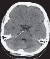
A hyperdense basilar artery (white arrow) can be seen on the axial unenhanced CT scan, with no definite infarction.
Fig. 5
Fig. 5
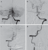
a Angiography via the left vertebral artery demonstrated a mid-basilar occlusion. b After successful mechanical thrombectomy there was complete restoration of flow. c After retracting the coaxial system into the proximal segment of the right vertebral artery, an extracranial pseudoaneurysm was noted at the site of the dissection. d Due to the stenosis and the high risk of early thrombus formation at the site of the wall injury, the decision was made to cover the dissection with a flow diverter stent. A weight-adapted bolus of eptifibatide was given, followed by careful placement of a compatible Marksman 0.027-inch microcatheter (Medtronic) distal to the dissected segment; a 3.5 × 16 mm Pipeline Flex Shield flow diverter stent (Medtronic) was successfully deployed with full coverage of the dissection and almost complete cessation of blood flow into the pseudoaneurysm.
Fig. 6
Fig. 6
Several small foci of infarction are seen in the cerebellar hemispheres (a), but the pons and medulla have a normal appearance (b).

References

    1. Sharshar T, Lamy C, Mas JL. Incidence and causes of strokes associated with pregnancy and puerperium. A study in public hospitals of Ile de France. Stroke in Pregnancy Study Group. Stroke. 1995;26:930–936. - PubMed
    1. Wiebers DO, Whisnant JP. The incidence of stroke among pregnant women in Rochester, Minn, 1955 through 1979. JAMA. 1985;254:3055–3057. - PubMed
    1. Kittner SJ, Stern BJ, Feeser BR, et al. Pregnancy and the risk of stroke. N Engl J Med. 1996;335:768–774. - PMC - PubMed
    1. Jaigobin C, Silver FL. Stroke and pregnancy. Stroke. 2000;31:2948–2951. - PubMed
    1. Skidmore FM, Williams LS, Fradkin KD, et al. Presentation, etiology, and outcome of stroke in pregnancy and puerperium. J Stroke Cerebrovasc Dis. 2001;10:1–10. - PubMed

LinkOut - more resources