Review
. 2017 May 29:7:217.
doi: 10.3389/fcimb.2017.00217. eCollection 2017.

Multiple Phenotypic Changes Define Neutrophil Priming

Affiliations
Review

Multiple Phenotypic Changes Define Neutrophil Priming

Irina Miralda et al. Front Cell Infect Microbiol. .

Abstract

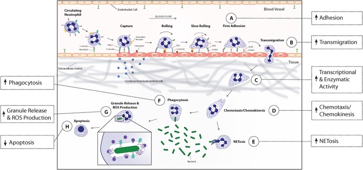
Exposure to pro-inflammatory cytokines, chemokines, mitochondrial contents, and bacterial and viral products induces neutrophils to transition from a basal state into a primed one, which is currently defined as an enhanced response to activating stimuli. Although, typically associated with enhanced generation of reactive oxygen species (ROS) by the NADPH oxidase, primed neutrophils show enhanced responsiveness of exocytosis, NET formation, and chemotaxis. Phenotypic changes associated with priming also include activation of a subset of functions, including adhesion, transcription, metabolism, and rate of apoptosis. This review summarizes the breadth of phenotypic changes associated with priming and reviews current knowledge of the molecular mechanisms behind those changes. We conclude that the current definition of priming is too restrictive. Priming represents a combination of enhanced responsiveness and activated functions that regulate both adaptive and innate immune responses.

Keywords: apoptosis; chemotaxis; cytokines; exocytosis; neutrophils; phagocytosis; priming; respiratory burst.


Figures

Figure 1
Figure 1
Priming-associated phenotypic changes and their effect on neutrophil functional responses. Neutrophils in circulating blood are in a resting state, characterized by a round morphology, non-adherence, minimal transcriptional activity, and a limited capacity to respond to activating stimuli. Microbial entry into tissues or tissue injury induces local immune cells to release pro-inflammatory cytokines that modify endothelial cell adhesion molecule profile and enter the bloodstream to begin priming neutrophils. Upon exposure to these priming agents, neutrophils undergo an increase in enzymatic and transcriptional activity that results in activation and synthesis of inflammatory mediators and enzymes that mediate downstream phenotypic and functional changes. Immediately, neutrophils begin to change their adhesion receptor pattern by shedding selectins, fusing secretory vesicles with the plasma membrane which leads to increased integrin expression, and a rapid increase in the gene expression of several surface receptors that allows newly primed cells to more rapidly adhere to endothelial cells (A). This phenotypic change coupled with the release of granules containing matrix metalloproteases, promotes neutrophil migration into inflamed tissues (B). The priming process continues when neutrophils bind to extracellular matrix proteins (C). Binding of neutrophil extracellular matrix receptors leads to an increase in actin polymerization, available receptors from secretory vesicle degranulation, and intracellular signaling that results in enhanced chemotaxis and chemokinesis (D). When primed neutrophils encounter bacteria, their phagocytic capacity is increased due to the upregulation in the number and affinity of receptors on the plasma membrane (F). By then, ROS production, granule release (G), and NET formation (E) have been primed to augment microbicidal activities. Finally, priming prolongs neutrophil lifespan by activating anti-apoptotic signal transduction pathways and transcription factors that decrease transcription of pro-apoptotic factors (H).

References

    1. Aas V., Lappegard K. T., Siebke E. M., Benestad H. B. (1996). Modulation by interferons of human neutrophilic granulocyte migration. J. Interferon Cytokine Res. 16, 929–935. 10.1089/jir.1996.16.929 - DOI - PubMed
    1. Acorci M. J., Dias-Melicio L. A., Golim M. A., Bordon-Graciani A. P., Peracoli M. T., Soares A. M. (2009). Inhibition of human neutrophil apoptosis by Paracoccidioides brasiliensis: role of interleukin-8. Scand. J. Immunol. 69, 73–79. 10.1111/j.1365-3083.2008.02199.x - DOI - PubMed
    1. Afonso P. V., Janka-Junttila M., Lee Y. J., McCann C. P., Oliver C. M., Aamer K. A., et al. (2012). LTB4 is a signal-relay molecule during neutrophil chemotaxis. Dev. Cell 22, 1079–1091. 10.1016/j.devcel.2012.02.003 - DOI - PMC - PubMed
    1. Aglietta M., Monzeglio C., Apra F., Mossetti C., Stern A. C., Giribaldi G., et al. (1990). In vivo priming of human normal neutrophils by granulocyte-macrophage colony stimulating factor: effect on the production of platelet activating factor. Br. J. Haematol. 75, 333–339. 10.1111/j.1365-2141.1990.tb04345.x - DOI - PubMed
    1. Andreasson E., Onnheim K., Forsman H. (2013). The subcellular localization of the receptor for platelet-activating factor in neutrophils affects signaling and activation characteristics. Clin. Dev. Immunol. 2013:456407. 10.1155/2013/456407 - DOI - PMC - PubMed

MeSH terms