. 2017 May 29:4:27.
doi: 10.3389/fsurg.2017.00027. eCollection 2017.

Management of Orbital and Periorbital Venous Malformation

Affiliations

Management of Orbital and Periorbital Venous Malformation

Lara A Benoiton et al. Front Surg. .

Abstract

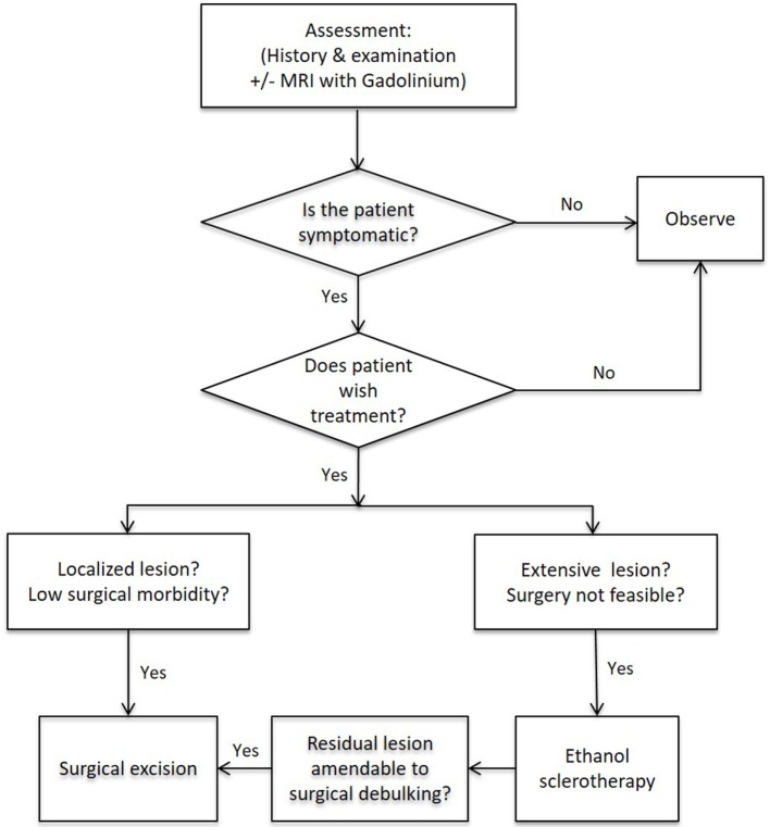
Background: To review our management of common venous malformation (VM) affecting the orbit and/or periorbital area.

Methods: Consecutive patients with orbital and/or periorbital VM were identified from our vascular anomalies database. Demographic details of the patients, anatomic site(s) affected, symptoms and signs, presence of a family history of VM, and types of treatment(s) were collected, supplemented by chart review.

Results: A total of 24 patients' age 1-68 (mean, 30) years with orbital and/or periorbital VM presented with cosmetic concerns (n = 17, 71%), distensibility (n = 15, 63%), pain (n = 9, 38%), diplopia (n = 4, 17%), and spontaneous thrombosis (n = 1, 8%). The VM caused globe dystopia (n = 13, 54%), enophthalmos (n = 6, 25%), proptosis (n = 3, 12%), exotropia (n = 3, 12%), and pseudoptosis with visual obstruction (n = 3, 13%). A total of 11 (46%) patients were managed conservatively. 13 (54%) patients underwent active treatment. Ethanol sclerotherapy (ES) was performed in six patients with extensive facial VM associated with orbital/periorbital involvement, resulting in symptomatic improvement in five patients, one of whom developed skin necrosis and another patient developed reduced infraorbital nerve sensation. Surgery was performed for localized lesion (n = 3, 23%), for extensive lesions (n = 4, 31%) and as an adjunct to ES (n = 6, 46%) resulting in symptomatic improvement in all patients. One patient required correction of lower lid ectropion.

Conclusion: Orbital and/or periorbital VMs are heterogeneous, and management needs to be individualized. Surgery is used for localized lesions aiming for complete excision, as a debulking procedure for extensive orbital/periorbital VM when ES was not possible, or following ES for extensive facial VM with orbital and/or periorbital involvement.

Keywords: management; orbital; periorbital; treatment; venous malformation.


Figures

Figure 1
Figure 1
A 17-year-old female with a left orbital venous malformation causing ocular dystopia. The visible temporal scar resulted from a previous failed attempt of excision elsewhere. (A) A n MRI (B) and a CT scan (C) showed a 2 cm × 2 cm soft tissue mass on the floor of the left orbit displacing the globe superiorly and a bony ridge at the inferior orbital margin (C). The lesion was excised through a subciliary incision with removal of the bony lip and reduction of the hypertrophic orbital floor, restoring the position of the globe. Photograph 2 years postoperatively (D). Reproduced with permission from the British Journal of Oral and Maxillofacial Surgery (5).
Figure 2
Figure 2
A 62-year-old female with right eye proptosis resulting from an intraconal venous malformation (VM) (A,B). Axial (C) and coronal (D) T2-weighted magnetic resonance imaging showing a 2.8 cm × 2.4 cm VM within the right orbital cone. The lesion (E) was removed through a lateral orbitotomy via a coronal incision. The orbitotomy was plated (E). Photograph of the lesion following removal (F). The position of the right globe was restored and remained satisfactory with no visual dysfunction 5 years postoperatively (G,H).
Figure 3
Figure 3
A newborn male with an extensive orbital and periorbital venous malformation (VM) (A,B) and small cutaneous lesions in the feet (C). The lesion massively expanded the upper lid, occluded the visual axis, displaced the globe, and involved the lower lid to a lesser extent (A,B,D). Axial (E) and coronal (F) T2-weighted magnetic resonance imaging showing extensive orbital/periorbital involvement with intracranial communication, and affecting the temporal area. A debulking procedure was performed (G) with significant reduction of the involved upper lid with improvement postoperatively (H). There was gradual expansion of the VM required further debulking procedure 28 months later. The globe remained displaced with minimal vision (I).
Figure 4
Figure 4
A 15-year-old female with a venous malformation (VM) affecting her left cheek and peri-orbital area (A). A T2-weighted magnetic resonance imaging showing the VM within the left buccal space, extending into the temporal region, deep to the temporalis muscle (B). Ethanol sclerotherapy of the VM led to improved facial symmetry (C). The residual VM over the lateral canthus was debulked and remained unchanged over 2 years (D). Reproduced with permission from the Australian and New Zealand Journal of Surgery (27).
Figure 5
Figure 5
A proposed management algorithm for orbital and/or periorbital venous malformation.

References

    1. van der Vleuten CJ, Kater A, Wijnen MH, Schultze Kool LJ, Rovers MM. Effectiveness of sclerotherapy, surgery, and laser therapy in patients with venous malformations: a systematic review. Cardiovasc Intervent Radiol (2014) 37(4):977–89.10.1007/s00270-013-0764-2 - DOI - PubMed
    1. Boon LM, Mulliken JB, Enjolras O, Vikkula M. Glomuvenous malformation (glomangioma) and venous malformation: distinct clinicopathologic and genetic entities. Arch Dermatol (2004) 140(8):971–6.10.1001/archderm.140.8.971 - DOI - PubMed
    1. Boon LM, Mulliken JB, Vikkula M, Watkins H, Seidman J, Olsen BR, et al. Assignment of a locus for dominantly inherited venous malformations to chromosome 9p. Hum Mol Genet (1994) 3(9):1583–7.10.1093/hmg/3.9.1583 - DOI - PubMed
    1. Limaye N, Wouters V, Uebelhoer M, Tuominen M, Wirkkala R, Mulliken JB, et al. Somatic mutations in angiopoietin receptor gene TEK cause solitary and multiple sporadic venous malformations. Nat Genet (2009) 41(1):118–24.10.1038/ng.272 - DOI - PMC - PubMed
    1. Tan ST, Bialostocki A, Brasch H, Fitzjohn T. Venous malformation of the orbit. J Oral Maxillofac Surg (2004) 62(10):1308–11.10.1016/j.joms.2003.12.039 - DOI - PubMed

LinkOut - more resources