

Moving from millifluidic to truly microfluidic sub-100-μm cross-section 3D printed devices
- PMID: 28612085
- PMCID: PMC5542000
- DOI: 10.1007/s00216-017-0398-3
Moving from millifluidic to truly microfluidic sub-100-μm cross-section 3D printed devices
Abstract
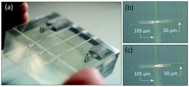
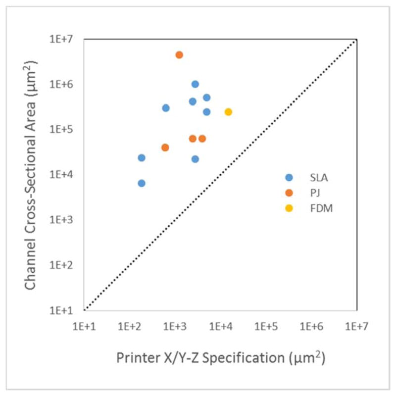
Three-dimensional (3D) printing has generated considerable excitement in recent years regarding the extensive possibilities of this enabling technology. One area in which 3D printing has potential, not only for positive impact but also for substantial improvement, is microfluidics. To date many researchers have used 3D printers to make fluidic channels directed at point-of-care or lab-on-a-chip applications. Here, we look critically at the cross-sectional sizes of these 3D printed fluidic structures, classifying them as millifluidic (larger than 1 mm), sub-millifluidic (0.5-1.0 mm), large microfluidic (100-500 μm), or truly microfluidic (smaller than 100 μm). Additionally, we provide our prognosis for making 10-100-μm cross-section microfluidic features with custom-formulated resins and stereolithographic printers. Such 3D printed microfluidic devices for bioanalysis will accelerate research through designs that can be easily created and modified, allowing improved assays to be developed.
Keywords: Bioanalytical methods; Microfluidics/microfabrication; Separations/instrumentation.
Conflict of interest statement
The authors declare no conflicts.
Figures

References
-
- Amin R, Knowlton S, Hart A, Yenilmez B, Ghaderinezhad F, Katebifar S, Messina M, Khademhosseini A, Tasoglu S. 3D-printed microfluidic devices. Biofabrication. 2016;8:022001. - PubMed
-
- Lee KG, Park KJ, Seok S, Shin S, Kim DH, Park JY, Heo YS, Lee SJ, Lee TJ. 3D printed modules for integrated microfluidic devices. RSC Adv. 2014;4:32876–80.
MeSH terms
Grants and funding
LinkOut - more resources
Full Text Sources
Other Literature Sources
Miscellaneous
