Comparative Study
. 2017 Aug;91(8):2849-2863.
doi: 10.1007/s00204-017-2002-1. Epub 2017 Jun 13.

Utility of spherical human liver microtissues for prediction of clinical drug-induced liver injury

Affiliations
Comparative Study

Utility of spherical human liver microtissues for prediction of clinical drug-induced liver injury

William R Proctor et al. Arch Toxicol. 2017 Aug.

Abstract

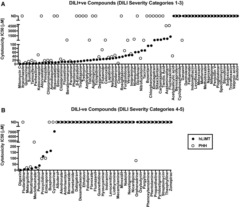
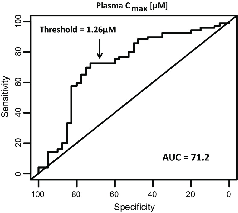
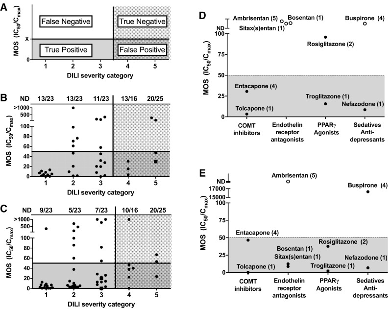
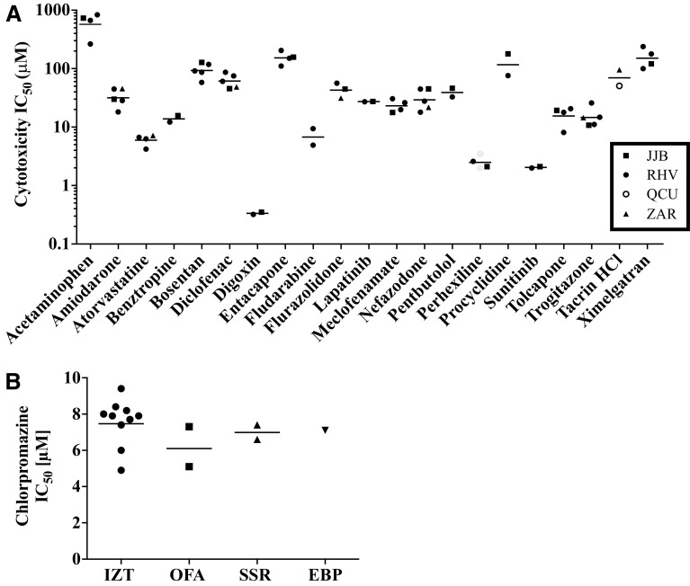
Drug-induced liver injury (DILI) continues to be a major source of clinical attrition, precautionary warnings, and post-market withdrawal of drugs. Accordingly, there is a need for more predictive tools to assess hepatotoxicity risk in drug discovery. Three-dimensional (3D) spheroid hepatic cultures have emerged as promising tools to assess mechanisms of hepatotoxicity, as they demonstrate enhanced liver phenotype, metabolic activity, and stability in culture not attainable with conventional two-dimensional hepatic models. Increased sensitivity of these models to drug-induced cytotoxicity has been demonstrated with relatively small panels of hepatotoxicants. However, a comprehensive evaluation of these models is lacking. Here, the predictive value of 3D human liver microtissues (hLiMT) to identify known hepatotoxicants using a panel of 110 drugs with and without clinical DILI has been assessed in comparison to plated two-dimensional primary human hepatocytes (PHH). Compounds were treated long-term (14 days) in hLiMT and acutely (2 days) in PHH to assess drug-induced cytotoxicity over an 8-point concentration range to generate IC50 values. Regardless of comparing IC50 values or exposure-corrected margin of safety values, hLiMT demonstrated increased sensitivity in identifying known hepatotoxicants than PHH, while specificity was consistent across both assays. In addition, hLiMT out performed PHH in correctly classifying hepatotoxicants from different pharmacological classes of molecules. The hLiMT demonstrated sufficient capability to warrant exploratory liver injury biomarker investigation (miR-122, HMGB1, α-GST) in the cell-culture media. Taken together, this study represents the most comprehensive evaluation of 3D spheroid hepatic cultures up to now and supports their utility for hepatotoxicity risk assessment in drug discovery.

Keywords: DILI; Drug discovery; Hepatocyte; Hepatotoxicity; Microtissue; Spheroid culture.


Conflict of interest statement

M. K., S. S., J. M. K. and S. M. are employees of InSphero AG. All cytotoxicity studies in hLiMT were performed at InSphero AG under conditions where the identities of the 110 compounds were blinded to them.

Figures

Fig. 1
Fig. 1
3D hLiMT were more sensitive to overall drug-induced cytotoxicity than 2D PHH. Cytotoxicity IC50 values for 110 drugs in PHH treated for 48 h (open symbols) and hLiMT treated for 14 days (closed symbols) for compounds classified as DILI+ve (DILI severity categories 1–3) (a) and DILI–ve (DILI severity categories 4–5) (b), respectively. Data represent IC50 value estimate for each compound listed on the x-axis. For compounds where no IC50 value converged, these were plotted at the top and classified as not determined (ND)
Fig. 2
Fig. 2
Optimized receiver operating curve (ROC) for total plasma concentration (C max) alone as predictor of clinical hepatotoxicity. Optimized ROC curve for total plasma concentration (C max) for 110 drugs associated with and without clinical hepatotoxicity. ROC curve was generated from total plasma C max data for the test set and an optimized threshold (in bold) was determined
Fig. 3
Fig. 3
Exposure-corrected cytotoxicity (MOS) of 110 marketed drugs stratified across the five DILI severity categories. A compound was considered to be DILI+ve if classified in the following: DILI severity category 1 (Severe clinical DILI), severity category 2 (High clinical DILI concern, cases of liver failure), and severity category 3 (low clinical DILI concern, isolated and infrequent cases of DILI). Conversely, a drug was considered DILI−ve if classified as DILI severity category 4 (enzyme elevations in clinic) and severity category 5 (No DILI). a Schematic of the four quadrants identifying true and false positives and true and false negatives across the 5 DILI severity categories in relation to optimized MOS threshold. b Plated primary human hepatocytes, dotted line threshold of 50× MOS, c hLiMT, dotted line threshold of 50× MOS. DILI severity category is indicated in brackets with the number of compounds that did not have an IC50 value converge per DILI severity category listed at top. d, e Comparison of the MOS values for structurally related hepatotoxic and non-hepatotoxic compounds in hLiMT (d) and PHH (e) for catechol-O-methyltransferase (COMT) inhibitors (tolcapone and entacapone), endothelin-receptor antagonists (sitax(s)entan, bosentan and ambrisentan), insulin sensitizers (troglitazone and rosiglitazone), and sedatives/antidepressants (nefazodone and buspirone). Filled circles margin of safety (MOS) = IC50/C max value; open circles not detected (ND) value and filled square <IC50/C max value
Fig. 4
Fig. 4
Reproducibility of the IC50 values from independent experiments following incubation of the 3D hLiMT to test compounds for 14 days. Cytotoxic IC50 values for subset of compounds were plotted for hLiMT were prepared using either: the same cryopreserved primary human hepatocyte lot (IZT) but four different NPC lots: filled square JJB; filled circle RHV; filled triangle ZAR; open circle QCU (a) or the same NPC lot but different lots of cryopreserved primary human hepatocytes (IZT, OFA, SSR or EBP) (b)
Fig. 5
Fig. 5
POC study demonstrating the utility of hLiMTs to detect biomarkers of hepatocellular injury in vitro. Representative changes in biomarker levels are depicted following exposure to chlorpromazine for 14 days (a) or 5 days (b and c). a In relation to decreases in total cellular ATP (filled circle) as determined on day 14. Filled square α-GST levels were determined on days 5, 9, and 14 and the values combined to give a fold change relative to control. b, c In relation to decreases in total cellular ATP as determined on day 5. Filled circle cell viability (% control); b open triangle HMGB-1 ng/ml, c open inverted triangle miRNA-122 relative expression. Data are from single experiments in triplicate

Comment in

References

    1. Aleo MD, Luo Y, Swiss R, Bonin PD, Potter DM, Will Y. Human drug-induced liver injury severity is highly associated with dual inhibition of liver mitochondrial function and bile salt export pump. Hepatology. 2014;60(3):1015–1022. doi: 10.1002/hep.27206. - DOI - PubMed
    1. Altman DG, Bland JM. Diagnostic tests 2: predictive values. BMJ. 1994;309(6947):102. doi: 10.1136/bmj.309.6947.102. - DOI - PMC - PubMed
    1. Altman DG, Bland JM. Diagnostic tests 3: receiver operating characteristic plots. BMJ. 1994;309(6948):188. doi: 10.1136/bmj.309.6948.188. - DOI - PMC - PubMed
    1. Antoine DJ, Williams DP, Kipar A, et al. High-mobility group box-1 protein and keratin-18, circulating serum proteins informative of acetaminophen-induced necrosis and apoptosis in vivo. Toxicol Sci. 2009;112(2):521–531. doi: 10.1093/toxsci/kfp235. - DOI - PubMed
    1. Bell CC, Hendriks DF, Moro SM, et al. Characterization of primary human hepatocyte spheroids as a model system for drug-induced liver injury, liver function and disease. Sci Rep. 2016;6:25187. doi: 10.1038/srep25187. - DOI - PMC - PubMed

Publication types

MeSH terms