

Comparison of preoperative imaging and pathological findings for pancreatic head adenocarcinoma: A retrospective analysis by the Association Française de Chirurgie
- PMID: 28614269
- PMCID: PMC5478354
- DOI: 10.1097/MD.0000000000007214
Comparison of preoperative imaging and pathological findings for pancreatic head adenocarcinoma: A retrospective analysis by the Association Française de Chirurgie
Abstract
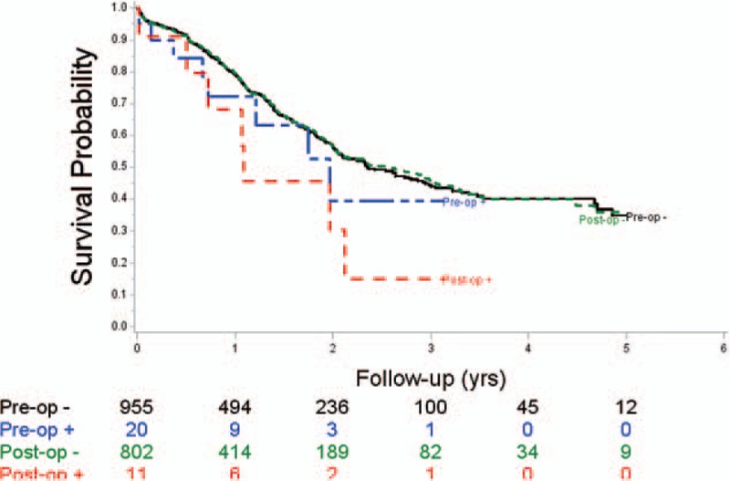
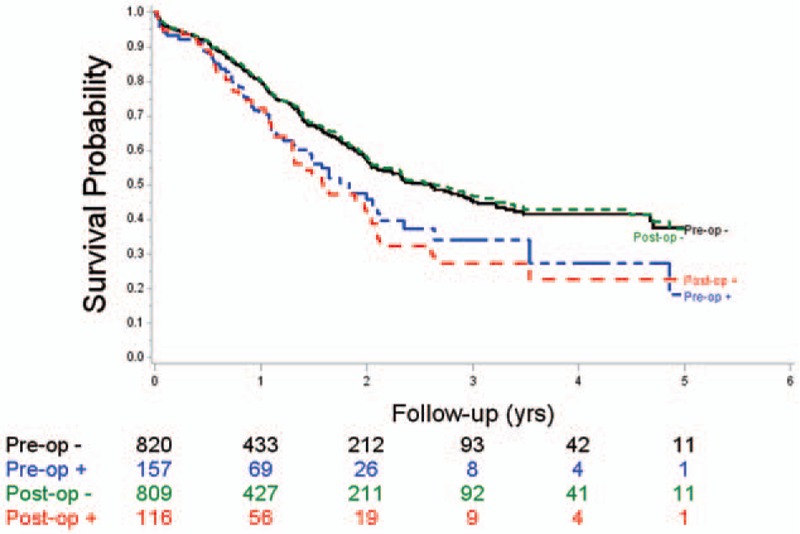
Initial imaging of pancreatic ductal adenocarcinoma is of crucial importance in the decision-making process. The aim of this study was to compare preoperative imaging, pathological data, and outcomes in a series of patients who underwent resection for pancreatic head cancer.From January 2004 to December 2009, data were collected by the Association Française de Chirurgie on 1044 patients who received first-line R0 resection of pancreatic head cancer.On imaging (computed tomography scan 97%, echoendoscopic ultrasound 61.3%, magnetic resonance imaging 46.5%), arterial, venous, or lymph node invasion was suspected in 20, 161, and 197 patients, respectively; arterial, venous, or lymph node invasion was observed histologically in 11, 116, and 736 cases, respectively. In the patients for whom both imaging and pathological data were available, the concordance, sensitivity, specificity, positive predictive value, and negative predictive value were as follows: 97.5%, 27.3%, 98%, 20%, and 99%, for arterial invasion; 86.5%, 54%, 91%, 47.8%, and 93.2%, for venous invasion; and 38%, 21%, 86%, 78%, and 41%, respectively, for lymph node invasion. Imaging of arterial invasion had no prognostic value, while histological evidence of invasion was associated with a poor prognosis. Venous and lymph node invasion, as demonstrated by imaging and by pathological analysis, had an adverse prognostic value.Imaging gives a fair positive predictive value for venous or arterial invasion; venous invasion on imaging and histology was associated with a poor prognosis; arterial invasion on imaging does not have any significant prognostic value.
Conflict of interest statement
The authors have no conflicts of interest to disclose.
Figures
References
-
- De Angelis R, Sant M, Coleman MP, et al. Cancer survival in Europe 1999–2007 by country and age: results of EUROCARE-5-a-population-based study. Lancet Oncol 2014;15:23–34. - PubMed
-
- Jemal A, Siegel R, Xu J, et al. Cancer statistics 2010. CA Cancer J Clin 2010;60:277–300. - PubMed
-
- Hartwig W, Jager D, Debus J, et al. Improvement of surgical results for pancreatic cancer. Lancet Oncol 2013;14:e476–85. - PubMed
-
- Al-Hawary MM, Francis IR, Chari ST, et al. Pancreatic ductal adenocarcinoma radiology reporting template: consensus of the Society of Abdominal Radiology and the American Pancreatic Association. Radiology 2014;270:248–60. - PubMed
-
- Delpero JR, Paye F, Bachelier P, et al. Cancer du Pancréas. Monographies de l’Association Française de Chirurgie, Arnette, Wolters Kluwer, France; 2010.
Publication types
MeSH terms
LinkOut - more resources
Full Text Sources
Other Literature Sources
Medical
