. 2017 Jun 29;546(7660):671-675.
doi: 10.1038/nature22820. Epub 2017 Jun 14.

ERF mutations reveal a balance of ETS factors controlling prostate oncogenesis

Collaborators, Affiliations

ERF mutations reveal a balance of ETS factors controlling prostate oncogenesis

Rohit Bose et al. Nature. .

Abstract

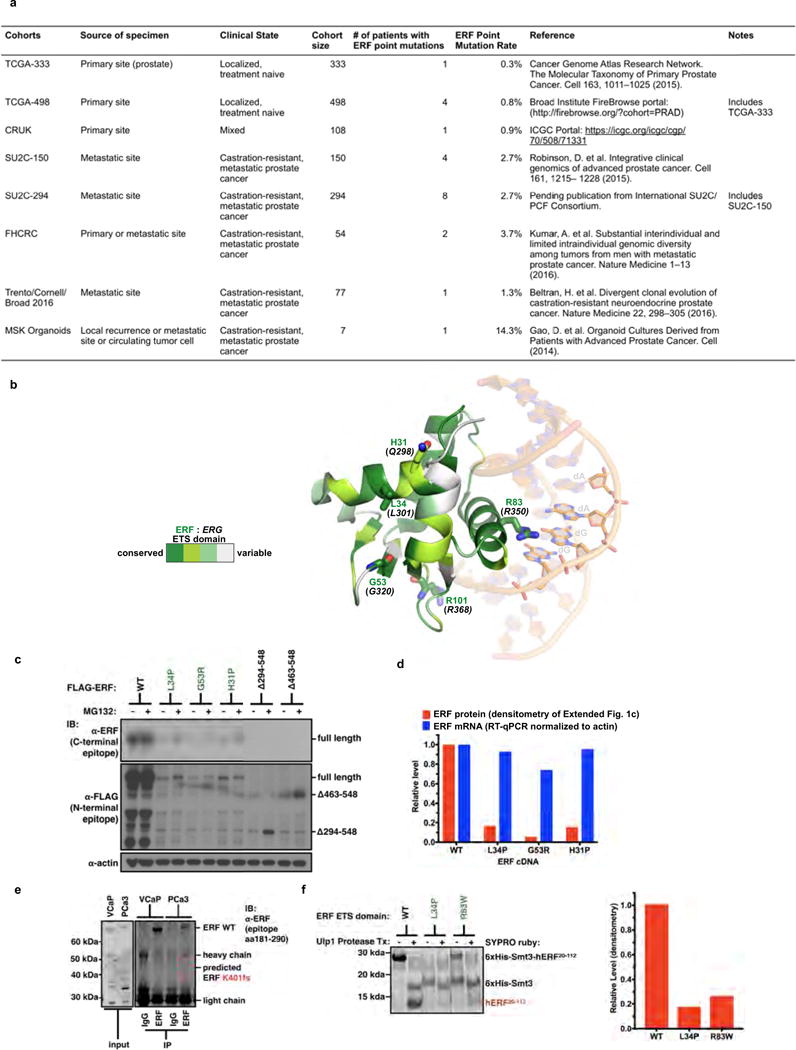
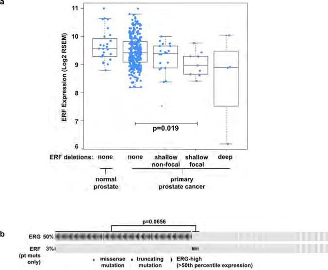
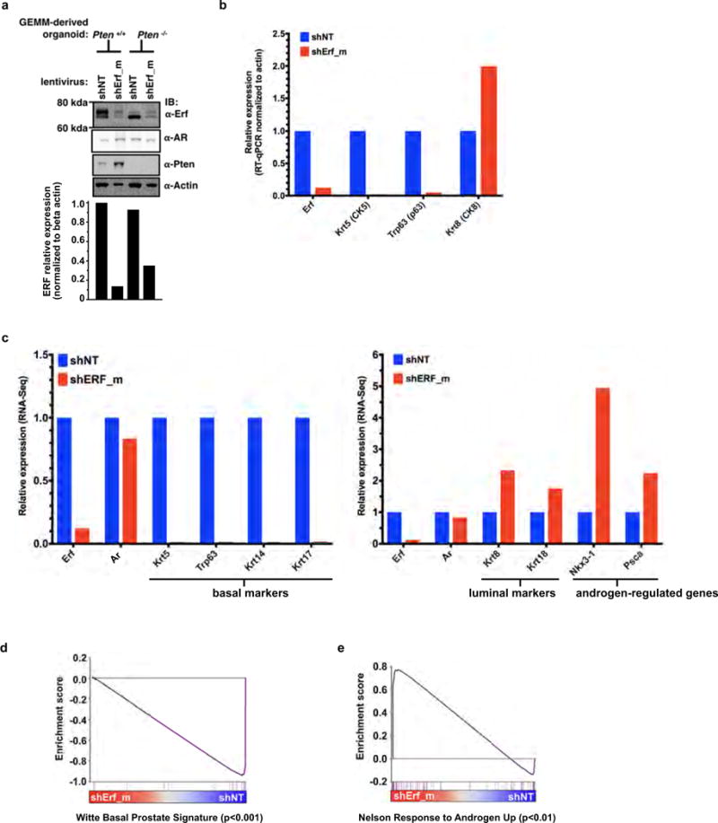
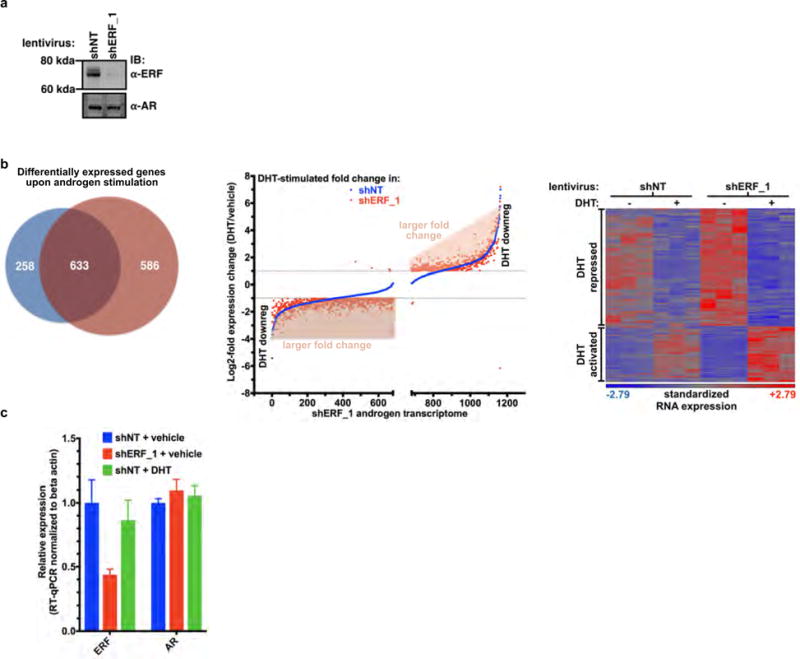
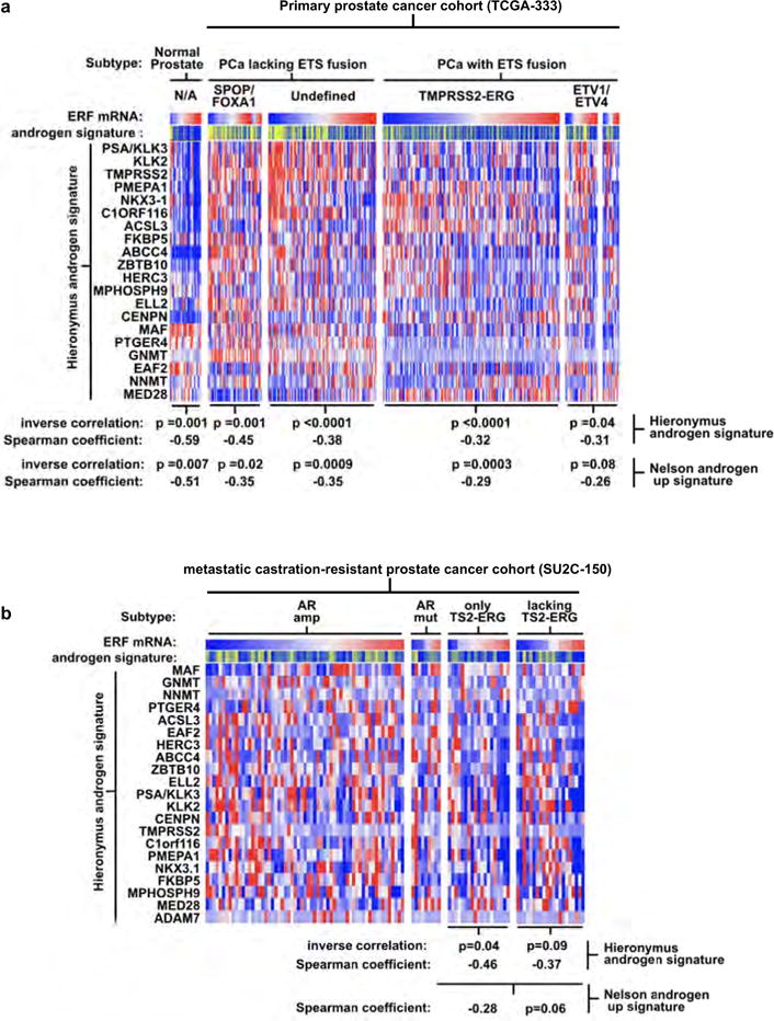
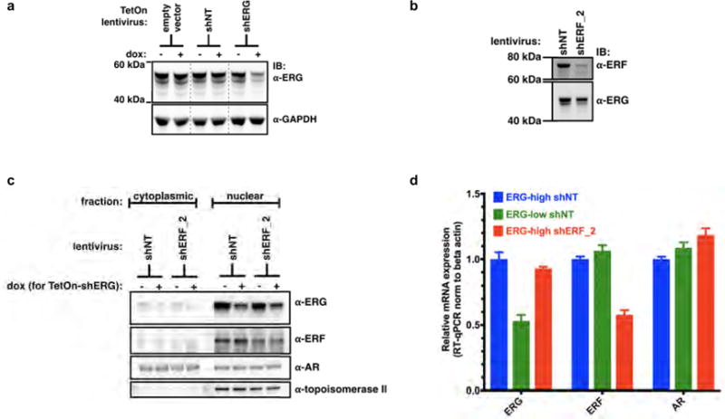
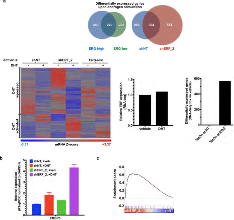
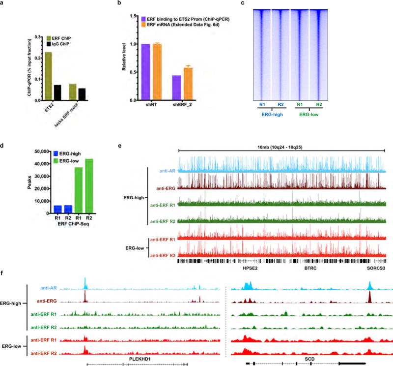
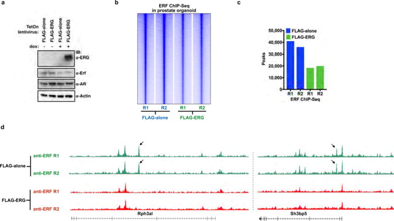
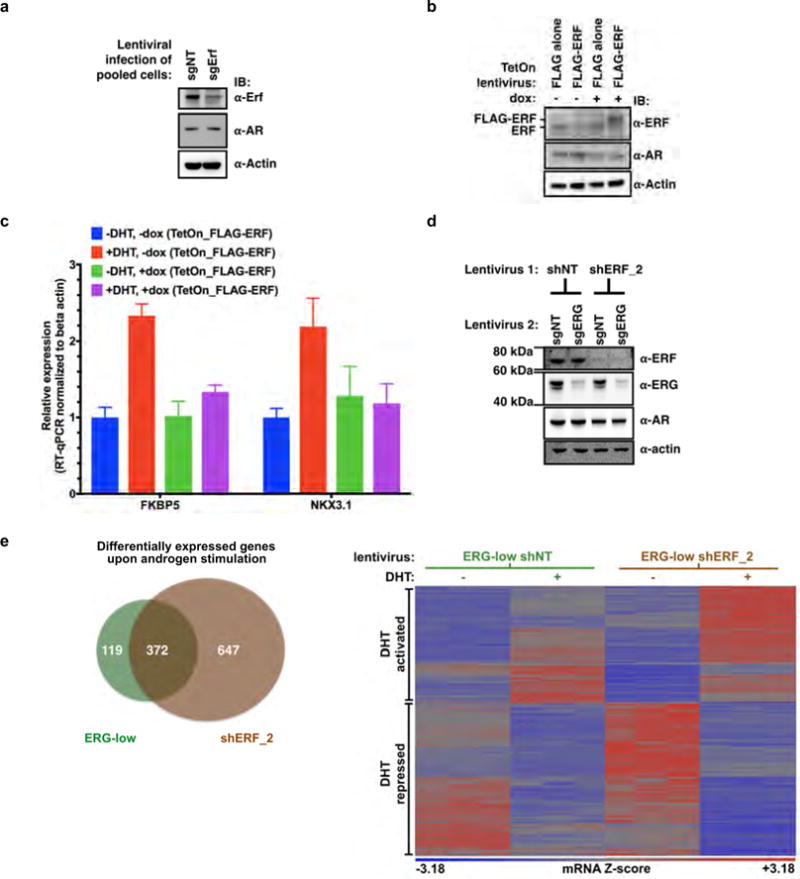
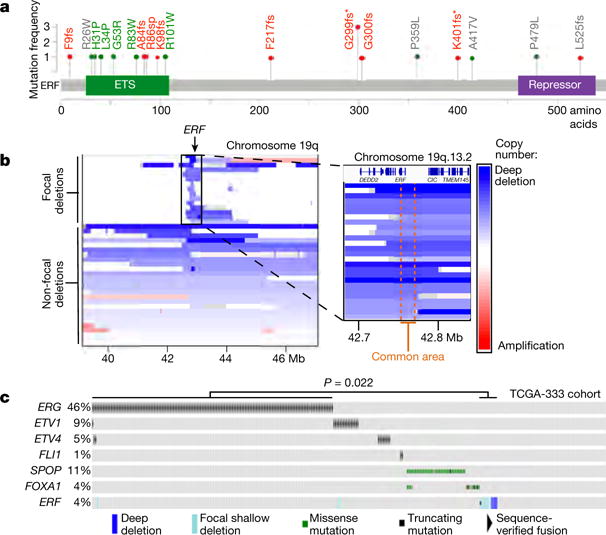
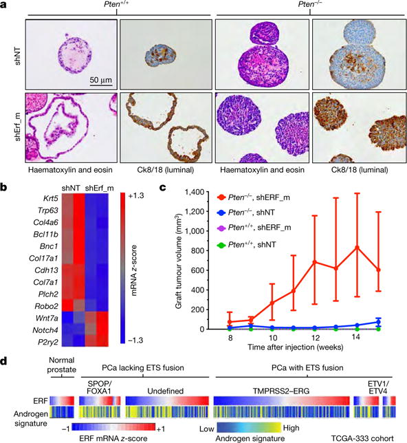
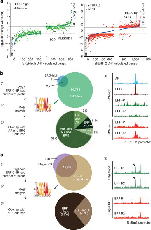
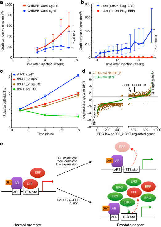
Half of all prostate cancers are caused by the TMPRSS2-ERG gene-fusion, which enables androgens to drive expression of the normally silent E26 transformation-specific (ETS) transcription factor ERG in prostate cells. Recent genomic landscape studies of such cancers have reported recurrent point mutations and focal deletions of another ETS member, the ETS2 repressor factor ERF. Here we show these ERF mutations cause decreased protein stability and mostly occur in tumours without ERG upregulation. ERF loss recapitulates the morphological and phenotypic features of ERG gain in normal mouse prostate cells, including expansion of the androgen receptor transcriptional repertoire, and ERF has tumour suppressor activity in the same genetic background of Pten loss that yields oncogenic activity by ERG. In the more common scenario of ERG upregulation, chromatin immunoprecipitation followed by sequencing indicates that ERG inhibits the ability of ERF to bind DNA at consensus ETS sites both in normal and in cancerous prostate cells. Consistent with a competition model, ERF overexpression blocks ERG-dependent tumour growth, and ERF loss rescues TMPRSS2-ERG-positive prostate cancer cells from ERG dependency. Collectively, these data provide evidence that the oncogenicity of ERG is mediated, in part, by competition with ERF and they raise the larger question of whether other gain-of-function oncogenic transcription factors might also inactivate endogenous tumour suppressors.


Conflict of interest statement

The authors declare no competing financial interests. Readers are welcome to comment on the online version of the paper. Publisher’s note: Springer Nature remains neutral with regard to jurisdictional claims in published maps and institutional affiliations.

Figures

Extended Data Figure 1
Extended Data Figure 1. Recurrent ERF loss-of-function mutations are found in prostate cancer and are destabilizing
Related to Fig. 1. a, Description of the prostate cancer patient cohorts. b, ERG ETS domain (Protein Data Bank accession number 4IRI) illustrating sequence conservation with ERF. ERF missense mutations indicated by sticks. c, Expression of ERF mutant proteins in LNCaP cells. For gel source data, see Supplementary Fig. 1. d, ERF mutant protein densitometry from immunoblot in Extended Data Fig. 1c compared with mRNA RT–qPCR. e, ERF immunoprecipitation in VCaP and MSK-PCa3 cells. f, Left, bacterially expressed ETS domain (hERF20–112) with ERF tumour mutations before (−) or after (+) Ulp1 protease cleavage. Right, densitometry of in vitro ETS domains.
Extended Data Figure 2
Extended Data Figure 2. Recurrent ERF loss-of-function mutations and focal deletions are found in prostate cancer and are mostly exclusive to tumours without TMPRSS2–ERG
Related to Fig. 1. a, ERF expression in TCGA-333 cohort (n = 333 patients) segregated by copy number loss. Data are Tukey box-and-whisker plots; P value calculated by two-tailed t-test of log2(RSEM values). b, cBio Oncoprint of the SU2C-294 (n = 294) metastatic prostate cancer cohort. Unlike Fig. 1c, focal ERF deletions could not be ascertained because of differences between TCGA and SU2C copy number data,. P value calculated by Fisher’s exact two-tailed test.
Extended Data Figure 3
Extended Data Figure 3. ERF is a negative regulator of androgen signalling
Related to Fig. 2. a, Mouse prostate organoids infected with non-targeting shRNA (shNT) or targeting ERF (shErf_m), grown in three-dimensional culture. For RT–qPCR, n = 2 biological replicates. For gel source data, see Supplementary Fig. 1. b, RT–qPCR analysis of the Pten+/+ organoids; n = 2 biological replicates. c, RNA-seq analysis of organoids derived from Pten+/+ and Pten−/− mouse prostates infected with non-targeting shNT or shErf_m; n = 2 biological replicates. d, Pten+/+ organoid RNA-seq (n = 2 biological replicates) interrogated by GSEA for expression signature of Witte basal prostate cancer. e, Same data interrogated by GSEA for Nelson androgen up expression signature.
Extended Data Figure 4
Extended Data Figure 4. ERF is a negative regulator of androgen signalling
Related to Fig. 2. a, CWR22Pc cells infected with shRNA targeting human ERF (shERF_1) or a non-targeting shRNA (shNT). For gel source data, see Supplementary Fig. 1. b, Androgen-regulated genes (at least a twofold change, FDR < 0.05 by RNA-seq with 1 nM DHT for 16 h) in CWR22PC cells infected with either shERF_1 or shNT, analysed by the number (left, Venn diagram), the magnitude of expression change (centre, graph), and heat map (right); n = 3 biological replicates. c, RT–qPCR from CWR22Pc cells infected with shERF_1 or shNT. Data are mean ± s.e.m.; n = 3 biological replicates.
Extended Data Figure 5
Extended Data Figure 5. ERF is a negative regulator of androgen signalling
Related to Fig. 2. a, Full version of Fig. 2d. Expression profiles of the TCGA-333 primary prostate cancer cohort (n = 333 patients) were interrogated for correlation between the ERF mRNA level and two androgen transcriptional activity signatures,. P values were calculated by the Spearman correlation test. b, The same analysis as a was applied to the SU2C-150 (n = 150 patients) metastatic castration-resistant prostate cancer cohort (mCRPC).
Extended Data Figure 6
Extended Data Figure 6. ERF and ERG knockdown do not affect androgen receptor levels or its subcellular localization
Related to Fig. 3. a, VCaP cells infected with doxycycline (dox)-inducible shRNA targeting ERG (+dox, ERG-low; −dox, ERG-high). For gel source data, see Supplementary Fig. 1. b, VCaP cells infected with shRNA targeting ERF (shERF_2) or a non-targeting shRNA (shNT). c, Nuclear/cytoplasmic fractionation of VCaP cells infected with shNT or shERF_2, and the doxycycline-inducible shRNA targeting ERG (shERG). d, RT–qPCR with ERG-low VCaP cells compared with those infected with shERF_2. Data are mean ± s.e.m.; n = 3 biological replicates.
Extended Data Figure 7
Extended Data Figure 7. ERF and TMPRSS2–ERG have opposing effects on the androgen transcriptome
Related to Fig. 3. a, Androgen-regulated genes (at least a twofold change, FDR < 0.05 by RNA-seq with 1 nM DHT for 16 h) in VCaP cells infected with a doxycycline-inducible shRNA targeting ERG (with doxycycline, ERG-low; without doxycycline, ERG-high) or a constitutive shRNA targeting ERF (shERF_2) and analysed by number (top) and heat map (bottom left); n = 3 biological replicates. Bottom centre, RNA-seq analysis evaluating the effect of dihydrotestosterone on ERF expression. Bottom right, the effect of doxycycline alone on RNA-seq differential expression analysis. b, RT–qPCR of shERF_2-infected VCaP cells treated ± DHT. Data are mean ± s.e.m.; n = 3 biological replicates. c, Interrogation of RNA-seq in shERF_2 VCaP cells (n = 3 biological replicates) by GSEA for Nelson androgen up expression signature.
Extended Data Figure 8
Extended Data Figure 8. ERF and TMPRSS2–ERG have opposing effects on the androgen receptor cistrome
Related to Fig. 3. a, ChIP–qPCR (n = 2 biological replicates) in VCaP cells, amplifying either the ETS2 promoter region that contains a known ERF binding site, or an upstream element of PSA lacking the ERF binding motif noted in (2) in Fig. 3b. b, The effect of ERF shRNA knockdown on its binding to the ETS2 promoter as assessed by ChIP–qPCR (n = 2 biological replicates), compared with its effect on ERF mRNA by RT–qPCR. Data are mean ± s.e.m.; n = 3 biological replicates. c, ERF ChIP–seq in ERG-high or ERG-low (n = 2 biological replicates: R1, R2) analysed by heat maps. d, Comparison of ERF ChIP–seq peak numbers, n = 2 biological replicates: R1, R2. e, A 10-Mb region illustrating ChIP–seq of ERF binding in ERG-high condition compared with ERG-low. f, ChIP–seq signals for the SCD and PLEKHD1 loci. In both e and f, ChIP–seq signals at the y axis were normalized by read depths.
Extended Data Figure 9
Extended Data Figure 9. ERG expression decreases the ERF cistrome in normal prostate organoids
Related to Fig. 3. a, Mouse normal prostate organoids were infected with a TetOn doxycycline-inducible Flag–ERG or empty vector (Flag-alone) and treated with or without doxycycline. For gel source data, see Supplementary Fig. 1. b, ERF ChIP–seq in normal prostate organoids infected with either Flag-alone or Flag–ERG lentivirus, both treated with doxycycline (n = 2 biological replicates: R1, R2), and analysed with heat maps. c, Comparison of ERF ChIP–seq peak numbers, n =2 biological replicates (R1, R2). d, ERF ChIP–seq signals for the Rph3a1 and Sh3bp5 loci, normalized by read depths.
Extended Data Figure 10
Extended Data Figure 10. TMPRSS2–ERG activity is mediated, in part, by inactivation of ERF function
Related to Fig. 4. a, Pooled mouse Pten−/−; R26ERG/ERG organoids infected with CRISPR–Cas9 targeting AAVS1 (sgNT) or ERF (sgErf). For gel source data, see Supplementary Fig. 1. b, Pten−/−; R26ERG/ERG organoids infected instead with a doxycycline-inducible Flag–ERF or empty vector (Flag alone). c, RT−qPCR of mRNA isolated from the Flag−ERF-infected organoids and treated with or without doxycycline, with or without dihydrotestosterone 1 nM for 16 h. Data are mean ± s.e.m.; n = 3 biological replicates. d, Pooled VCaP cells were first infected with non-targeting shRNA (shNT) or ERF (shERF_2), followed by sgNT or sgERF CRISPR–Cas9. e, Androgen-regulated genes (at least a twofold change, FDR < 0.05 by RNA-seq with 1 nM DHT for 16 h) in VCaP cells infected with a doxycycline-inducible shRNA targeting ERG (with doxycycline, ERG-low) and either a constitutive shRNA targeting ERF (shERF_2) or a non-targeting shRNA (shNT). Data analysed by number (left, Venn diagram) and heat map (right); n = 3 biological replicates.
Figure 1
Figure 1. Recurrent ERF loss-of-function mutations and focal deletions are found in prostate cancer and are mostly exclusive to tumours without TMPRSS2–ERG
a, ERF point mutations identified in all prostate cancer cohorts. * Mutations shared with patients having craniosynostosis. Green: ETS missense mutations of residues conserved with ERG. Red: frameshift (fs)/splice-site (sp) mutations upstream of repressor domain. b, ERF copy number deletions in TCGA-333 human primary prostate cancer cohort. c, cBio Oncoprint of patients with primary prostate cancer (n = 333 patients). Each column corresponds to a unique patient’s tumour profile. P value calculated by Fisher’s exact two-tailed test.
Figure 2
Figure 2. ERF is a tumour suppressor, a negative regulator of androgen signalling, and its loss phenocopies TMPRSS2-ERG gain
a, Immunohistochemistry of mouse prostate organoids infected with non-targeting shRNA (shNT) or targeting ERF (shErf_m). b, Basal signature applied to Pten+/+ organoid RNA-seq (n = 2 biological replicates). c, Tumour volumes of organoid grafts. Data are median ± interquartile range; n = 10 tumours per condition. d, Human TCGA primary prostate cancer (PCa) cohort (n = 333 patients) interrogated for ERF mRNA level and the Hieronymus androgen transcriptional activity signature,,.
Figure 3
Figure 3. ERF and TMPRSS2–ERG have opposing effects on the androgen receptor transcriptome and cistrome
a, Androgen-regulated genes (as in Extended Data Fig. 4b) analysed by magnitude of change in VCaP cells infected with a doxycycline-inducible shRNA targeting ERG (with doxycycline, ERG-low; without doxycycline, ERG-high) or a constitutive shRNA targeting ERF (shERF_2); n = 3 biological replicates. DHT downreg., DHT downregulated. b, ERF ChIP–seq in VCaP cells (n = 2 biological replicates: R1, R2) analysed by (1) peak overlap, (2) motif analysis, (3) overlap with ERG and androgen receptor ChIP–seq, and (4) example ChIP–seq signals. c, ERF ChIP–seq in normal prostate organoids infected with either a Flag–ERG lentivirus or Flag alone, both treated with doxycycline (n = 2 biological replicates: R1, R2) and analysed as in b.
Figure 4
Figure 4. TMPRSS2-ERG activity is mediated, in part, by inactivation of ERF function
a, Tumour volumes of grafts derived from Pten−/−;R26ERG/ERG organoids infected with CRISPR–Cas9 targeting ERF (sgERF). Data are median ± interquartile range; n = 10 tumours per condition, P value via Mann-Whitney exact two-tailed test. b, Similar to a but infected instead with doxycycline-inducible Flag–ERF. c, Cell viability assay in pooled VCaP cells infected first with shRNA targeting ERF (shERF_2) followed by sgERF. Data are mean ± s.e.m.; n = 3 biological replicates. d, Androgen-regulated genes (as in Extended Data Fig. 4b) in ERG-low VCaPs and shERF_2 analysed by magnitude of change; n = 3 biological replicates. DHT downreg., DHT downregulated. e, ERF inactivation model for genes with androgen receptor and ETS binding sites.

Comment in

References

    1. Tomlins SA, et al. Recurrent fusion of TMPRSS2 and ETS transcription factor genes in prostate cancer. Science. 2005;310:644–648. - PubMed
    1. Carver BS, et al. Aberrant ERG expression cooperates with loss of PTEN to promote cancer progression in the prostate. Nat Genet. 2009;41:619–624. - PMC - PubMed
    1. Robinson D, et al. Integrative clinical genomics of advanced prostate cancer. Cell. 2015;161:1215–1228. - PMC - PubMed
    1. Cancer Genome Atlas Research Network. The molecular taxonomy of primary prostate cancer. Cell. 2015;163:1011–1025. - PMC - PubMed
    1. Gao D, et al. Organoid cultures derived from patients with advanced prostate cancer. Cell. 2014;159:176–187. - PMC - PubMed

Publication types

MeSH terms