

Unlocking the vital role of host cells in hair follicle reconstruction by semi-permeable capsules
- PMID: 28614369
- PMCID: PMC5470686
- DOI: 10.1371/journal.pone.0179279
Unlocking the vital role of host cells in hair follicle reconstruction by semi-permeable capsules
Abstract
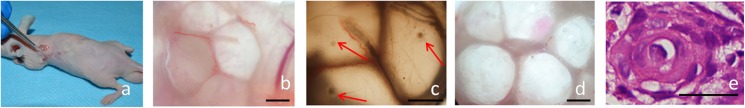
Organ regeneration is becoming a promising choice for many patients; however, many details about the mechanisms underlying organ regeneration remain unknown. As regenerative organs, hair follicles offer a good model to study the mechanisms associated with regenerative medicine. The relevant studies have mainly focused on donor cells, and there are no systematic studies involving the effect of host factors on hair follicle reconstruction. Thus, we intend to explore the effect of host cells on hair follicle reconstruction. Epidermal and dermal cells from red fluorescent protein (RFP) transgenic newborn mice were injected into green fluorescent protein (GFP) transgenic mice. In addition, we wrapped the mixed dermal and epidermal cells from GFP transgenic and RFP transgenic mice by the Cell-in-a-Box kit to form "capsules," so that the cells within would be isolated from host cells. These capsules were cultured in vitro and transplanted in vivo. Fully developed reconstructed hair follicles were observed after the injection of mixed cells. These reconstructed follicles mainly consisted of donor cells, as well as a small number of host cells. The encapsulated cells gradually aggregated into cell spheres in vitro without apparent differentiation towards hair follicles. With respect to the transplanted capsules, concentric circle structures were observed, but no hair follicles or hair shafts formed. When the concentric circle structures were transplanted in vivo, mature hair follicles were observed 30 days later. Host cells were found in the reconstructed hair follicles. Thus, we conclude that host cells participate in the process of hair follicle reconstruction, and they play a vital role in the process, especially for the maturation of reconstructed hair follicles. Furthermore, we established a special hair follicle reconstruction system with the help of capsules: transplant cells were isolated from host, but other factors from host could exchange with cells inside.
Conflict of interest statement
Figures


Similar articles
-
[CO-TRANSPLANTATION OF MOUSE EPIDERMIS AND DERMIS CELLS IN INDUCING HAIR FOLLICLE REGENERATION].Zhongguo Xiu Fu Chong Jian Wai Ke Za Zhi. 2016 Apr;30(4):485-90. Zhongguo Xiu Fu Chong Jian Wai Ke Za Zhi. 2016. PMID: 27411280 Chinese.
-
[Hair follicle regeneration by injection of follicular cells].Zhonghua Zheng Xing Wai Ke Za Zhi. 2012 Jan;28(1):44-9. Zhonghua Zheng Xing Wai Ke Za Zhi. 2012. PMID: 22497189 Chinese.
-
Cultured peribulbar dermal sheath cells can induce hair follicle development and contribute to the dermal sheath and dermal papilla.J Invest Dermatol. 2003 Dec;121(6):1267-75. doi: 10.1111/j.1523-1747.2003.12568.x. J Invest Dermatol. 2003. PMID: 14675169
-
Bioengineering the hair follicle: fringe benefits of stem cell technology.Curr Opin Biotechnol. 2005 Oct;16(5):493-7. doi: 10.1016/j.copbio.2005.08.002. Curr Opin Biotechnol. 2005. PMID: 16098737 Review.
-
Cryopreservation of Hair-Follicle Associated Pluripotent (HAP) Stem Cells Maintains Differentiation and Hair-Growth Potential.Adv Exp Med Biol. 2016;951:191-198. doi: 10.1007/978-3-319-45457-3_16. Adv Exp Med Biol. 2016. PMID: 27837565 Review.
References
-
- Ellis JA, Sinclair RD. Male pattern baldness: current treatments, future prospects. Drug Discov Today. 2008;13: 791–797. doi: 10.1016/j.drudis.2008.05.010 - DOI - PubMed
-
- Ikeda E, Morita R, Nakao K, Ishida K, Nakamura T, Takano-Yamamoto T, et al. Fully functional bioengineered tooth replacement as an organ replacement therapy. Proc Natl Acad Sci U S A. 2009;106: 13475–13480. doi: 10.1073/pnas.0902944106 - DOI - PMC - PubMed
-
- Orlando G, Soker S, Stratta RJ. Organ bioengineering and regeneration as the new Holy Grail for organ transplantation. Ann Surg. 2013;258: 221–232. doi: 10.1097/SLA.0b013e31829c79cf - DOI - PubMed
-
- Bolton EM, Bradley JA. Avoiding immunological rejection in regenerative medicine. Regen Med. 2015;10: 287–304. doi: 10.2217/rme.15.11 - DOI - PubMed
-
- Mao AS, Mooney DJ. Regenerative medicine: current therapies and future directions. Proc Natl Acad Sci U S A. 2015;112: 14452–14459. doi: 10.1073/pnas.1508520112 - DOI - PMC - PubMed
MeSH terms
Substances
LinkOut - more resources
Full Text Sources
Other Literature Sources
Research Materials
