. 2017 Sep 1;196(5):609-620.
doi: 10.1164/rccm.201701-0104OC.

Severe Pneumococcal Pneumonia Causes Acute Cardiac Toxicity and Subsequent Cardiac Remodeling

Affiliations

Severe Pneumococcal Pneumonia Causes Acute Cardiac Toxicity and Subsequent Cardiac Remodeling

Luis F Reyes et al. Am J Respir Crit Care Med. .

Abstract

Rationale: Up to one-third of patients hospitalized with pneumococcal pneumonia experience major adverse cardiac events (MACE) during or after pneumonia. In mice, Streptococcus pneumoniae can invade the myocardium, induce cardiomyocyte death, and disrupt cardiac function following bacteremia, but it is unknown whether the same occurs in humans with severe pneumonia.

Objectives: We sought to determine whether S. pneumoniae can (1) translocate the heart, (2) induce cardiomyocyte death, (3) cause MACE, and (4) induce cardiac scar formation after antibiotic treatment during severe pneumonia using a nonhuman primate (NHP) model.

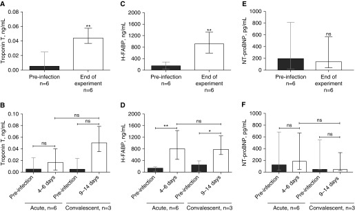
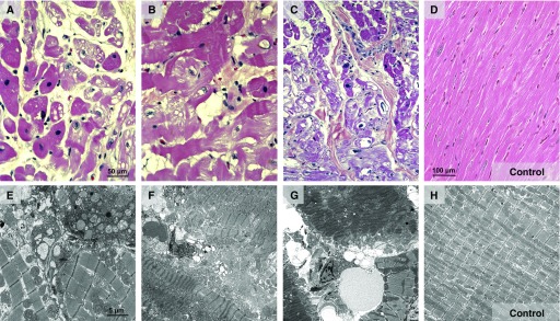
Methods: We examined cardiac tissue from six adult NHPs with severe pneumococcal pneumonia and three uninfected control animals. Three animals were rescued with antibiotics (convalescent animals). Electrocardiographic, echocardiographic, and serum biomarkers of cardiac damage were measured (troponin T, N-terminal pro-brain natriuretic peptide, and heart-type fatty acid binding protein). Histological examination included hematoxylin and eosin staining, immunofluorescence, immunohistochemistry, picrosirius red staining, and transmission electron microscopy. Immunoblots were used to assess the underlying mechanisms.

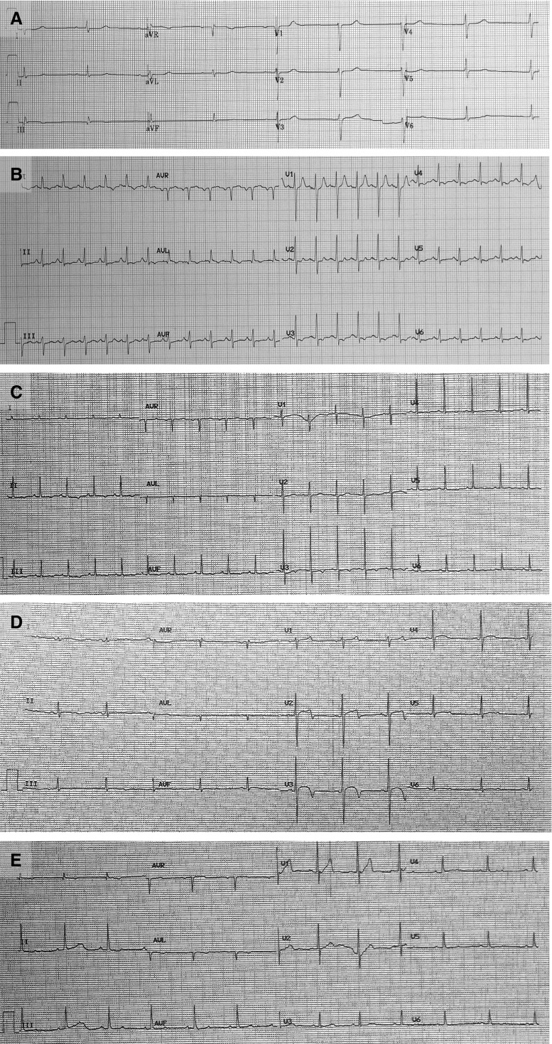
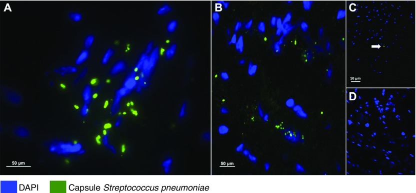
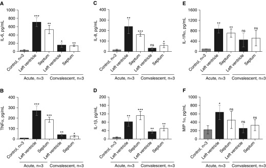
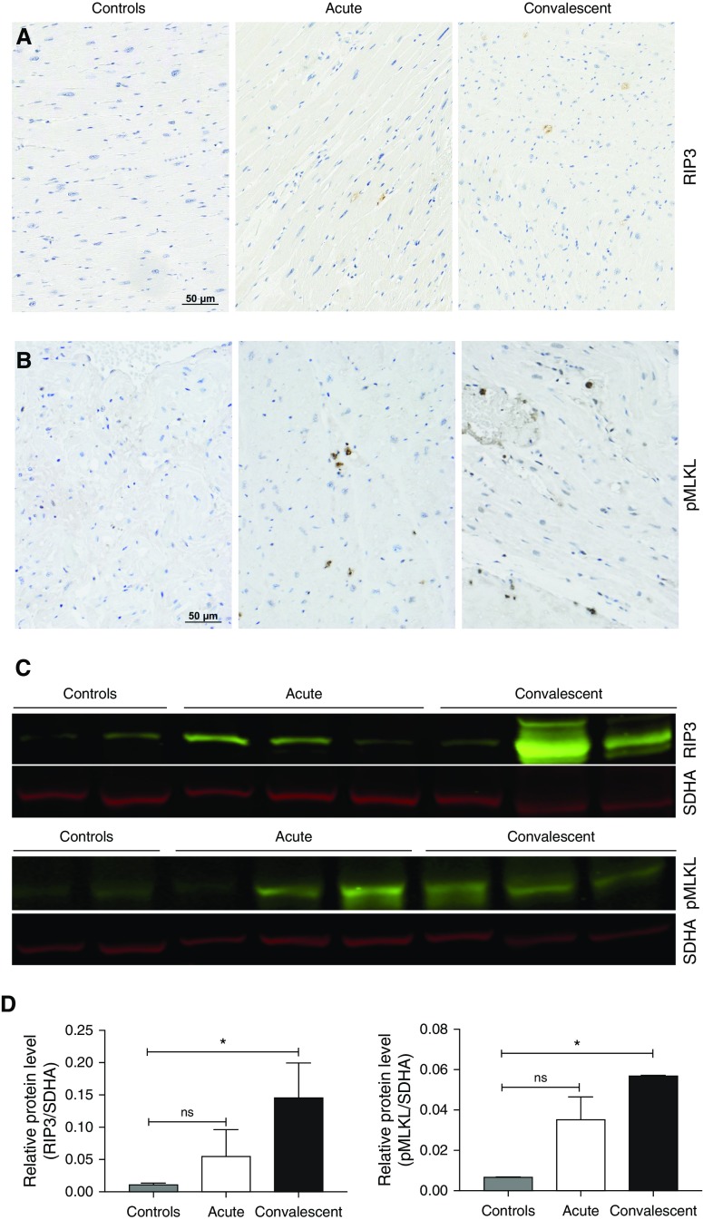
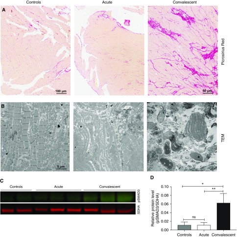
Measurements and main results: Nonspecific ischemic alterations were detected by electrocardiography and echocardiography. Serum levels of troponin T and heart-type fatty acid binding protein were increased (P < 0.05) after pneumococcal infection in both acutely ill and convalescent NHPs. S. pneumoniae was detected in the myocardium of all NHPs with acute severe pneumonia. Necroptosis and apoptosis were detected in the myocardium of both acutely ill and convalescent NHPs. Evidence of cardiac scar formation was observed only in convalescent animals by transmission electron microscopy and picrosirius red staining.

Conclusions: S. pneumoniae invades the myocardium and induces cardiac injury with necroptosis and apoptosis, followed by cardiac scarring after antibiotic therapy, in an NHP model of severe pneumonia.

Keywords: Streptococcus pneumoniae; cardiovascular complications; community-acquired pneumonia; pneumococcal pneumonia.


Figures

Figure 1.
Figure 1.
Nonhuman primates with severe pneumococcal pneumonia developed diffuse, nonspecific repolarization abnormalities. Twelve-lead ECGs were recorded (A) at baseline and (BE) at the end of the experiment. (A) At baseline, all animals were in sinus rhythm with no repolarization abnormalities. After development of pneumococcal pneumonia, the ECGs consistently showed (B and C) sinus tachycardia and (BE) abnormal repolarization (i.e., diffuse T-wave and ST-segment flattening).
Figure 2.
Figure 2.
Serum concentrations of cardiac damage biomarkers were elevated in animals with acute and convalescent pneumococcal pneumonia. The serum biomarkers of cardiac damage, troponin T, heart-type fatty acid binding protein (H-FABP), and N-terminal pro-brain natriuretic peptide (NT-proBNP) were assessed before infection, at Days 4–6 for the acute group, and at Days 9–14 for the convalescent group. Median (n = 6) serum concentrations of (A) troponin T, (C) H-FABP, and (E) NT-proBNP are shown before infection and at the end of the experiment (i.e., before animals were killed). To test whether antibiotic treatment could prevent cardiac damage, the cohort was stratified into nonhuman primates with acute pneumonia (i.e., without antibiotics; n = 6) and nonhuman primates with convalescent pneumonia (i.e., with antibiotics; n = 3). Serum levels of (B) troponin T, (D) H-FABP, and (F) NT-proBNP were assessed for each group before infection, at Days 4–6 for the acute group, and at Days 9–14 for the convalescent group. Values are shown as medians with interquartile ranges. A paired nonparametric Wilcoxon signed-rank test was used to analyze statistical differences among the groups. *P < 0.05; **P < 0.01. ns = not significant.
Figure 3.
Figure 3.
Streptococcus pneumoniae translocates into the heart during pneumonia. Representative images from (A and C) left ventricular and (B) interventricular septal sections from the six nonhuman primates (NHPs) with pneumococcal pneumonia and (D) three uninfected control animals. S. pneumoniae was visualized using immunofluorescent staining with antiserum against serotype 4 capsular polysaccharide (green) and 4′,6-diamidino-2-phenylindole (DAPI; blue) to reveal nucleated tissue cells. (A and B) Bacterial aggregates with diplococci morphology are seen within the left ventricles and septa of NHPs with acute pneumonia. (A) Bacterial cluster conformation indicates active replication. (C) Single-capsule debris (marked with the white arrow) were scattered throughout the left ventricles and in the interventricular septa of convalescent NHPs. (D) Pneumococcal capsules were not identified in the hearts of uninfected control NHPs.
Figure 4.
Figure 4.
Hearts of nonhuman primates (NHPs) with severe pneumonia developed severe cardiac injury with variable pathological characteristics. Representative hematoxylin and eosin–stained and transmission electron microscopic images of heart specimens from (A, B, and E) NHPs with acute pneumococcal pneumonia and (C, F, and G) NHPs with convalescent pneumonia, as well as (D and H) uninfected control NHPs. (A and G) NHPs developed myocyte cytoplasm degenerative changes, including fatty change, vacuolization, and myofibrillar separation. (A and E) Perinuclear and interstitial edema are evident, and several myocyte nuclei contain condensed chromatin, a necrotic change. (B and F) Additionally, in the left ventricle, myocytes with cytoplasmic changes of contraction bands, focal vacuolization, and myocytolysis were identified. (B) The edematous interstitium contained increased small mononuclear cells and some neutrophils. (B and G) Nuclear edema and karyolysis are diffusely evident in cardiomyocytes. (C, E, and F) Widespread myocytolysis, contraction bands, striking nuclear pyknosis, and karyolysis are seen in the septum. (F and G) Finally, degradation of mitochondrial cristae and edema were consistently observed by transmission electron microscopy.
Figure 5.
Figure 5.
Cytokines and chemokines present in homogenized hearts of nonhuman primates (NHPs) with acute or convalescent pneumococcal pneumonia. Median concentrations of proinflammatory cytokines in homogenized left ventricles and interventricular septa of NHPs with acute and convalescent pneumococcal pneumonia are shown. NHPs with acute pneumonia had higher concentrations of (A) IL-6, (B) tumor necrosis factor (TNF)-⍺, (C) IL-8, (D) IL-1β, (E) IL-1R⍺, and (F) macrophage inflammatory protein (MIP)-1⍺ than uninfected NHPs. Convalescent NHPs had lower concentrations of cytokines than NHPs with acute pneumonia, but (A) IL-6, (B) TNF-⍺, (C) IL-8, and (D) IL-1β persisted at higher levels than in uninfected control animals. Values are shown as medians and interquartile ranges. An unpaired nonparametric Mann-Whitney U test was used to analyze statistical differences among the groups. All comparisons were made against uninfected control animals. *P < 0.05; **P < 0.01; ***P < 0.001. ns = not significant.
Figure 6.
Figure 6.
Necroptosis in heart tissue increases after antibiotic treatment. Immunohistochemistry was used to elucidate the cell pathways involved during cell death in nonhuman primates (NHPs) with pneumococcal pneumonia. Immunohistochemistry of heart sections stained for (A) receptor-interacting protein kinase 3 (RIP3) and (B) phosphorylated mixed lineage kinase domain-like pseudokinase (pMLKL). (C) Immunoblot analysis for RIP3, pMLKL, and succinate dehydrogenase complex, subunit A (SDHA; loading control), in heart tissue of infected NHPs versus uninfected control animals. (D) Relative levels of RIP3 and pMLKL protein expression determined by comparing the ratio of the detected band with total protein levels as determined using LI-COR Image Studio Lite software (LI-COR Biosciences, Lincoln, NE). Values are shown as medians and interquartile ranges. Unpaired nonparametric Mann-Whitney U tests were used to analyze statistical differences among the groups. *P < 0.05. ns = not significant.
Figure 7.
Figure 7.
Streptococcus pneumoniae induces de novo collagen deposition in nonhuman primates (NHPs) with convalescent pneumonia. Representative images of heart sections from the six NHPs infected with S. pneumoniae and three uninfected control animals. (A) Picrosirius red was used to identify collagen deposition. NHPs with convalescent pneumonia developed collagen deposition throughout the left ventricle and interventricular septum. (B) Transmission electron microscopy (TEM) was used to confirm collagen deposition, identifying areas with conglomerates of collagen fibers in NHPs that survived their acute pneumonia. (C) The upregulation of pSMAD3 in convalescent NHPs was identified by immunoblot analysis, and (D) relative protein levels (pSMAD3/succinate dehydrogenase complex, subunit A [SDHA; loading control]) were determined using LI-COR Image Studio Lite software. Values are shown as medians and interquartile ranges. Unpaired nonparametric Mann-Whitney U tests were used to analyze statistical differences among the groups. *P < 0.05; **P < 0.01; ns = not significant.

Comment in

References

    1. Wunderink RG, Waterer GW. Community-acquired pneumonia. N Engl J Med. 2014;370:1863. - PubMed
    1. Prina E, Ranzani OT, Torres A. Community-acquired pneumonia. Lancet. 2015;386:1097–1108. - PMC - PubMed
    1. GBD 2015 Mortality and Causes of Death Collaborators. Global, regional, and national life expectancy, all-cause mortality, and cause-specific mortality for 249 causes of death, 1980-2015: a systematic analysis for the Global Burden of Disease Study 2015. Lancet. 2016;388:1459–1544. - PMC - PubMed
    1. Torres A, Sibila O, Ferrer M, Polverino E, Menendez R, Mensa J, Gabarrús A, Sellarés J, Restrepo MI, Anzueto A, et al. Effect of corticosteroids on treatment failure among hospitalized patients with severe community-acquired pneumonia and high inflammatory response: a randomized clinical trial. JAMA. 2015;313:677–686. - PubMed
    1. Musher DM, Thorner AR. Community-acquired pneumonia. N Engl J Med. 2014;371:1619–1628. - PubMed

Publication types