. 2017 Jun 15;2(12):e93683.
doi: 10.1172/jci.insight.93683.

Human vaccination against Plasmodium vivax Duffy-binding protein induces strain-transcending antibodies

Affiliations

Human vaccination against Plasmodium vivax Duffy-binding protein induces strain-transcending antibodies

Ruth O Payne et al. JCI Insight. .

Abstract

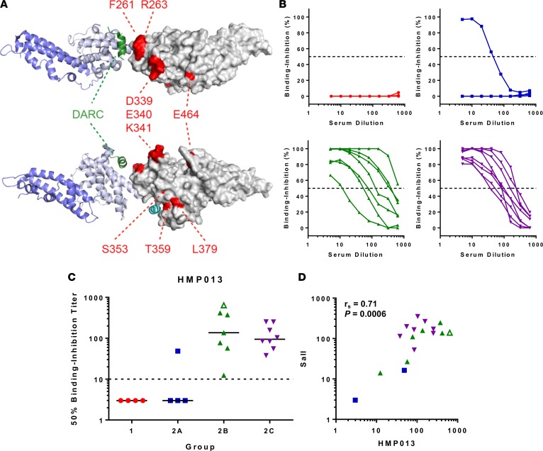
Background: Plasmodium vivax is the most widespread human malaria geographically; however, no effective vaccine exists. Red blood cell invasion by the P. vivax merozoite depends on an interaction between the Duffy antigen receptor for chemokines (DARC) and region II of the parasite's Duffy-binding protein (PvDBP_RII). Naturally acquired binding-inhibitory antibodies against this interaction associate with clinical immunity, but it is unknown whether these responses can be induced by human vaccination.

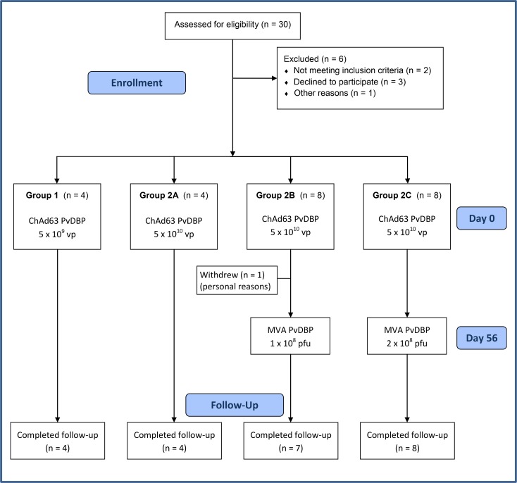
Methods: Safety and immunogenicity of replication-deficient chimpanzee adenovirus serotype 63 (ChAd63) and modified vaccinia virus Ankara (MVA) viral vectored vaccines targeting PvDBP_RII (Salvador I strain) were assessed in an open-label dose-escalation phase Ia study in 24 healthy UK adults. Vaccines were delivered by the intramuscular route in a ChAd63-MVA heterologous prime-boost regimen using an 8-week interval.

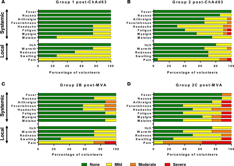
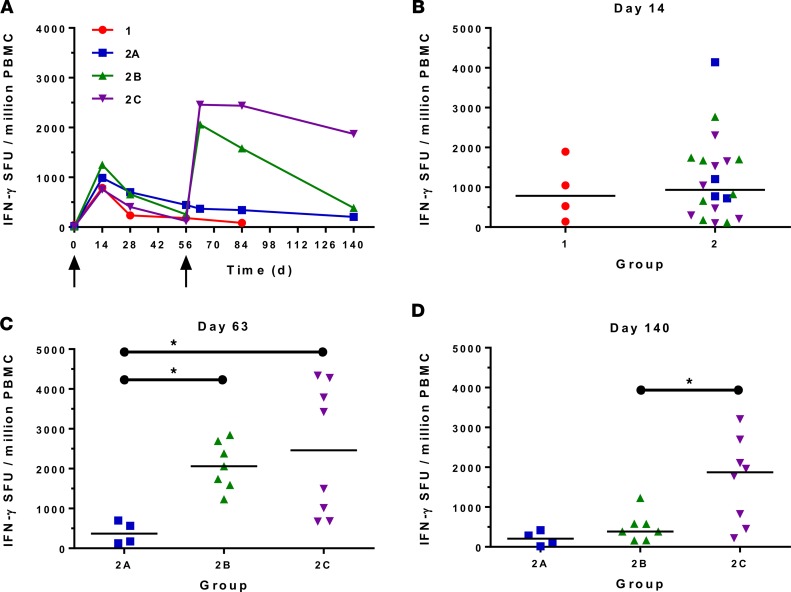
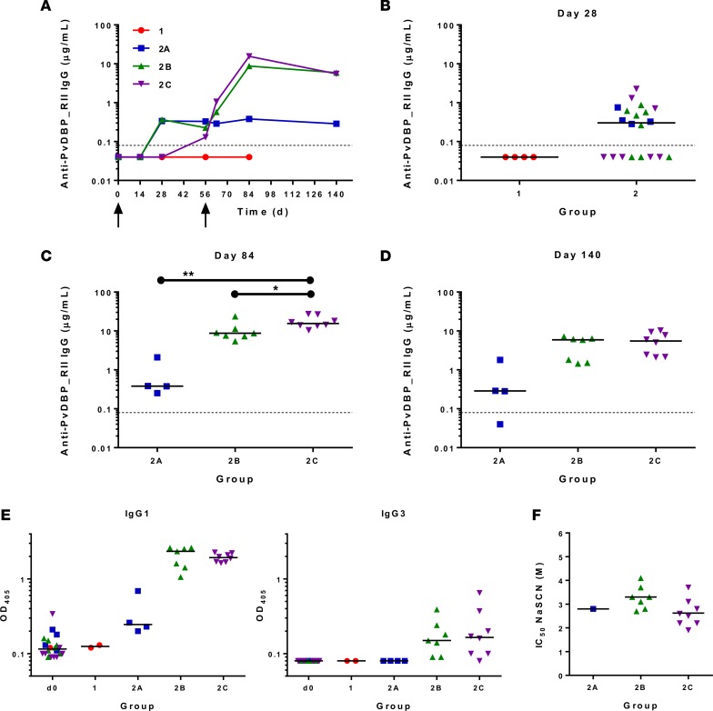
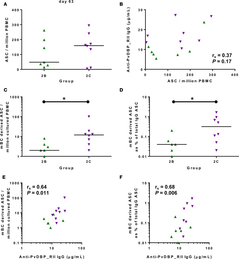
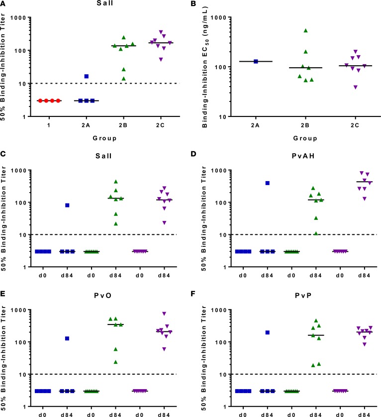
Results: Both vaccines were well tolerated and demonstrated a favorable safety profile in malaria-naive adults. PvDBP_RII-specific ex-vivo IFN-γ T cell, antibody-secreting cell, memory B cell, and serum IgG responses were observed after the MVA boost immunization. Vaccine-induced antibodies inhibited the binding of vaccine homologous and heterologous variants of recombinant PvDBP_RII to the DARC receptor, with median 50% binding-inhibition titers greater than 1:100.

Conclusion: We have demonstrated for the first time to our knowledge that strain-transcending antibodies can be induced against the PvDBP_RII antigen by vaccination in humans. These vaccine candidates warrant further clinical evaluation of efficacy against the blood-stage P. vivax parasite.

Trial registration: Clinicaltrials.gov NCT01816113.

Funding: Support was provided by the UK Medical Research Council, UK National Institute of Health Research Oxford Biomedical Research Centre, and the Wellcome Trust.

Keywords: Infectious disease; Vaccines.


Conflict of interest statement

Conflict of interest: S.C. de Cassan, M.K. Higgins, A.V.S. Hill, and S.J. Draper are named inventors on patent applications (patent nos. GB1413530.5, GB1016471.3, and WO/2008/122811) covering malaria vaccines and immunization regimens. A. Nicosia was an employee of and shareholder in Okairòs (since acquired by GlaxoSmithKline), which is developing vectored vaccines for a number of diseases. T. Jørgensen and W.A. de Jongh are employees of, and W.A. de Jongh is a shareholder in, ExpreS2ion Biotechnologies, which has developed and is marketing the ExpreS2 cell expression platform. C.E. Chitnis is a named inventor on a patent covering PvDBP_RII (patent no. WO/1996/040766).

Figures

Figure 1
Figure 1. VAC051 flow chart of study design and volunteer recruitment.
Recruitment for the VAC051 study took place between May 2013 and February 2014. The final follow-up visit took place in July 2014. All immunizations were administered intramuscularly, with sequential vaccines administered into the deltoid of alternating arms.
Figure 2
Figure 2. Solicited AEs following vaccination with ChAd63 and MVA PvDBP_RII.
The solicited local and systemic adverse events (AEs) recorded for 14 days following ChAd63 PvDBP_RII and for 7 days following MVA PvDBP_RII are shown at the maximum severity reported by all volunteers. (A) Four volunteers received 5 × 10⁹ viral particles (vp) ChAd63 PvDBP_RII (group 1), and (B) 20 received 5 × 10¹⁰ vp (group 2). (C) Seven of the group 2 volunteers went on to receive MVA PvDBP_RII 1 × 10⁸ PFU (group 2B), and (D) 8 received 2 × 10⁸ PFU (group 2C). ChAd63, replication-deficient chimpanzee adenovirus serotype 63; MVA, modified vaccinia virus Ankara; PvDBP_RII, region II of the P. vivax Duffy-binding protein.
Figure 3
Figure 3. Ex-vivo IFN-γ T cell response to vaccination.
(A) Median ex vivo IFN-γ ELISPOT responses in peripheral blood mononuclear cells (PBMCs) to the PvDBP_RII insert (summed response across all the individual peptide pools) shown for all groups. Individual responses are shown in Supplemental Figure 1. Median and individual responses are shown at (B) day 14, (C) day 63, and (D) day 140. Symbols are coded according to group. *P < 0.05. Responses between groups 1 (n = 4) and 2 (n = 20) at day 14, and between groups 2B (n = 7) and 2C (n = 8) at day 140 were assessed by Mann-Whitney test (B and D); responses between groups 2A (n = 4), 2B (n = 7), and 2C (n = 8) at day 63 were assessed by Kruskal-Wallis test with Dunn’s multiple comparison test (C). SFU, spot-forming units; PvDBP_RII, region II of the P. vivax Duffy-binding protein.
Figure 4
Figure 4. Serum antibody response to vaccination.
(A) Median anti–PvDBP_RII serum total IgG responses shown for all groups over time. Individual responses are shown in Supplemental Figure 3. Median and individual responses are shown at (B) day 28, (C) day 84, and (D) day 140. The horizontal dotted line indicates the limit of detection of the assay. (E) Isotype profiles of serum antibody responses were assessed by ELISA. Responses are shown at baseline (d0) and for all groups at day 84. Individual and median responses are shown for IgG1 and IgG3; results for IgG2, IgG4, IgA, and IgM are shown in Supplemental Figure 6. (F) Avidity of serum IgG responses at day 84 was assessed by NaSCN-displacement PvDBP_RII ELISA and is reported as the molar (M) concentration of NaSCN required to reduce the starting OD in the ELISA by 50% (IC50). Symbols are coded according to group. *P < 0.05, **P < 0.01. Responses in groups 2A (n = 4), 2B (n = 7), and 2C (n = 8) were assessed by Kruskal-Wallis test with Dunn’s multiple comparison test; responses between groups 2B and 2C were assessed by Mann-Whitney test (C). PvDBP_RII, region II of the P. vivax Duffy-binding protein.
Figure 5
Figure 5. B cell response to vaccination.
(A) PvDBP_RII–specific antibody-secreting cell (ASC) responses were assessed by ex-vivo ELISPOT using PvDBP_RII protein and frozen peripheral blood mononuclear cells (PBMCs) from the day 63 time point. Individual and median responses are shown for each group and reported as PvDBP_RII–specific ASCs per million PBMCs used in the assay. (B) Correlation of the ASC response versus the concentration of serum anti–PvDBP_RII IgG measured at day 84. (C) PvDBP_RII–specific memory B cell (mBC) responses were assessed by ELISPOT assay using PvDBP_RII protein. Frozen PBMCs were thawed and underwent a 6-day polyclonal restimulation during which ASCs were derived from mBCs, before testing in the assay. Individual and median responses are shown from the day 84 time point and are reported as mBC-derived PvDBP_RII–specific ASCs per million cultured PBMCs or as (D) percentage of total number of IgG-secreting ASCs. (E and F) Correlations of the mBC response versus the concentration of serum anti–PvDBP_RII IgG at day 84. For all correlations, Spearman’s rank correlation coefficient (rs) and P value are shown. *P < 0.05. Responses between groups 2B (n = 7) and 2C (n = 8) were assessed by Mann-Whitney test. PvDBP_RII, region II of the P. vivax Duffy-binding protein.
Figure 6
Figure 6. PvDBP_RII–DARC in vitro binding inhibition.
(A) Day 84 sera from volunteers in groups 1 (n = 4), 2A (n = 4), 2B (n = 7), and 2C (n = 8) were tested for their ability to inhibit binding of recombinant PvDBP_RII (SalI) to the Duffy antigen receptor for chemokines (DARC) using an ELISA-based assay in Oxford. Samples were titrated starting at 1:5 dilution down to 1:640 (Supplemental Figure 7A). Data show the interpolated dilution for each sample that gave 50% binding inhibition. (B) For positive samples in A (n = 16), the concentration of anti–PvDBP_RII (SalI) serum IgG that gives 50% binding inhibition (EC50) was calculated by dividing the serum ELISA μg/ml by the 50% binding-inhibition serum titer. The result is reported in ng/ml. (CF) Day 0 and day 84 sera were assessed as in A using the assay established at ICGEB, India, using 4 recombinant alleles of PvDBP_RII: SalI, PvAH, PvO, and PvP. In all panels, the individual and median results are shown for each group. The dashed line shows an arbitrary cutoff below which negative samples are plotted. PvDBP_RII, region II of the P. vivax Duffy-binding protein; SalI, Salvador I reference strain.
Figure 7
Figure 7. Binding inhibition of the P. vivax HMP013 strain DBP_RII.
(A) The location of polymorphic residues in PvDBP_RII (HMP013 strain) have been marked on a structure of the PvDBP_RII (SalI strain) dimer bound to the Duffy antigen receptor for chemokines (DARC) aa 19–30 (PDB code 4NVU) (24). Two views of the dimer are shown, rotated by 90 degrees around the horizontal axis. One molecule of PvDBP_RII is shown in gray surface representation with polymorphic residues colored in red. The second molecule of PvDBP_RII is in blue cartoon representation with SD3 in a darker blue. The 2 helices from DARC are shown in green and cyan, respectively. (B) Day 84 sera from volunteers in groups 1 (n = 4), 2A (n = 4), 2B (n = 7), and 2C (n = 8) were tested for their ability to inhibit binding of recombinant PvDBP_RII (HMP013) to DARC using the ELISA-based assay in Oxford. Samples were titrated starting at 1:5 dilution down to 1:640. Dashed line indicates 50% binding inhibition. Groups coded by color and symbol. (C) Data show the interpolated dilution for each sample that gave 50% binding inhibition. One sample in group 2B did not reach 50% binding inhibition by 1:640 dilution and is plotted at this final titer with open triangle symbol. (D) Correlation of 50% binding-inhibition titers for the SalI and HMP013 alleles of PvDBP_RII measured using the assay in Oxford. Spearman’s rank correlation coefficient (rs) and P value are shown (n = 19). PvDBP_RII, region II of the P. vivax Duffy-binding protein.

References

    1. Halbroth BR, Draper SJ. Recent developments in malaria vaccinology. Adv Parasitol. 2015;88:1–49. - PubMed
    1. Gething PW, et al. A long neglected world malaria map: Plasmodium vivax endemicity in 2010. PLoS Negl Trop Dis. 2012;6(9):e1814. doi: 10.1371/journal.pntd.0001814. - DOI - PMC - PubMed
    1. Howes RE, et al. Plasmodium vivax transmission in Africa. PLoS Negl Trop Dis. 2015;9(11):e0004222. doi: 10.1371/journal.pntd.0004222. - DOI - PMC - PubMed
    1. Baird JK. Evidence and implications of mortality associated with acute Plasmodium vivax malaria. Clin Microbiol Rev. 2013;26(1):36–57. doi: 10.1128/CMR.00074-12. - DOI - PMC - PubMed
    1. Moorthy VS, Newman RD, Okwo-Bele JM. Malaria vaccine technology roadmap. Lancet. 2013;382(9906):1700–1701. - PubMed

Associated data