. 2017 Jun 15;2(12):e93222.
doi: 10.1172/jci.insight.93222.

Robust memory responses against influenza vaccination in pemphigus patients previously treated with rituximab

Affiliations

Robust memory responses against influenza vaccination in pemphigus patients previously treated with rituximab

Alice Cho et al. JCI Insight. .

Abstract

Rituximab is a therapeutic anti-CD20 monoclonal antibody widely used to treat B cell lymphoma and autoimmune diseases, such as rheumatic arthritis, systemic lupus erythematosus, and autoimmune blistering skin diseases (AIBD). While rituximab fully depletes peripheral blood B cells, it remains unclear whether some preexisting B cell memory to pathogens or vaccines may survive depletion, especially in lymphoid tissues, and if these memory B cells can undergo homeostatic expansion during recovery from depletion. The limited data available on vaccine efficacy in this setting have been derived from rituximab-treated patients receiving concomitant chemotherapy or other potent immunosuppressants. Here, we present an in-depth analysis of seasonal influenza vaccine responses in AIBD patients previously treated with rituximab, who generally did not receive additional therapeutic interventions. We found that, despite a lack of influenza-specific memory B cells in the blood, patients mount robust recall responses to vaccination, comparable to healthy controls, both at a cellular and a serological level. Repertoire analyses of plasmablast responses suggest that they likely derive from a diverse pool of tissue-resident memory cells, refractory to depletion. Overall, these data have important implications for establishing an effective vaccine schedule for AIBD patients and the clinical care of rituximab-treated patients in general and contribute to our basic understanding of maintenance of normal and pathogenic human B cell memory.

Keywords: Immunology; Vaccines.


Conflict of interest statement

Conflict of interest: The authors have declared that no conflict of interest exists.

Figures

Figure 1
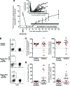
Figure 1. Reconstitution of the B cell compartment after rituximab treatment.
(A) Average kinetics (mean ± SEM) of peripheral blood CD19+ B cells after rituximab therapy. Dotted line represents depletion at 1% B cells of total lymphocytes. The inset graph depicts B cell depletion kinetics for each individual patient. (B) Representative flow plots of CD3CD19+ peripheral B cells, gated on lymphocytes, at time of enrollment. (C) Overall, median frequency and total counts of B cells were comparable between healthy controls and patients. (D) Median frequency and total number of transitional B cells, defined as CD24hiCD38hi, were significantly higher in patients than in healthy controls. Mann-Whitney U test was used to analyze data. *P ≤ 0.05; ***P ≤ 0.001. Black circles represent data from the 2014/15 influenza season; red circles represent data from the 2015/16 influenza season.
Figure 2
Figure 2. Lack of memory B cells in peripheral blood in rituximab-treated patients.
(A) Representative flow cytometry plots showing CD27+ memory B cells (MBCs), gated on CD19+ lymphocytes, at time of enrollment in a healthy control and a patient. (B) The median frequency and total number of MBCs were significantly lower in patients than in healthy controls. (C) Antigen-specific MBCs were stimulated in vitro and detected using an ELISPOT-based assay, as previously described (39, 41). The median frequency of influenza-specific IgG+ MBCs was significantly lower in patients than in healthy controls prior to vaccination. Mann-Whitney U test was used to analyze data. ****P ≤ 0.0001. Black circles represent data from the 2014/15 influenza season; red circles represent data from the 2015/16 influenza season.
Figure 3
Figure 3. Robust vaccine-induced plasmablast responses likely originating from memory recall responses.
(A) Representative flow cytometry plots showing transient expansion of CD27hiCD38hi plasmablasts, gated on CD19+ B cells, induced at day 7 after vaccination. (B) Representative ELISPOT of influenza-specific plasmablasts at day 7 after vaccination. (C) Influenza-specific antibody-secreting cells (ASCs) were measured by ELISPOT at day 0, 7, and 28 after vaccination. The average number of influenza-specific ASCs per million PBMCs was plotted by IgG, IgA, and IgM isotypes. Dotted line represents the limit of detection of the ELISPOT assay. Number in parentheses represents the number of vaccinees who did not have a detectable influenza-specific plasmablast response. (D) Patients who had no history of rituximab treatment prior to enrollment had a significantly higher IgG and IgA plasmablast response to vaccination compared with patients who had received a range of 2–4 cycles of rituximab. Mann-Whitney U test was used to analyze data. *P ≤ 0.05. Black lines represent data from the 2014/15 influenza season; red lines represent data from the 2015/16 influenza season.
Figure 4
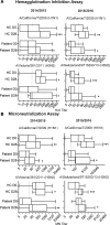
Figure 4. Comparable serological responses to vaccination in patients and healthy controls.
(A) Influenza-specific serum antibody titers determined by HAI. HAI titers were plotted, comparing serum samples from day 0 to day 28 after vaccination. The dotted line represents the limit of detection of the assay at an HAI titer of 5 and seroprotection at an HAI titer of 40. (B) Influenza-neutralizing serum antibody titers determined by microneutralization assay. Titers are graphed as a box-and-whisker plots. Horizontal lines within boxes indicate medians, while widths of boxes represent interquartile ranges. Whiskers show the highest and lowest titer values measured. The experiments were performed in duplicate and reproduced twice. One representative experiment in shown. Wilcoxon paired t test was used to compare day 0 to day 28 after vaccination within cohorts. *P ≤ 0.05; **P ≤ 0.01; ***P ≤ 0.005.
Figure 5
Figure 5. Previous rituximab depletion has no effect on the generation of new influenza-specific memory B cells.
The frequency of influenza-vaccine specific MBCs was measured at baseline and day 28 after vaccination. (A) Representative memory B cell assay. PBMCs were stimulated with a mitogen cocktail, and the frequency of antigen-specific IgG+ MBCs of total IgG+ MBCs was determined by ELISPOT. (B) The frequency of influenza-specific IgG+ MBCs at day 0 and day 28 after vaccination. Only vaccinees that responded to the polyclonal stimulation were included. One-way ANOVA was used to analyze data. *P ≤ 0.05; ***P ≤ 0.001. Black circles represent data from the 2014/15 influenza season; red circles represent data from the 2015/16 influenza season.
Figure 6
Figure 6. Vaccine-induced plasmablasts display comparable repertoire breadth in patients and healthy controls.
Variable genes from plasmablasts induced by the seasonal influenza vaccine were amplified by single-cell PCR and analyzed for (A) number of somatic mutations, (B) CDR3 length, and (C) clonality (shared identical VH gene, JH gene, and CDR3 junction) of class-switched sequences. Each dot represents one individual donor, averaged from 22–46 sequences. (D) Frequency of isotypes in patients and healthy controls. A total of 172 sequences were analyzed from 5 healthy controls, and 286 sequences were analyzed from 8 patients. (E) Overall VH gene usage of class-switched sequences, reported as a frequency of total sequences analyzed from each cohort. (F) VH gene usage of antibodies in clonal groups reported as a frequency of total number of antibodies involved in clonal groups identified. Black circles represent data from the 2014/15 influenza season; red circles represent data from the 2015/16 influenza season.

References

    1. Thompson WW, et al. Influenza-associated hospitalizations in the United States. JAMA. 2004;292(11):1333–1340. doi: 10.1001/jama.292.11.1333. - DOI - PubMed
    1. Thompson WW, et al. Mortality associated with influenza and respiratory syncytial virus in the United States. JAMA. 2003;289(2):179–186. doi: 10.1001/jama.289.2.179. - DOI - PubMed
    1. Kunisaki KM, Janoff EN. Influenza in immunosuppressed populations: a review of infection frequency, morbidity, mortality, and vaccine responses. Lancet Infect Dis. 2009;9(8):493–504. doi: 10.1016/S1473-3099(09)70175-6. - DOI - PMC - PubMed
    1. Govaert TM, Thijs CT, Masurel N, Sprenger MJ, Dinant GJ, Knottnerus JA. The efficacy of influenza vaccination in elderly individuals. A randomized double-blind placebo-controlled trial. JAMA. 1994;272(21):1661–1665. - PubMed
    1. Potter CW, Oxford JS. Determinants of immunity to influenza infection in man. Br Med Bull. 1979;35(1):69–75. doi: 10.1093/oxfordjournals.bmb.a071545. - DOI - PubMed